Acute Sinusitis (Acute Rhinosinusitis): Classification, Clinical Manifestations, Diagnosis, and Treatment
A detailed review of rhinosinusitis, including classification, symptoms, diagnostic approaches, and current treatment strategies.
Anesthesia
Pain management and sedation techniques
Angiology
Arterial and venous pathologies
Cardiology
Acquired and congenital heart diseases
Dentistry
Diseases of teeth, gums, and the oral cavity
Dermatology
Disorders of the skin and subcutaneous tissue
Endocrinology
Disorders of the glands and hormonal imbalance
Gastroenterology
Stomach, intestinal, and digestive diseases
Gynecology
Diseases of female reproductive organs
Hepatology
Liver, gallbladder, and biliary tract diseases
Neurology
Brain, spinal cord, and peripheral nerve disorders
Obstetrics
Pregnancy complications and abnormal fetal positions
Oncology
Cancer types, benign and malignant tumors
Ophthalmology
Conditions affecting the eyes and vision
Otorhinolaryngology
Ear, nose, and throat diseases
Pediatrics
Child health, development, and clinical conditions
Physiology
Biological processes within organs and systems
Pulmonology
Lung and respiratory tract diseases
Traumatology
Acute injuries and musculoskeletal trauma
Urology
Urinary tract and male reproductive disorders

This article is for informational purposes only
The content on this website, including text, graphics, and other materials, is provided for informational purposes only. It is not intended as advice or guidance. Regarding your specific medical condition or treatment, please consult your healthcare provider.
The nasal septum is a bony cartilaginous plate dividing the nasal cavity into two halves. Its pathologic changes, such as nasal septal hematomas, abscesses, or perforations, are serious complications of trauma or infections. These conditions require timely diagnosis because they can lead to serious consequences, including irreversible nasal deformation.
In otorhinolaryngology, three main groups of post-traumatic septal pathologies are distinguished:
In most cases, the common cause of the aforementioned pathologies is mechanical damage, such as household injuries, falls, and traffic accidents.
In rare cases, the cause is surgical interventions in the nasal cavity. These conditions can be caused by chronic slow infections or autoimmune diseases in the nasal cavity, for which patients do not seek help from otorhinolaryngologists for a long time.
Nasal septal hematomas can arise from blunt trauma or a viral infection. Signs of it are hyperemia and increased fragility of the vessels of the mucous membrane, which contribute to spontaneous hemorrhages.
Nasal abscesses are most often a consequence of the suppuration of a hematoma. It can also be formed:
A nasal septal perforation develops due to:
Idiopathic perforations, i.e., when the exact cause cannot be established, also occur.
A hematoma is an accumulation of blood between the cartilage and supracartilage. It occurs within the first 24 hours after an injury. In the nasal cavity on the side of the lesion, a fluctuating, tense bulge of the mucous membrane of a cyanotic (bluish gray) color can be identified.

The hematoma may involve the cartilaginous part or extend into the posterior bony part, although this is extremely rare. It’s also common to encounter a bilateral lesion, in which blood accumulates on both sides.
A nasal septal abscess is an accumulation of purulent contents under the mucous membrane, more often in the area of the septal cartilage. It occurs as a complication of an undrained nasal hematoma or after trauma and intranasal interventions.
In the absence of treatment, the purulent inflammation spreads deeper. The cartilage, deprived of nutrition and in a purulent environment, quickly degrades via chondrolysis. Complication of the condition includes the development of a nasal septal perforation, deformation of the back of the nose (its depression), and spread of inflammation to the periorbital area and intracerebral structures.
When the nasal septum is perforated, the anterior parts in the area of the septal cartilage and Kiesselbach’s plexus are affected. At the site of ulceration of the mucous membrane, a perforation forms in the area of the cartilage, which increases in size over time. Granulations may form along the edges of the perforation.
The appearance of the perforation changes the natural laminar airflow to a turbulent airflow. This leads to the drying of the mucous membrane, the formation of crusts, and impaired ventilation of the nasal cavity and paranasal sinuses.
Perforations are categorized according to their size. The size of the perforation affects treatment tactics and prognosis.
| Perforation type | Perforation size (diameter) |
|---|---|
| Small perforations | up to 0.5 cm |
| Medium perforations | 0.5 to 2 cm |
| Large perforations | >2 cm |
Symptoms depend on the type of pathology and the stage of the process.
A thorough history and routine examination via anterior rhinoscopy are sufficient to clarify the diagnosis in this group of diseases.
Therapeutic tactics are determined by the type of pathological process and are aimed at evacuating the contents, preventing purulent complications, and restoring the integrity of the nasal structures.
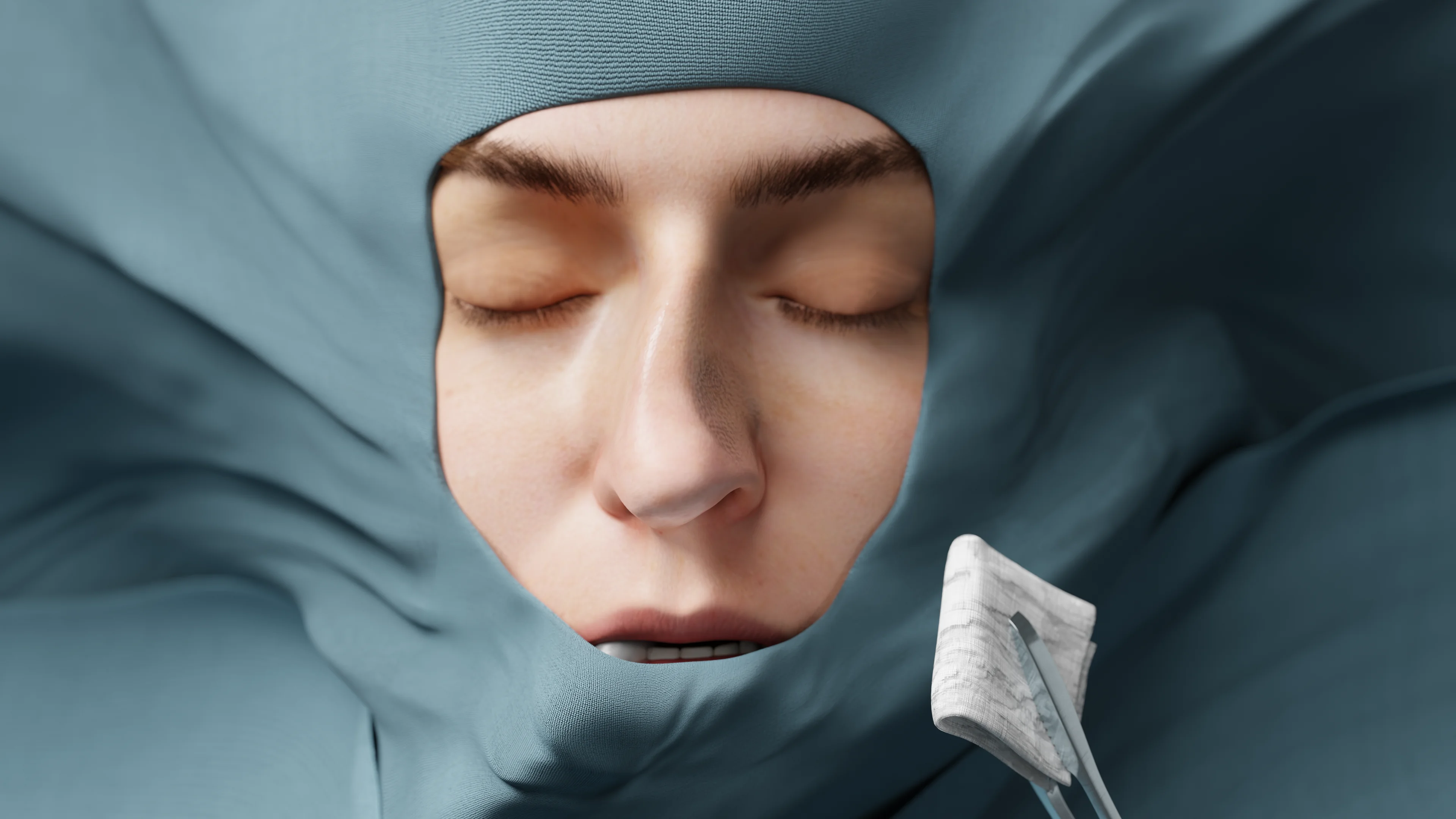
The nasal septal hematoma is punctured with a needle to aspirate the contents, then a tight anterior tamponade is performed to fix and graft the mucous membrane to the underlying structures. When indicated, oral antibacterial therapy is prescribed, and antiseptic solutions are applied to the nasal packing to prevent purulent complications.
With nasal septal abscesses, only surgical treatment is used. A wide incision of the mucous membrane is performed where the swelling is largest, and the pathologic contents are removed.
Closing a nasal septal perforation is currently a difficult task for nose surgeons. To achieve the best effect, the first thing that should be done is identifying the cause of the perforation and attempting to eliminate or minimize it.
1. Non-surgical therapy
To reduce clinical manifestations, it’s recommended to regularly moisten the nasal cavity with saline solutions and soften crusts with ointment preparations. This only prevents the perforation from increasing in size. Doing so does not close it. Silicone implants are also placed in the site of perforation. However, they sometimes contribute to an increase in its size.
2. Surgical Therapy
Rhinoplastyof the nasal septal perforation is performed using autochartilage and mucous membrane on a vascular pedicle. The tissues are then fixed with nasal protectants until it heals.
However, even with surgical correction, it is currently rarely possible to completely eliminate the defect. In some cases, surgical interventions can even contribute to the perforation growing larger.
1. Why does the nasal septum hurt?
2. What is the difference between a hematoma and a nasal septal abscess?
3. What is the danger of a perforated nasal septum?
4. Does a perforation affect the shape of the nose?
5. Can a perforation in the nasal septum heal?
References
1.
VOKA Catalog. [Electronic resource].
https://catalog.voka.io/
2.
Total Otolaryngology—Head and Neck Surgery, Anthony P. Sclafani, Robin A. Dyleski, Michael J. Pitman, Stimson P. Schantz. Thieme Medical Publishers, Inc., 2015. ISBN 978-1-60406-646-3.
3.
Бербом Х. Болезни уха, горла и носа / Ханс Бербом, Оливер Кашке, Тадеус Навка, Эндрю Свифт; пер. с англ. – 2-е изд. – М. : МЕДпреcс-информ, 2016. – 776 с. : ил. ISBN 978-5-00030- 322-1.
4.
Gupta G, Mahajan K. Nasal Septal Hematoma. 2023 Aug 8. In: StatPearls [Internet]. Treasure Island (FL): StatPearls Publishing; 2025 Jan–. PMID: 29261856.
5.
Zielnik-Jurkiewicz B, Olszewska-Sosińska O, Rapiejko P. Leczenie pourazowych krwiaków i ropni przegrody nosa u dzieci [Treatment of the nasal septal hematoma and abscess in children]. Otolaryngol Pol. 2008;62(1):71-5. Polish. doi: 10.1016/S0030-6657(08)70212-9. PMID: 18637425.
6.
Nanu DP, Adelsberg D, Nguyen SA, Radulovich NP, Carr MM. Unmasking Nasal Septal Hematoma/Abscess: A Systematic Review and Meta-analysis. OTO Open. 2024 Oct 8;8(4):e174. doi: 10.1002/oto2.174. PMID: 39381799; PMCID: PMC11460746.
7.
Shaari AL, Patil D, Youssef V, Bebawy G, Shaari DS, Schwartz C, Eloy JA. Nasal Septal Abscesses: A Systematic Review. J Craniofac Surg. 2025 Jan 3. doi: 10.1097/SCS.0000000000011029. Epub ahead of print. PMID: 39750551.
8.
Lanier B, Kai G, Marple B, Wall GM. Pathophysiology and progression of nasal septal perforation. Ann Allergy Asthma Immunol. 2007 Dec;99(6):473-9; quiz 480-1, 521. doi: 10.1016/S1081-1206(10)60373-0. PMID: 18219827.
9.
Downs BW, Sauder HM. Septal Perforation. 2023 Jul 31. In: StatPearls [Internet]. Treasure Island (FL): StatPearls Publishing; 2025 Jan–. PMID: 30725893.
Loading test 6 questions
Table of Contents


Summarize article with AI
Choose your preferable AI assistant:
Link successfully copied to clipboard
Thank you!
Your message is sent!
Our experts will contact you shortly. If you have any additional questions, please contact us at info@voka.io