Odontogenic Sinusitis: Classification, Etiology, Clinical Presentation, Diagnosis, and Treatment
A detailed overview of odontogenic maxillary sinusitis: causes, symptoms of acute and chronic inflammation, diagnosis, and modern treatment approaches.


Anesthesia
Pain management and sedation techniques

Angiology
Arterial and venous pathologies

Cardiology
Acquired and congenital heart diseases

Dentistry
Diseases of teeth, gums, and the oral cavity

Dermatology
Disorders of the skin and subcutaneous tissue

Embryology
Stages of embryogenesis and congenital anomalies

Endocrinology
Disorders of the glands and hormonal imbalance

Gastroenterology
Stomach, intestinal, and digestive diseases

Gynecology
Diseases of female reproductive organs

Hematology
Hematopoiesis and blood-related disorders

Hepatology
Liver, gallbladder, and biliary tract diseases

Histology
Microscopic tissue and cell structures

Infectious diseases
Bacterial, viral, and parasitic infections

Neurology
Brain, spinal cord, and peripheral nerve disorders

Obstetrics
Pregnancy complications and abnormal fetal positions

Oncology
Cancer types, benign and malignant tumors

Ophthalmology
Conditions affecting the eyes and vision

Orthopedics
Bone, joint, and soft tissue disorders

Otorhinolaryngology
Ear, nose, and throat diseases

Pediatrics
Child health, development, and clinical conditions

Physiology
Biological processes within organs and systems

Pulmonology
Lung and respiratory tract diseases

Traumatology
Acute injuries and musculoskeletal trauma

Urology
Urinary tract and male reproductive disorders
Anesthesia
Pain management and sedation techniques
Angiology
Arterial and venous pathologies
Cardiology
Acquired and congenital heart diseases
Dentistry
Diseases of teeth, gums, and the oral cavity
Dermatology
Disorders of the skin and subcutaneous tissue
Embryology
Stages of embryogenesis and congenital anomalies
Endocrinology
Disorders of the glands and hormonal imbalance
Gastroenterology
Stomach, intestinal, and digestive diseases
Gynecology
Diseases of female reproductive organs
Hematology
Hematopoiesis and blood-related disorders
Hepatology
Liver, gallbladder, and biliary tract diseases
Histology
Microscopic tissue and cell structures
Infectious diseases
Bacterial, viral, and parasitic infections
Neurology
Brain, spinal cord, and peripheral nerve disorders
Obstetrics
Pregnancy complications and abnormal fetal positions
Oncology
Cancer types, benign and malignant tumors
Ophthalmology
Conditions affecting the eyes and vision
Orthopedics
Bone, joint, and soft tissue disorders
Otorhinolaryngology
Ear, nose, and throat diseases
Pediatrics
Child health, development, and clinical conditions
Physiology
Biological processes within organs and systems
Pulmonology
Lung and respiratory tract diseases
Traumatology
Acute injuries and musculoskeletal trauma
Urology
Urinary tract and male reproductive disorders
Specialties
AnesthesiaAngiologyCardiologyDentistryDermatologyEmbryologyEndocrinologyGastroenterologyGynecologyHematologyHepatologyHistologyInfectious diseasesNeurologyObstetricsOncologyOphthalmologyOrthopedicsOtorhinolaryngologyPediatricsPhysiologyPulmonologyTraumatologyUrology
This article is for informational purposes only
The content on this website, including text, graphics, and other materials, is provided for informational purposes only. It is not intended as advice or guidance. Regarding your specific medical condition or treatment, please consult your healthcare provider.
Thermal nasal injuries may develop when extremely high or low temperatures damage the skin and underlying tissues of the nose.
Burns are caused by exposure of the nasal area to hot liquids, steam, or high doses of UV radiation (sunburns). The higher the temperature and exposure time of the liquid (steam) is, the more damage a patient can incur.
Nasal burns may be classified by depth of injury:
The first two types of burns heal when the wound surface heals through epithelialization, whereas third- and fourth-degree lesions require scar tissue formation.
Any type of burn triggers severe pain in the affected area.
In first-degree burns, the skin typically appears hyperemic. The burn site is painful upon palpation and turns pale when pressed.
In second-degree burns, hyperemic skin develops serous blisters filled with clear fluid; the wound bed is pink. In such cases, blisters may form within 24 hours from the moment of injury. Tenderness on palpation is also observed.
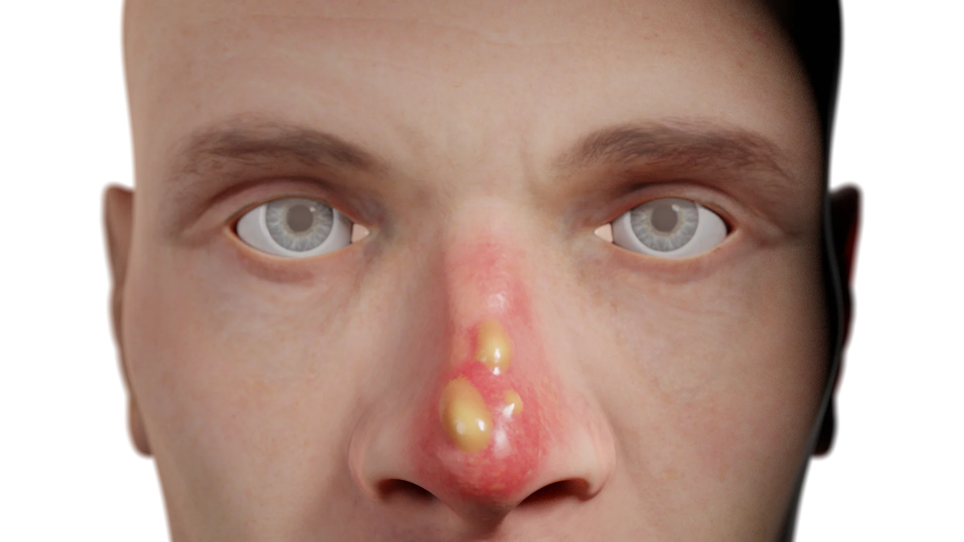
Third-degree burns are characterised by serous hemorrhagic blisters on hyperemic and infiltrated skin. Coagulation necrosis may also be observed. The lesion is moderately painful or painless, hairs can easily be pulled from their roots.
Fourth-degree deep full-thickness burns are characterized by black scab. An exposed bone becomes the wound bed, which is otherwise painless.
Note that nasal burns are rarely observed alone. Other body parts are typically damaged as well.
Classification of nasal burns by depth of injury
| Grade | Lesion depth | Local changes | Soreness | Healing |
|---|---|---|---|---|
| First | Epidermis only | Hyperemic (red) skin. On palpation, there is soreness. The area blanches when pressed | Moderate pain when touched | Epithelization, without scarring |
| Second | Epidermis and superficial layers of dermis (papillary layer) | Hyperemic skin develops serous blisters filled with clear fluid. The bottom of the bubbles are pink. Blisters may appear within 24 hours |
Pronounced | Epithelization, without scarring |
| Third | Epididymis, dermis, fatty layer of subcutaneous tissue, skin appendages | Serous or hemorrhagic blisters, coagulation necrosis. The skin is hyperemic and infiltrated. Hairs are easy to remove |
Reduced or none | Scarring |
| Fourth | All layers of skin, muscles, tendons, cartilages, possibly bones | Black crusts, an exposed bone becomes the wound bed. Extensive necrosis | None | Thick scarring |
Diagnosis is based on a detailed history of the trauma and an ENT exam. During a prehospital check-up, the patient’s airway patency must be assessed. Subsequently, the nasopharyngeal mucosa is regularly reassessed for early detection of burns, reactive mucosal edema, and prevention of respiratory failure. To devise a treatment plan, a healthcare professional should evaluate the depth of the lesion and the percentage of the total body surface area (TBSA) affected. In cases of extensive burns, blood tests, urinalysis, and an ECG are performed to anticipate any systemic complications.
First and foremost, any contact with the damaging factor should be terminated. If a patient has inhaled smoke, humidified oxygen is administered at a prehospital stage.
Whenever possible, the damaged body area should be cooled down and an adequate level of analgesia should be provided. For superficial burns, local therapy is recommended. Dressings with antibacterial ointments are prescribed. Blisters are managed differently depending on their size: small lesions should remain as they are, while larger blisters require excision. Deep burns necessitate skin graft transplantation.
Patients with large and deep burns must be admitted to a specialized burn unit.
Frostbites typically develop on exposed skin areas subjected to low temperatures for extended periods. However, frostbites may occur even at temperatures above zero if the weather conditions are unfavorable (high humidity and strong wind).
Moreover, cold exposure triggers vasoconstriction of peripheral blood vessels to provide more blood flow to the vital organs. The tissues become ischemic, leading to cell death. In some cases, it may be challenging to assess the degree of a frostbite immediately, as typical alterations may develop only a few days later. For instance, blisters are observed within the first 4–8 hours, and necrosis borders become demarcated several days after the event.
Initially, frostbites cause burning sensations and numbness in the affected area. The frostbitten skin is pale, cold to the touch, hard, and mottled. Upon warming, patients start to feel severe pain. Pain intensity correlates with the lesion degree. The skin becomes highly hyperemic and edematous. As described earlier, depending on the degree, frostbites present with specific local skin alterations.
Classification of Nasal Frostbites by Lesion Severity
| Grade | Lesion depth | Local changes | Soreness | Healing |
|---|---|---|---|---|
| First | Superficial layers of the dermis | Skin pallor, numbness, burning, cold area. Hyperemia and edema develop after warming. No blisters form | Moderate pain which intensifies upon warming | Rapid with no scars left |
| Second | Epidermis and superficial layers of dermis (papillary layer) | Hyperemic skin develops serous blisters filled with clear fluid within 4–8 hours. The skin is pale and cold. Edema and hyperemia develop later | Pronounced | Complete, possible hyperpigmentation |
| Third | Deep skin layers | Blisters with hemorrhagic contents, the skin is cyanotic, marbled. The hair on the affected area falls out. Eventual necrosis | Severe, then decreased | Slow, with scarring |
| Fourth | All skin layers and underlying tissues (fatty layer of subcutaneous tissue, muscles, bones) | Dry gangrene, black dry scab, autoamputation It is often accompanied by second- and third-degree signs. Demarcation after a few days | Absent due to nerve endings dying | Slow, with tissue loss and thick scarring |
Diagnosis is based on medical history and patient’s complaints. A healthcare professional should conduct an assessment of both general condition and local changes.
Find more scientifically accurate content on our social media



The damaged body area should be warmed up. Under no circumstances should the skin be massaged. Direct exposure to open flame should also be avoided, as frostbites reduce skin sensitivity to stimuli, which can lead to burns. The affected body part may be immersed in warm water or covered with a dry, warm dressing.
An adequate level of analgesia should be provided; sometimes opioids may be indicated. In cases of general hypothermia, warm fluids and thermal blankets may be used. Locally, blisters may be treated with antibacterial ointments and sterile dressings. To improve tissue perfusion, anticoagulants, antiplatelet agents, and vasodilators are indicated. If dry gangrene is present, amputation is performed after a few days.
1. Why is frostbite degree not always immediately identifiable?
2. Why should frostbitten skin not be rubbed?
3. How does the treatment of burns and frostbite differ?
4. Is it possible to sustain both a burn and frostbite simultaneously?
5. What complications can arise from nasal burns and frostbites?
References
1.
VOKA 3D Anatomy & Pathology – Complete Anatomy and Pathology 3D Atlas. VOKA 3D Anatomy & Pathology.
Available from: https://catalog.voka.io/
2.
Sclafani AP, Dyleski RA, Pitman MJ, Schantz SP. Total otolaryngology—head and neck surgery. New York: Thieme Medical Publishers; 2015. ISBN: 978-1-60406-646-3.
3.
Behrbohm H, Kaschke O, Nawka T, Swift A. Bolezni ukha, gorla i nosa [Ear, nose, and throat diseases]. 2nd ed. Moscow: MEDpress-inform; 2016. 776 p. [In Russian.] ISBN 978-5-00030-322-1.
4.
Zatriqi V, Arifi H, Zatriqi S, Duci S, Rrecaj Sh, Martinaj M. Facial burns – our experience. Mater Sociomed. 2013;25(1):26–7. doi: 10.5455/msm.2013.25.26-27. PMID: 23687458; PMCID: PMC3655731.
5.
Clark C, Ledrick D, Moore A. Facial Burns. [Updated 2023 Jul 3]. In: StatPearls [Internet]. Treasure Island (FL): StatPearls Publishing; 2025 Jan-.
Available from: https://www.ncbi.nlm.nih.gov/books/NBK559290/
6.
Basit H, Wallen TJ, Dudley C. Frostbite. [Updated 2023 Jun 26]. In: StatPearls [Internet]. Treasure Island (FL): StatPearls Publishing; 2025 Jan-.
Available from: https://www.ncbi.nlm.nih.gov/books/NBK536914/
Make VOKA your preferred source
See more VOKA articles in Google Search
Table of Contents









Summarize article with AI
Choose your preferable AI assistant:
Link successfully copied to clipboard
Thank you!
Your message is sent!
Our experts will contact you shortly. If you have any additional questions, please contact us at info@voka.io