Congenital Anomalies of External Ear: Symptoms, Progress, Treatment
Afanasyeva D.Otorhinolaryngologist, MD
20 min read·April 01, 2025
This article is for informational purposes only
The content on this website, including text, graphics, and other materials, is provided for informational purposes only. It is not intended as advice or guidance. Regarding your specific medical condition or treatment, please consult your healthcare provider.
Congenital anomalies of the external ear refer to developmental abnormalities of the auricle or the external acoustic meatus (EAM), also known as the external auditory canal (EAC). This group of pathologies may give rise to a range of dysfunctions and esthetic concerns. The functional issues are often associated with developmental speech delays, while the cosmetic concerns can affect an individual’s satisfaction with their appearance.
3D Animation: Protruding Ears
Definition
In complete external auditory canal atresia (EACA), the EAM is absent.
EAC stenosis refers to the narrowing of the EAM lumen to less than 4 mm in diameter.
Microtia is a hypoplastic malformation of the auricle.
Macrotia, on the other hand, represents an excessively large ear.
Anotia (type 4 microtia) is defined by the complete absence of the auricle.
3D Animation: Type 4 Microtia (Anotia)
Ears may be described as protruding when the angle between the auricle and scalp increases by more than 30 °.
The tubercle of Darwin is a rudimentary auricular tubercle.
Periauricular fistulas appear as blind pathways that open near the auricle or along its helix.
Periauricular skin appendages, or accessory auricles, are nodules located anterior to the auricle. These structures appear as skin outgrowths and may contain cartilaginous tissue.
Classification of Congenital Anomalies of External Ear
External auditory canal atresia — partial (stenosis) and complete.
The auricles start to form at week 5 of pregnancy. They originate from branchial arches 1 and 2 (mandibular and hyoid, respectively), each contributing to the development of three hillocks of His. These hillocks later fuse together, and by week 22, form the auricle has developed its characteristic features, such as the tragus, antitragus, crus of the helix, scaphoid fossa, and antihelix. By the age of 5–6, the auricle is almost fully grown.
Between weeks 12 and 28, the EAM develops. The ectodermal epithelium of the first branchial cleft, located between branchial arches 1 and 2, invaginates to form the EAM.
Epidemiology
The incidence rate of congenital auricular deformities is 1 : 7,000–15,000 newborns, with a higher prevalence in males (2–2.5 times higher). Unilateral alterations account for 85 % of cases, with the right ear being affected in 60 % of patients. There are also some ethnic variations: the prevalence of auricular deformities is higher among populations of Latin American, Asian, and Navajo ancestry.
Microtia is observed in 1 in 7000 live births, with type 3 microtia accounting for 75 % of cases.
3D Animation: Type 3 Microtia
EACA develops in 1 in 10,000–20,000 neonates, with the condition being twice as common in males as in females. Additionally, atresia can be associated with microtia.
Protruding ears affect 0.5–15 % of the population, with approximately 5 % of all cases reported in Europeans. In 66 % of cases, the condition is inherited as an autosomal dominant trait with incomplete penetrance.
Periauricular fistulas may affect 15–43 individuals per 100,000 population. In 27 % of cases, this condition is inherited as an autosomal recessive disorder. Periauricular skin appendages are the most common external ear anomaly, with a prevalence of 5–10 : 1000 newborns.
The majority of congenital ear malformations are associated with genetic disorders, such as Konigsmark syndrome, Moebius syndrome, Treacher — Collins syndrome, Down syndrome, etc.
Etiology and Pathogenesis
Congenital anomalies of the external ear may be caused by genetic mutations (approximately 15 %) and teratogenic factors, however the majority of cases are idiopathies. Teratogenic factors include substances that can affect the auricle include isotretinoin, thalidomide, and ethanol.
Various etiological factors can lead to impaired epithelium invagination of the ectoderm of the first branchial cleft. As a result, atresia develops. However, atresia of the bony portion of the EAC is always secondary and is typically associated with developmental anomalies of the temporal bone. Microtia, on the other hand, occurs when the hillocks of His fail to merge, preventing proper development of the auricle. Furthermore, periauricular fistulas may form due to delayed migration of the epithelium and cartilaginous tissue or excessive tissue growth. Last but not least, periauricular skin appendages may be observed when one of the three hillocks of His from branchial arch 1 fails to migrate. They are typically located on the skin within a triangle formed by the angle of the mouth and the base of the auricle (between the helix and the lobule of the auricle).
3D Animation: Periauricular Skin Appendages
Clinical Manifestations
Congenital anomalies of the external ear typically raise esthetic concerns and/or may affect an individual’s hearing. Patients may feel self-conscious about their appearance if their auricles have abnormal sizes and positioning. Periauricular skin formations are of particular concern. By the age of 5 to 6, patients often start to experience psychological pressure from peers who may comment on or tease them about their distinctive features. All of this may trigger self-esteem issues and hinder social adjustment. Type 3–4 microtia and EACA are associated with impaired hearing of varying degrees on the affected side. However, a unilateral disorder may remain asymptomatic for years due to compensatory mechanisms in the healthy ear. Unilateral EAC stenosis may not affect hearing function or appearance. The main reason patients consult an ENT in such cases is earwax buildup. Due to the anatomically narrow EAM, the cleaning process becomes challenging.
In addition to cosmetic concerns, periauricular fistulas may become inflamed. In such cases, a fistula may exude discharge, and the affected area becomes painful and hyperemic.
3D Animation: Periauricular fistula
Periauricular skin appendages may be located in the buccal region or anterior to the ear. Their shape varies from spherical to lobulated, nodulated, or oval. Unlike other conditions, they do not impair any function and are considered purely a cosmetic imperfection.
Diagnosis is based on physical examination and investigations. They provide extensive data on the location and extent of tissue damage, the nature and severity of hearing impairment, and the middle and internal ear structures affected (if any).
Physical Examination
During a check-up, a healthcare professional evaluates the size and shape of the auricles, their position relative to the skull, and any accessory periauricular formations (fistulas, appendages). The auricle should be examined and compared to the contralateral side. Type 1 microtia is characterized by a smaller auricle, but all identifiable anatomical landmarks (cartilaginous structures) are present.
3D Animation: Type 1 Microtia
In type 2 microtia, the auricle is deformed and reduced in size to varying degrees, with some of the anatomical structures of the auricle not fully differentiated.
3D Animation: Type 2 Microtia
In type 3 microtia, the auricles appear as small, rudimentary, cartilaginous, or skin processes that resemble a peanut. Type 4 microtia is represented by the complete absence of the external ear (both the auricle and the EAC). In rare cases only, the EAC is present. Protruding ears are characterized by an increased auriculocephalic angle (more than 30 °). Macrotia refers to an enlargement of the vertical dimension of the auricle by more than 6 cm compared to the normal size.
3D Animation: Macrotia
The tubercle of Darwin is a rudimentary enlargement of the cartilage in the superior part of the helix.
3D Animation: Tubercle of Darwin
Otoscopy
This method helps determine whether the EAM is present. If not, this is a sign of complete EACA. If the EAM is preserved, a healthcare professional should assess its size, shape, and visibility of the tympanic membrane. Stenosis may be diagnosed if the diameter of the meatus at its broadest part is less than 4 mm. The tympanic membrane is also assessed by its identifying edges, color, and transparency. Note that microtia types 2–4 may be combined with EACA.
These include pure-tone audiometry, audiometry, short-latency auditory evoked potentials (SAEP), and otoacoustic emissions (OAE). Upon examination, the type and degree of hearing loss are determined. Isolated EACA is often characterized by mild to profound conductive hearing loss. It is important to note that both ears must be examined.
Temporal Bone CT
This examination helps determine the severity of EACA, assess the condition of the middle and internal ear, and identify any additional abnormalities. CT is also useful for evaluating the feasibility of surgery using the Jahrsdoerfer grading scale (see Treatment of Congenital Anomalies of External Ear). However, this procedure is associated with high radiation exposure. Since surgery can only be performed later in life, it is not recommended for children under 6 years old who present with a unilateral condition.
Treatment of Congenital Anomalies of External Ear
Regarding the severity of the condition, external ear anomalies may be addressed through the use of a prosthetic ear (external prosthesis), hearing aids, or surgery.
External Auditory Canal Atresia
Hearing Aids
EAC stenosis combined with conductive hearing loss may be corrected with behind-the-ear (BTE) or in-the-ear (ITE) devices. These aids amplify the audible signal and transmit it to the middle ear structures. Bone-anchored hearing aids (BAHA) are implanted in cases of complete EACA and conductive hearing loss. Such devices consist of two parts. The first part is a titanium implant placed into the temporal bone. Over time, the bone integrates with the implant in a process known as osseointegration. The second part is a removable audio processor that clips onto the implant to amplify sounds. The device transmits sound waves from the external environment directly to the cochlea receptors through the skull bone. The process is known as bone conduction. Children typically wear their BAHA with an elastic head band. Children with complete bilateral EACA require early bone conduction hearing aids (around 3–4 months of age). This helps them normally adapt to the society they live in and develop their language skills properly.
Surgery in EACA aims to restore the normal anatomy of the ear. One type of ear surgery is meatoplasty (canaloplasty), which can be combined with tympanoplasty (if necessary), either as a single-stage or a two-stage procedure. When the condition is combined with malformations of the middle ear, the management strategy should be at physician’s discretion. In such cases, a healthcare professional should consider the feasibility of ossiculoplasty.
Indications:
Bilateral EACA (either complete or partial)
Intact cochlea function (conductive hearing loss)
A Jahrsdoerfer score of 7 or higher is associated with better outcomes postsurgery, while a score of 6 is considered a boundary value. This scale is a sum of scores, with 10 being the highest, derived from the patient’s temporal bone CT results on the affected side.
The Jahrsdoerfer grading scale:
Anatomical structure present
Score
Incudomallear joint
1
Incudostapedial joint
1
Oval window
1
Round window
1
Mastoid process pneumatization
1
Middle ear cavity pneumatization
1
External ear
1
Facial nerve
1
Stapes
2
Contraindications:
Unilateral atresia
Age under 5–6 years
Step-by-Step Method Description: The temporal bone is exposed via a postauricular or anterior access, while the temporal fascia is harvested and preserved. Intraoperative neuromonitoring of the facial nerve is recommended throughout the procedure. In complete EACA, a new pathway toward the tympanic cavity is created using a burr. The resulting EAM should have a diameter of 12 mm. Next, the preserved auditory ossicles are exposed. A thin layer donor cartilage and temporal fascia are laid over the malleus. At the same time, a full-thickness skin graft is harvested from the anterior thigh. After the new EAC has been formed, the skin graft is placed around its circumference to cover the exposed bone and the neotympanic membrane (temporal fascia).
A stenotic EAC may require enlargement to achieve an adequate diameter. The skin is harvested in advance to later cover the walls of the newly formed canal. Finally, the EAC is tightly packed for the next 10–14 days.
Outcomes: An anatomical, sound-conducting pathway is formed, improving the patient’s hearing.
Complications and Disadvantages: Restenosis. Possible failure to improve hearing.
Find more scientifically accurate content on our social media
Subscribe and don’t miss out the latest resources
Microtia
A common nonsurgical option for managing microtia types 3–4 is a prosthetic ear (external prosthesis). During this procedure, specialists shape a silicone auricular prosthesis based on an impression of the healthy ear. Prosthetic ears can look very natural since they are made of materials that look similar to the color and structure of the patient’s skin. They can be attached to the affected site with either medical grade glue, clips, or osseointegrated magnets. This option is not age-limited, with material intolerance being the only contraindication. The main advantage is that the technique is simple and time-efficient.
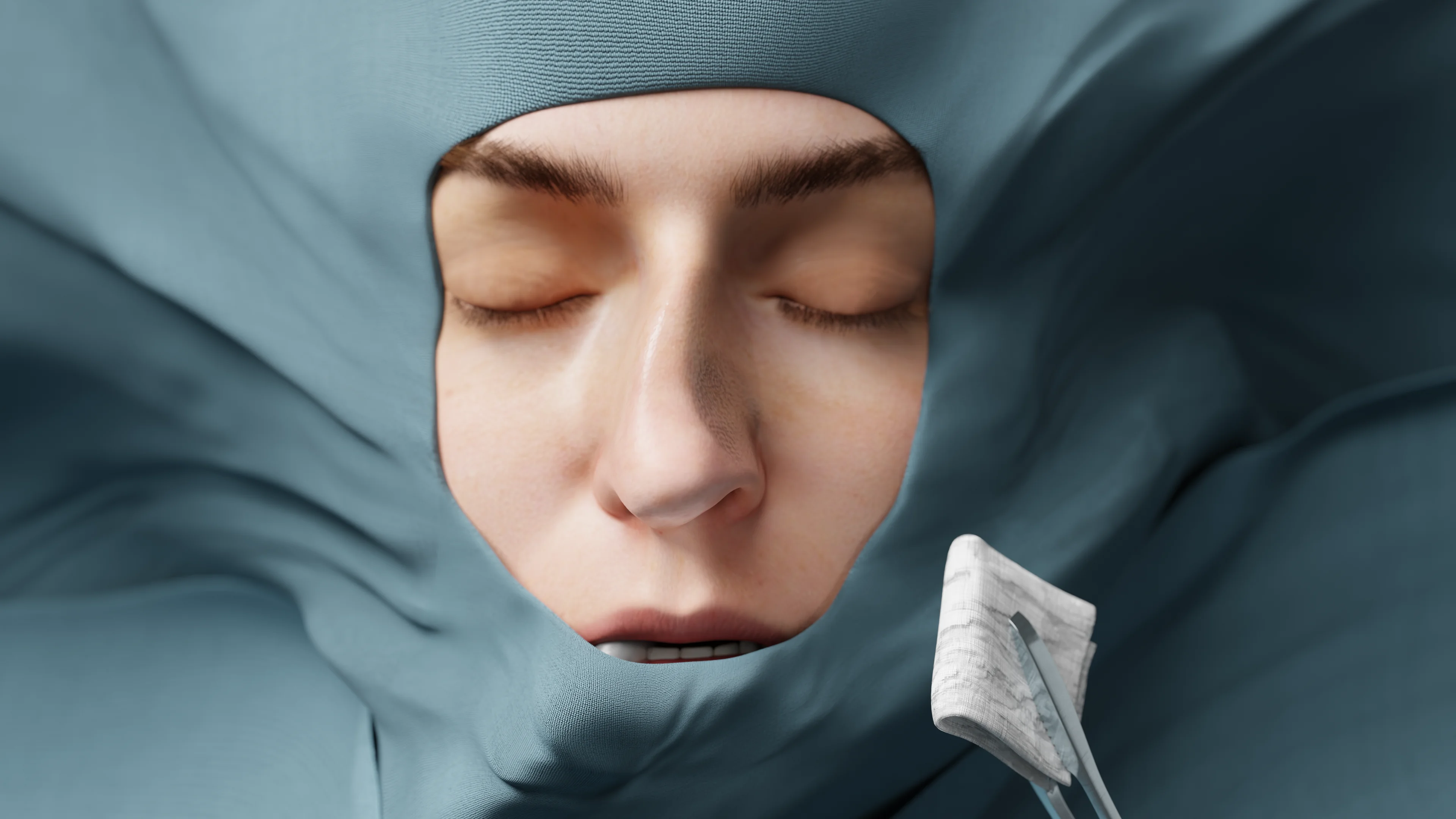
Sometimes, microtia may necessitate surgical ear correction, known as auriculoplasty. During the procedure, a person’s own cartilage (autografting) or an artificial ear model is used to restore the auricle. Indications: Microtia types 3–4. Contraindications: Age under 5–6 years. This limitation is based on the assumption that by this age, ears have reached approximately 85 % of their maximal size, making it possible to model the most suitable transplant. Furthermore, there is enough costal cartilage available for the procedure.
Step-by-Step Method Description: First, autografting requires an impression of the healthy ear, which is used to shape the absent auricle. A surgeon removes fragments 6, 7, and 8 of the costal cartilage, along with the perichondrium (7–9 cm in length). These tissues are then used to form the framework of the future auricle. Sutures are placed in different directions to create anatomical curves (such as helix, antihelix, etc.). The skin in the temporal region, where the new auricle will be positioned, is carefully separated from the underlying tissues. The transplant is inserted into the pocket formed by the skin to reapproximate it. The second stage happens 3–4 months later. The skin is incised along the posterior edge of the helix, and the auricle as well as the skin is separated along the medial surface, The structure is then positioned as needed. A full-thickness skin graft, harvested from the thigh region, is placed onto the posterior surface of the newly formed auricle and the resulting defect in the temporal region. A pressure dressing is applied to the postauricular area and should be changed regularly until full recovery. The next steps may include the formation of the tragus and EAC, if needed.
Medical artificial implants are also placed where the future auricle should be and covered with a skin graft. Outcomes: A properly shaped auricle with no visible cosmetic defect. Complications and Disadvantages: Possible hypertrophic scars, transplant deformation. Cellulitis or chondritis, though extremely rare. An ideal copy of the healthy ear may be challenging to create, which can also be seen as a drawback.
Otoplasty may be performed to manage macrotia and protruding ears.
3D Animation: Otoplasty for Protruding Ear
Indications: Macrotia; protruding ears.
Contraindications: A concomitant medical condition; age under 6 years.
Step-by-Step Method Description: The skin is separated along the posterior surface of the auricle. Depending on the condition of the auricle, the cartilage is dissected and/or sutured in different directions to shape its curves. The skin is then closed using cosmetic sutures. A pressure dressing is applied. The procedure may be performed on the other ear if necessary.
Outcomes: An auricle of a desired shape with no visible cosmetic defect.
Complications and Disadvantages: Possible hypertrophic scars, auricle deformation. Cellulitis or chondritis, though extremely rare.
Periauricular formations (fistulas, appendages) are typically treated surgically. Indications: Esthetic dissatisfaction. Recurrent inflammation of the fistulous passage . Contraindications: A concomitant medical condition. Acute inflammation of the fistula . Step-by-Step Method Description: In cases of a fistula, an elliptical skin incision around its outlet is performed. The fistula is dissected along its entire length within the healthy tissue. Sometimes, to better localize the fistula, a contrast medium (such as methylene blue) may be injected. The wound edges are then sutured. Appendages are removed with a scalpel or laser. Outcomes: No anatomical defects Complications and Disadvantages: A possibility of incomplete removal of the fistulous passage and subsequent infection.
FAQ
1. Which types of hearing aids are used in EAC stenosis?
Behind-the-ear (BTE) or in-the-ear (ITE) devices are used in EAC stenosis. These aids amplify the audible signal and transmit it to the middle ear structures, compensating for conductive hearing loss.
2. How do implanted bone-anchored hearing aids (BAHA) function?
An implanted hearing aid consists of two parts: a titanium implant placed into the temporal bone and an audio processor. The bone integrates with the implant in a process known as osseointegration. The audio processor amplifies sounds and transmits them through the bone to the cochlea.
3. When is surgery indicated in EACA?
Surgery is typically indicated in cases of bilateral EACA (either complete or partial) when the cochlea function is preserved and the score according to the Jahrsdoerfer grading scale is at least 7.
4. What complications may develop following surgery for EACA?
The major complications may be restenosis (recurrent narrowing of the EAM) and a lack of significant hearing improvement.
List of Sources
1.
VOKA Catalog.
https://catalog.voka.io/
2.
Georgakopoulos B, Zafar Gondal A. Embryology, Ear Congenital Malformations. [Updated 2023 May 1]. In: StatPearls [Internet]. Treasure Island (FL): StatPearls Publishing; 2025 Jan-. PMID: 31424840
3.
Jeffrey Liaw, Vijay A. Patel, Michele M. Carr, Congenital anomalies of the external ear, Operative Techniques in Otolaryngology-Head and Neck Surgery, Volume 28, Issue 2, 2017, Pages 72-76, ISSN 1043-1810, doi.org/10.1016/j.otot.2017.03.012.
4.
Total Otolaryngology-Head and Neck Surgery, Anthony P. Sclafani, Robin A. Dyleski, Michael J. Pitman, Stimson P. Schantz. Thieme Medical Publishers, Inc, 2015. ISBN 978-1-60406-646-3
5.
Luquetti DV, Heike CL, Hing AV, Cunningham ML, Cox TC. Microtia: epidemiology and genetics. Am J Med Genet A. 2012 Jan;158A(1):124-39. doi.org/10.1002/ajmg.a.34352. Epub 2011 Nov 21. PMID: 22106030; PMCID: PMC3482263
6.
Park H, Seong J, Park H, Yeo H. Standardized surgical strategy for the treatment of preauricular sinus to reduce recurrence. Arch Craniofac Surg. 2023 Oct;24(5):223-229. doi: doi.org/10.7181/acfs.2023.00423 Epub 2023 Oct 20. PMID: 37919909; PMCID: PMC10622947.
7.
Ear Microtia James Andrews; Avery A. Kopacz; Marc H. Hohman. StatPearls. March 1, 2024. PMID: 33085390