Sternum anatomy quiz
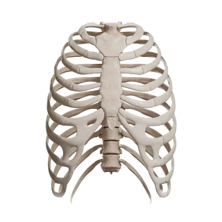
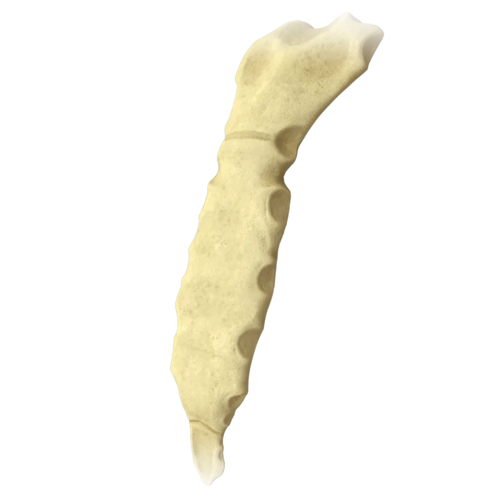
The quiz covers the anatomy of the sternum: its parts, joints, synchondroses, rib attachments, ligaments, and blood supply.
1/20
bold
text
1. What three parts are distinguished in the sternum?
-
Manubrium, body and xiphoid process
The sternum (sternum) consists of three parts: the manubrium (manubrium sterni), the body (corpus sterni), and the xiphoid process (processus xiphoideus). This is the classical division accepted in Terminologia Anatomica.
-
Base, body, and apex
The sternum (sternum) consists of three parts: the manubrium (manubrium sterni), the body (corpus sterni), and the xiphoid process (processus xiphoideus). This is the classical division accepted in Terminologia Anatomica.
-
Manubrium, neck, and xiphoid process
The sternum (sternum) consists of three parts: the manubrium (manubrium sterni), the body (corpus sterni), and the xiphoid process (processus xiphoideus). This is the classical division accepted in Terminologia Anatomica.
-
Head, body, and tail
The sternum (sternum) consists of three parts: the manubrium (manubrium sterni), the body (corpus sterni), and the xiphoid process (processus xiphoideus). This is the classical division accepted in Terminologia Anatomica.
-
I find it difficult to answer
The sternum (sternum) consists of three parts: the manubrium (manubrium sterni), the body (corpus sterni), and the xiphoid process (processus xiphoideus). This is the classical division accepted in Terminologia Anatomica.
2. What is the cartilaginous connection between the manubrium and the body of the sternum called?
-
Manubriosternal symphysis
The connection between the manubrium and the body of the sternum is called the manubriosternal synchondrosis (synchondrosis manubriosternalis). In adulthood, it frequently ossifies and becomes a synostosis; however, it is primarily classified as a synchondrosis.
-
Manubriosternal synchondrosis
The connection between the manubrium and the body of the sternum is called the manubriosternal synchondrosis (synchondrosis manubriosternalis). In adulthood, it frequently ossifies and becomes a synostosis; however, it is primarily classified as a synchondrosis.
-
Sternal syndesmosis
The connection between the manubrium and the body of the sternum is called the manubriosternal synchondrosis (synchondrosis manubriosternalis). In adulthood, it frequently ossifies and becomes a synostosis; however, it is primarily classified as a synchondrosis.
-
Sternocostal joint
The connection between the manubrium and the body of the sternum is called the manubriosternal synchondrosis (synchondrosis manubriosternalis). In adulthood, it frequently ossifies and becomes a synostosis; however, it is primarily classified as a synchondrosis.
-
I find it difficult to answer
The connection between the manubrium and the body of the sternum is called the manubriosternal synchondrosis (synchondrosis manubriosternalis). In adulthood, it frequently ossifies and becomes a synostosis; however, it is primarily classified as a synchondrosis.
3. Where is the jugular notch (incisura jugularis) located on the sternum?
-
On the superior border of the body of the sternum
The jugular notch (incisura jugularis) is located on the superior border of the manubrium of the sternum, along the midline between the clavicular notches. It serves as a landmark for determining the level of the second thoracic vertebra.
-
On the inferior border of the manubrium of the sternum
The jugular notch (incisura jugularis) is located on the superior border of the manubrium of the sternum, along the midline between the clavicular notches. It serves as a landmark for determining the level of the second thoracic vertebra.
-
On the superior border of the manubrium of the sternum, along the midline
The jugular notch (incisura jugularis) is located on the superior border of the manubrium of the sternum, along the midline between the clavicular notches. It serves as a landmark for determining the level of the second thoracic vertebra.
-
On the lateral surface of the body of the sternum
The jugular notch (incisura jugularis) is located on the superior border of the manubrium of the sternum, along the midline between the clavicular notches. It serves as a landmark for determining the level of the second thoracic vertebra.
-
I find it difficult to answer
The jugular notch (incisura jugularis) is located on the superior border of the manubrium of the sternum, along the midline between the clavicular notches. It serves as a landmark for determining the level of the second thoracic vertebra.
4. To what type of joint does the sternocostal joint of the second rib belong?
-
Synchondrosis
The sternocostal joint of the second rib comprises intra-articular cartilage (fibrocartilago interarticularis), which divides the joint cavity into two compartments. The joints of ribs III–VII are simple, without intra-articular cartilage.
-
Syndesmosis
The sternocostal joint of the second rib comprises intra-articular cartilage (fibrocartilago interarticularis), which divides the joint cavity into two compartments. The joints of ribs III–VII are simple, without intra-articular cartilage.
-
Diarthrosis (simple joint)
The sternocostal joint of the second rib comprises intra-articular cartilage (fibrocartilago interarticularis), which divides the joint cavity into two compartments. The joints of ribs III–VII are simple, without intra-articular cartilage.
-
Diarthrosis with intra-articular cartilage
The sternocostal joint of the second rib comprises intra-articular cartilage (fibrocartilago interarticularis), which divides the joint cavity into two compartments. The joints of ribs III–VII are simple, without intra-articular cartilage.
-
I find it difficult to answer
The sternocostal joint of the second rib comprises intra-articular cartilage (fibrocartilago interarticularis), which divides the joint cavity into two compartments. The joints of ribs III–VII are simple, without intra-articular cartilage.
5. Which rib articulates with the manubrium of the sternum at the level of its inferior border (angle of Louis)?
-
First rib
The angle of Louis (angulus Ludovici, or angulus sterni) is the angle between the manubrium and the body of the sternum. The cartilage of the second rib attaches to the sternum precisely at the level of this angle, making it an important anatomical landmark.
-
Second rib
The angle of Louis (angulus Ludovici, or angulus sterni) is the angle between the manubrium and the body of the sternum. The cartilage of the second rib attaches to the sternum precisely at the level of this angle, making it an important anatomical landmark.
-
Third rib
The angle of Louis (angulus Ludovici, or angulus sterni) is the angle between the manubrium and the body of the sternum. The cartilage of the second rib attaches to the sternum precisely at the level of this angle, making it an important anatomical landmark.
-
IV rib
The angle of Louis (angulus Ludovici, or angulus sterni) is the angle between the manubrium and the body of the sternum. The cartilage of the second rib attaches to the sternum precisely at the level of this angle, making it an important anatomical landmark.
-
I find it difficult to answer
The angle of Louis (angulus Ludovici, or angulus sterni) is the angle between the manubrium and the body of the sternum. The cartilage of the second rib attaches to the sternum precisely at the level of this angle, making it an important anatomical landmark.
6. The cartilages of how many ribs articulate directly with the sternum?
-
5 ribs (I–V)
The cartilages of the first seven ribs (I–VII) articulate directly with the sternum. These ribs are called true ribs (costae verae). The cartilages of ribs VIII–X articulate with the overlying costal cartilages rather than directly with the sternum.
-
6 ribs (I–VI)
The cartilages of the first seven ribs (I–VII) articulate directly with the sternum. These ribs are called true ribs (costae verae). The cartilages of ribs VIII–X articulate with the overlying costal cartilages rather than directly with the sternum.
-
7 ribs (I–VII)
The cartilages of the first seven ribs (I–VII) articulate directly with the sternum. These ribs are called true ribs (costae verae). The cartilages of ribs VIII–X articulate with the overlying costal cartilages rather than directly with the sternum.
-
8 ribs (I–VIII)
The cartilages of the first seven ribs (I–VII) articulate directly with the sternum. These ribs are called true ribs (costae verae). The cartilages of ribs VIII–X articulate with the overlying costal cartilages rather than directly with the sternum.
-
I find it difficult to answer
The cartilages of the first seven ribs (I–VII) articulate directly with the sternum. These ribs are called true ribs (costae verae). The cartilages of ribs VIII–X articulate with the overlying costal cartilages rather than directly with the sternum.
7. What is the notch on the manubrium of the sternum for articulation with the clavicle called?
-
Jugular notch
On each side of the manubrium of the sternum is a clavicular notch (incisura clavicularis), which forms the sternal component of the sternoclavicular joint. The jugular notch (incisura jugularis) is located between them along the midline.
-
Costal notch
On each side of the manubrium of the sternum is a clavicular notch (incisura clavicularis), which forms the sternal component of the sternoclavicular joint. The jugular notch (incisura jugularis) is located between them along the midline.
-
Clavicular notch
On each side of the manubrium of the sternum is a clavicular notch (incisura clavicularis), which forms the sternal component of the sternoclavicular joint. The jugular notch (incisura jugularis) is located between them along the midline.
-
Intercostal notch
On each side of the manubrium of the sternum is a clavicular notch (incisura clavicularis), which forms the sternal component of the sternoclavicular joint. The jugular notch (incisura jugularis) is located between them along the midline.
-
I find it difficult to answer
On each side of the manubrium of the sternum is a clavicular notch (incisura clavicularis), which forms the sternal component of the sternoclavicular joint. The jugular notch (incisura jugularis) is located between them along the midline.
8. What ligament connects the costal cartilage of the first rib directly to the sternum without forming a synovial joint?
-
Radiate sternocostal ligament
The connection of the first rib to the manubrium of the sternum is a synchondrosis (synchondrosis costosternalis I)—a continuous cartilaginous connection without a joint cavity. All the other true ribs form diarthroses with the sternum.
-
Intra-articular sternocostal ligament
The connection of the first rib to the manubrium of the sternum is a synchondrosis (synchondrosis costosternalis I)—a continuous cartilaginous connection without a joint cavity. All the other true ribs form diarthroses with the sternum.
-
Synchondrosis (synchondrosis costosternalis I)
The connection of the first rib to the manubrium of the sternum is a synchondrosis (synchondrosis costosternalis I)—a continuous cartilaginous connection without a joint cavity. All the other true ribs form diarthroses with the sternum.
-
Sternal membrane
The connection of the first rib to the manubrium of the sternum is a synchondrosis (synchondrosis costosternalis I)—a continuous cartilaginous connection without a joint cavity. All the other true ribs form diarthroses with the sternum.
-
I find it difficult to answer
The connection of the first rib to the manubrium of the sternum is a synchondrosis (synchondrosis costosternalis I)—a continuous cartilaginous connection without a joint cavity. All the other true ribs form diarthroses with the sternum.
9. The radiate sternocostal ligaments (ligg. sternocostalia radiata) are located on the:
-
Posterior surface of the sternum only
The radiate sternocostal ligaments are located on both the anterior and posterior surfaces of the sternocostal joints, interweaving with the periosteal fibers and forming the sternal membrane (membrana sterni) on the anterior surface of the sternum.
-
Anterior surface of the sternum only
The radiate sternocostal ligaments are located on both the anterior and posterior surfaces of the sternocostal joints, interweaving with the periosteal fibers and forming the sternal membrane (membrana sterni) on the anterior surface of the sternum.
-
Anterior and posterior surfaces of the sternum
The radiate sternocostal ligaments are located on both the anterior and posterior surfaces of the sternocostal joints, interweaving with the periosteal fibers and forming the sternal membrane (membrana sterni) on the anterior surface of the sternum.
-
Lateral surfaces of the manubrium of the sternum
The radiate sternocostal ligaments are located on both the anterior and posterior surfaces of the sternocostal joints, interweaving with the periosteal fibers and forming the sternal membrane (membrana sterni) on the anterior surface of the sternum.
-
I find it difficult to answer
The radiate sternocostal ligaments are located on both the anterior and posterior surfaces of the sternocostal joints, interweaving with the periosteal fibers and forming the sternal membrane (membrana sterni) on the anterior surface of the sternum.
10. To which bone is the xiphoid process of the sternum connected by a synchondrosis?
-
To the body of the sternum
The xiphoid process (processus xiphoideus) is connected to the body of the sternum by the xiphisternal synchondrosis (synchondrosis xiphosternalis). In older age, this synchondrosis usually ossifies.
-
To the cartilage of the seventh rib
The xiphoid process (processus xiphoideus) is connected to the body of the sternum by the xiphisternal synchondrosis (synchondrosis xiphosternalis). In older age, this synchondrosis usually ossifies.
-
To the manubrium of the sternum
The xiphoid process (processus xiphoideus) is connected to the body of the sternum by the xiphisternal synchondrosis (synchondrosis xiphosternalis). In older age, this synchondrosis usually ossifies.
-
To the sixth rib
The xiphoid process (processus xiphoideus) is connected to the body of the sternum by the xiphisternal synchondrosis (synchondrosis xiphosternalis). In older age, this synchondrosis usually ossifies.
-
I find it difficult to answer
The xiphoid process (processus xiphoideus) is connected to the body of the sternum by the xiphisternal synchondrosis (synchondrosis xiphosternalis). In older age, this synchondrosis usually ossifies.
11. Where does the sternocleidomastoid muscle (m. sternocleidomastoideus) originate from the sternum?
-
From the body of the sternum
The sternal head (caput sternale) of the sternocleidomastoid muscle originates from the anterior surface of the manubrium of the sternum. The clavicular head originates from the medial third of the clavicle. The two heads converge and attach to the mastoid process of the temporal bone.
-
From the manubrium of the sternum—from the manubrium
The sternal head (caput sternale) of the sternocleidomastoid muscle originates from the anterior surface of the manubrium of the sternum. The clavicular head originates from the medial third of the clavicle. The two heads converge and attach to the mastoid process of the temporal bone.
-
From the clavicular notch
The sternal head (caput sternale) of the sternocleidomastoid muscle originates from the anterior surface of the manubrium of the sternum. The clavicular head originates from the medial third of the clavicle. The two heads converge and attach to the mastoid process of the temporal bone.
-
From the xiphoid process
The sternal head (caput sternale) of the sternocleidomastoid muscle originates from the anterior surface of the manubrium of the sternum. The clavicular head originates from the medial third of the clavicle. The two heads converge and attach to the mastoid process of the temporal bone.
-
I find it difficult to answer
The sternal head (caput sternale) of the sternocleidomastoid muscle originates from the anterior surface of the manubrium of the sternum. The clavicular head originates from the medial third of the clavicle. The two heads converge and attach to the mastoid process of the temporal bone.
12. What is the relationship of the pectoralis major muscle to the sternum?
-
The pectoralis major muscle originates from the sternum rather than attaching to it
The pectoralis major muscle (m. pectoralis major) originates from the anterior surface of the sternum, the clavicles, and the costal cartilages. Its insertion is the crest of the greater tubercle of the humerus.
-
The pectoralis major muscle attaches to the sternum by tendons
The pectoralis major muscle (m. pectoralis major) originates from the anterior surface of the sternum, the clavicles, and the costal cartilages. Its insertion is the crest of the greater tubercle of the humerus.
-
The pectoralis major muscle has no relationship to the sternum
The pectoralis major muscle (m. pectoralis major) originates from the anterior surface of the sternum, the clavicles, and the costal cartilages. Its insertion is the crest of the greater tubercle of the humerus.
-
The pectoralis major muscle surrounds the sternum like a tendon sheath
The pectoralis major muscle (m. pectoralis major) originates from the anterior surface of the sternum, the clavicles, and the costal cartilages. Its insertion is the crest of the greater tubercle of the humerus.
-
I find it difficult to answer
The pectoralis major muscle (m. pectoralis major) originates from the anterior surface of the sternum, the clavicles, and the costal cartilages. Its insertion is the crest of the greater tubercle of the humerus.
13. Which artery provides the main blood supply to the sternum?
-
Internal thoracic artery (a. thoracica interna)
The main blood supply to the sternum is provided by the internal thoracic artery (a. thoracica interna), a paired branch of the subclavian artery. It gives rise to medial branches (rami sternales) and perforating branches (rami perforantes), which supply the periosteum and cancellous bone of the sternum.
-
Lateral thoracic artery (a. thoracica lateralis)
The main blood supply to the sternum is provided by the internal thoracic artery (a. thoracica interna), a paired branch of the subclavian artery. It gives rise to medial branches (rami sternales) and perforating branches (rami perforantes), which supply the periosteum and cancellous bone of the sternum.
-
Superior epigastric artery (a. epigastrica superior)
The main blood supply to the sternum is provided by the internal thoracic artery (a. thoracica interna), a paired branch of the subclavian artery. It gives rise to medial branches (rami sternales) and perforating branches (rami perforantes), which supply the periosteum and cancellous bone of the sternum.
-
Aortic arch (arcus aortae)
The main blood supply to the sternum is provided by the internal thoracic artery (a. thoracica interna), a paired branch of the subclavian artery. It gives rise to medial branches (rami sternales) and perforating branches (rami perforantes), which supply the periosteum and cancellous bone of the sternum.
-
I find it difficult to answer
The main blood supply to the sternum is provided by the internal thoracic artery (a. thoracica interna), a paired branch of the subclavian artery. It gives rise to medial branches (rami sternales) and perforating branches (rami perforantes), which supply the periosteum and cancellous bone of the sternum.
14. Where does the internal thoracic artery pass in relation to the sternum?
-
Along the posterior surface of the sternum, on the midline
The internal thoracic artery (a. thoracica interna) runs vertically along the posterior surface of the thoracic wall, approximately 1.5 cm lateral to the border of the sternum, between the costal cartilages and the transversus thoracis muscle. This is an important landmark when describing the sternum.
-
Along the lateral borders of the sternum on its posterior surface, approximately 1.5 cm from the border
The internal thoracic artery (a. thoracica interna) runs vertically along the posterior surface of the thoracic wall, approximately 1.5 cm lateral to the border of the sternum, between the costal cartilages and the transversus thoracis muscle. This is an important landmark when describing the sternum.
-
Beneath the periosteum on the anterior surface of the sternum
The internal thoracic artery (a. thoracica interna) runs vertically along the posterior surface of the thoracic wall, approximately 1.5 cm lateral to the border of the sternum, between the costal cartilages and the transversus thoracis muscle. This is an important landmark when describing the sternum.
-
Within the sternum, through the cancellous bone
The internal thoracic artery (a. thoracica interna) runs vertically along the posterior surface of the thoracic wall, approximately 1.5 cm lateral to the border of the sternum, between the costal cartilages and the transversus thoracis muscle. This is an important landmark when describing the sternum.
-
I find it difficult to answer
The internal thoracic artery (a. thoracica interna) runs vertically along the posterior surface of the thoracic wall, approximately 1.5 cm lateral to the border of the sternum, between the costal cartilages and the transversus thoracis muscle. This is an important landmark when describing the sternum.
15. What forms the sternal membrane (membrana sterni)?
-
Tendinous fibers of the diaphragm
The sternal membrane (membrana sterni) is a strong fibrous layer on the anterior surface of the sternum, formed by the interweaving of the periosteum with the radiate sternocostal ligaments (ligg. sternocostalia radiata) and the tendinous fibers of the pectoral muscles.
-
The periosteum interwoven with the radiate sternocostal ligaments and the tendons of the pectoral muscles
The sternal membrane (membrana sterni) is a strong fibrous layer on the anterior surface of the sternum, formed by the interweaving of the periosteum with the radiate sternocostal ligaments (ligg. sternocostalia radiata) and the tendinous fibers of the pectoral muscles.
-
Exclusively the fibers of the pectoralis major muscle
The sternal membrane (membrana sterni) is a strong fibrous layer on the anterior surface of the sternum, formed by the interweaving of the periosteum with the radiate sternocostal ligaments (ligg. sternocostalia radiata) and the tendinous fibers of the pectoral muscles.
-
The fibrous pericardium
The sternal membrane (membrana sterni) is a strong fibrous layer on the anterior surface of the sternum, formed by the interweaving of the periosteum with the radiate sternocostal ligaments (ligg. sternocostalia radiata) and the tendinous fibers of the pectoral muscles.
-
I find it difficult to answer
The sternal membrane (membrana sterni) is a strong fibrous layer on the anterior surface of the sternum, formed by the interweaving of the periosteum with the radiate sternocostal ligaments (ligg. sternocostalia radiata) and the tendinous fibers of the pectoral muscles.
16. What is the shape of the body of the sternum in transverse section?
-
Round
The body of the sternum (corpus sterni) is a flattened bony plate elongated in the vertical direction. In transverse section, it is flattened in the anteroposterior (sagittal) direction. The anterior surface is slightly convex, and the posterior surface is concave.
-
Triangular
The body of the sternum (corpus sterni) is a flattened bony plate elongated in the vertical direction. In transverse section, it is flattened in the anteroposterior (sagittal) direction. The anterior surface is slightly convex, and the posterior surface is concave.
-
Flattened (a sagittally flattened plate)
The body of the sternum (corpus sterni) is a flattened bony plate elongated in the vertical direction. In transverse section, it is flattened in the anteroposterior (sagittal) direction. The anterior surface is slightly convex, and the posterior surface is concave.
-
Trapezoidal
The body of the sternum (corpus sterni) is a flattened bony plate elongated in the vertical direction. In transverse section, it is flattened in the anteroposterior (sagittal) direction. The anterior surface is slightly convex, and the posterior surface is concave.
-
I find it difficult to answer
The body of the sternum (corpus sterni) is a flattened bony plate elongated in the vertical direction. In transverse section, it is flattened in the anteroposterior (sagittal) direction. The anterior surface is slightly convex, and the posterior surface is concave.
17. What are the horizontal lines on the body of the sternum that indicate its segmental origin called?
-
Arcuate line
Transverse lines (lineae transversae) are visible on the body of the sternum and represent traces of the fusion of individual bony segments. These lines reflect the embryonic segmental origin of the body of the sternum from several cartilaginous primordia.
-
Transverse lines (sternebrae)
Transverse lines (lineae transversae) are visible on the body of the sternum and represent traces of the fusion of individual bony segments. These lines reflect the embryonic segmental origin of the body of the sternum from several cartilaginous primordia.
-
Semilunar line
Transverse lines (lineae transversae) are visible on the body of the sternum and represent traces of the fusion of individual bony segments. These lines reflect the embryonic segmental origin of the body of the sternum from several cartilaginous primordia.
-
Intercostal grooves
Transverse lines (lineae transversae) are visible on the body of the sternum and represent traces of the fusion of individual bony segments. These lines reflect the embryonic segmental origin of the body of the sternum from several cartilaginous primordia.
-
I find it difficult to answer
Transverse lines (lineae transversae) are visible on the body of the sternum and represent traces of the fusion of individual bony segments. These lines reflect the embryonic segmental origin of the body of the sternum from several cartilaginous primordia.
18. To which type of bone does the sternum belong according to the classification?
-
Short cancellous bones
The sternum belongs to the flat (broad) bones (ossa plana). It consists of two thin plates of compact bone with cancellous bone containing red bone marrow between them. This is a typical feature of flat bones.
-
Long bones
The sternum belongs to the flat (broad) bones (ossa plana). It consists of two thin plates of compact bone with cancellous bone containing red bone marrow between them. This is a typical feature of flat bones.
-
Flat (broad) bones
The sternum belongs to the flat (broad) bones (ossa plana). It consists of two thin plates of compact bone with cancellous bone containing red bone marrow between them. This is a typical feature of flat bones.
-
Sesamoid bones
The sternum belongs to the flat (broad) bones (ossa plana). It consists of two thin plates of compact bone with cancellous bone containing red bone marrow between them. This is a typical feature of flat bones.
-
I find it difficult to answer
The sternum belongs to the flat (broad) bones (ossa plana). It consists of two thin plates of compact bone with cancellous bone containing red bone marrow between them. This is a typical feature of flat bones.
19. At the level of which thoracic vertebra does the sternal angle (angulus sterni) project?
-
Thoracic vertebrae I–II
The sternal angle (angulus sterni, angle of Louis) corresponds to the level of the intervertebral disc between the second and third thoracic vertebrae. At this level, the cartilage of the second rib articulates with the sternum, and the aortic arch continues as the descending aorta.
-
Thoracic vertebrae IV–V
The sternal angle (angulus sterni, angle of Louis) corresponds to the level of the intervertebral disc between the second and third thoracic vertebrae. At this level, the cartilage of the second rib articulates with the sternum, and the aortic arch continues as the descending aorta.
-
Thoracic vertebrae II–III
The sternal angle (angulus sterni, angle of Louis) corresponds to the level of the intervertebral disc between the second and third thoracic vertebrae. At this level, the cartilage of the second rib articulates with the sternum, and the aortic arch continues as the descending aorta.
-
Sixth thoracic vertebra
The sternal angle (angulus sterni, angle of Louis) corresponds to the level of the intervertebral disc between the second and third thoracic vertebrae. At this level, the cartilage of the second rib articulates with the sternum, and the aortic arch continues as the descending aorta.
-
I find it difficult to answer
The sternal angle (angulus sterni, angle of Louis) corresponds to the level of the intervertebral disc between the second and third thoracic vertebrae. At this level, the cartilage of the second rib articulates with the sternum, and the aortic arch continues as the descending aorta.
20. Which muscle originates from the posterior surface of the xiphoid process of the sternum?
-
Transversus thoracis muscle (m. transversus thoracis)
The sternal part of the diaphragm (pars sternalis diaphragmatis) originates from the posterior surface of the xiphoid process of the sternum. The transversus thoracis muscle originates from the posterior surface of the body of the sternum, whereas the rectus abdominis muscle originates from the anterior surface of the xiphoid process.
-
Diaphragm (diaphragma)—sternal part
The sternal part of the diaphragm (pars sternalis diaphragmatis) originates from the posterior surface of the xiphoid process of the sternum. The transversus thoracis muscle originates from the posterior surface of the body of the sternum, whereas the rectus abdominis muscle originates from the anterior surface of the xiphoid process.
-
Rectus abdominis muscle (m. rectus abdominis)
The sternal part of the diaphragm (pars sternalis diaphragmatis) originates from the posterior surface of the xiphoid process of the sternum. The transversus thoracis muscle originates from the posterior surface of the body of the sternum, whereas the rectus abdominis muscle originates from the anterior surface of the xiphoid process.
-
Subclavius muscle (m. subclavius)
The sternal part of the diaphragm (pars sternalis diaphragmatis) originates from the posterior surface of the xiphoid process of the sternum. The transversus thoracis muscle originates from the posterior surface of the body of the sternum, whereas the rectus abdominis muscle originates from the anterior surface of the xiphoid process.
-
I find it difficult to answer
The sternal part of the diaphragm (pars sternalis diaphragmatis) originates from the posterior surface of the xiphoid process of the sternum. The transversus thoracis muscle originates from the posterior surface of the body of the sternum, whereas the rectus abdominis muscle originates from the anterior surface of the xiphoid process.
Retake this quiz?
Your current progress will be reset.
