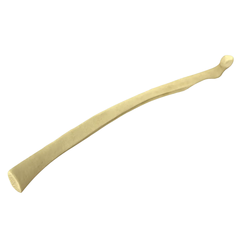
Second rib anatomy quiz
Systematic anatomy quiz on the second rib: structure, joints, muscles, vessels, nerves, and functional relationships.
1/20
bold
text
1. Which rib serves as a landmark for counting the ribs during clinical examination of the thorax and attaches to the manubrium of the sternum at the level of the angle of Louis?
-
First rib
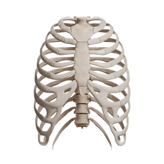
The second rib articulates with the sternum at the level of the angulus sterni (angle of Louis—the junction of the manubrium and body of the sternum), making it the principal bony landmark for counting the ribs.
-
Second rib
The second rib articulates with the sternum at the level of the angulus sterni (angle of Louis—the junction of the manubrium and body of the sternum), making it the principal bony landmark for counting the ribs.
-
Third rib
The second rib articulates with the sternum at the level of the angulus sterni (angle of Louis—the junction of the manubrium and body of the sternum), making it the principal bony landmark for counting the ribs.
-
Fourth rib
The second rib articulates with the sternum at the level of the angulus sterni (angle of Louis—the junction of the manubrium and body of the sternum), making it the principal bony landmark for counting the ribs.
-
I find it difficult to answer
The second rib articulates with the sternum at the level of the angulus sterni (angle of Louis—the junction of the manubrium and body of the sternum), making it the principal bony landmark for counting the ribs.
2. What is the cartilaginous part of the second rib that connects it to the sternum called?
-
Cartilago costalis prima
The cartilaginous part of the second rib is called the cartilago costalis secunda (second costal cartilage). It connects the bony part of the rib to the sternum.
-
Cartilago costalis secunda
The cartilaginous part of the second rib is called the cartilago costalis secunda (second costal cartilage). It connects the bony part of the rib to the sternum.
-
Cartilago epiphysialis
The cartilaginous part of the second rib is called the cartilago costalis secunda (second costal cartilage). It connects the bony part of the rib to the sternum.
-
Cartilago xiphoidea
The cartilaginous part of the second rib is called the cartilago costalis secunda (second costal cartilage). It connects the bony part of the rib to the sternum.
-
I find it difficult to answer
The cartilaginous part of the second rib is called the cartilago costalis secunda (second costal cartilage). It connects the bony part of the rib to the sternum.
3. In which part of the sternum is the complete articular facet for articulation with the second costal cartilage located?
-
Only on the manubrium of the sternum
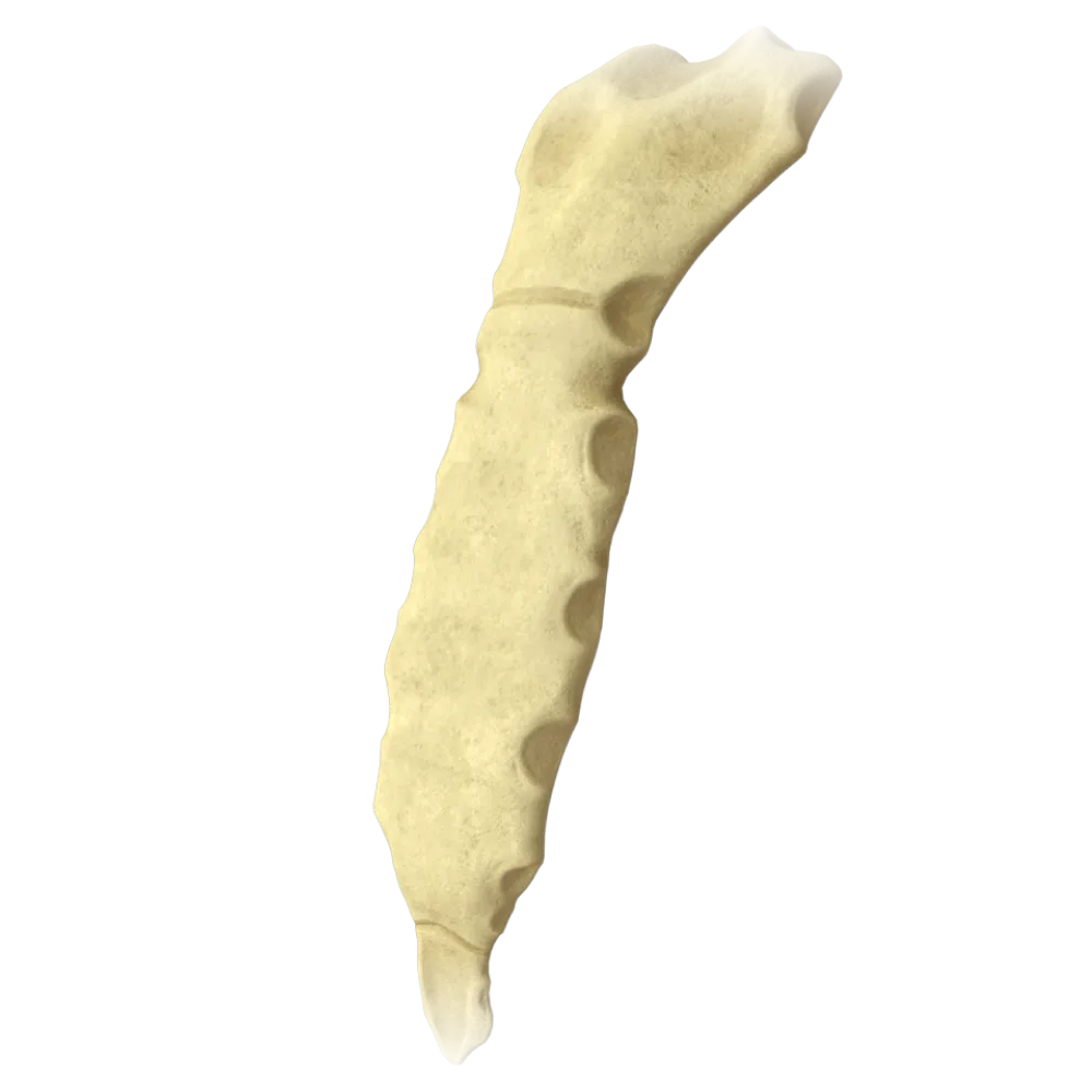
The second costal cartilage articulates at the incisura costalis II, which is located precisely at the junction of the manubrium and body of the sternum—this forms the angle of Louis.
-
At the junction of the manubrium and body of the sternum (incisura costalis II)
The second costal cartilage articulates at the incisura costalis II, which is located precisely at the junction of the manubrium and body of the sternum—this forms the angle of Louis.
-
Only on the body of the sternum
The second costal cartilage articulates at the incisura costalis II, which is located precisely at the junction of the manubrium and body of the sternum—this forms the angle of Louis.
-
On the xiphoid process
The second costal cartilage articulates at the incisura costalis II, which is located precisely at the junction of the manubrium and body of the sternum—this forms the angle of Louis.
-
I find it difficult to answer
The second costal cartilage articulates at the incisura costalis II, which is located precisely at the junction of the manubrium and body of the sternum—this forms the angle of Louis.
4. What type of joint is the articulation between the second costal cartilage and the sternum?
-
Synchondrosis
The articulations of the II–VII costal cartilages with the sternum are true joints (articulationes sternocostales), diarthroses with a joint cavity and capsule. The first rib forms a synchondrosis.
-
Syndesmosis
The articulations of the II–VII costal cartilages with the sternum are true joints (articulationes sternocostales), diarthroses with a joint cavity and capsule. The first rib forms a synchondrosis.
-
Diarthrosis (articulatio sternocostalis)
The articulations of the II–VII costal cartilages with the sternum are true joints (articulationes sternocostales), diarthroses with a joint cavity and capsule. The first rib forms a synchondrosis.
-
Symphysis
The articulations of the II–VII costal cartilages with the sternum are true joints (articulationes sternocostales), diarthroses with a joint cavity and capsule. The first rib forms a synchondrosis.
-
I find it difficult to answer
The articulations of the II–VII costal cartilages with the sternum are true joints (articulationes sternocostales), diarthroses with a joint cavity and capsule. The first rib forms a synchondrosis.
5. Which muscle attaches to the external surface of the second rib and is the largest superficial muscle of the thorax?
-
M. serratus posterior superior
M. serratus anterior arises by digitations from the external surfaces of ribs I–IX, including the second rib, and is the largest superficial muscle of the lateral thoracic wall.
-
M. pectoralis minor
M. serratus anterior arises by digitations from the external surfaces of ribs I–IX, including the second rib, and is the largest superficial muscle of the lateral thoracic wall.
-
M. serratus anterior
M. serratus anterior arises by digitations from the external surfaces of ribs I–IX, including the second rib, and is the largest superficial muscle of the lateral thoracic wall.
-
M. intercostalis externus
M. serratus anterior arises by digitations from the external surfaces of ribs I–IX, including the second rib, and is the largest superficial muscle of the lateral thoracic wall.
-
I find it difficult to answer
M. serratus anterior arises by digitations from the external surfaces of ribs I–IX, including the second rib, and is the largest superficial muscle of the lateral thoracic wall.
6. Which nerve innervates the intercostal muscles and skin in the region of the second intercostal space?
-
N. intercostalis I
The second intercostal nerve (n. intercostalis II)—the anterior ramus of the second thoracic spinal nerve (Th2)—innervates the muscles of the second intercostal space and the skin of the corresponding region.
-
N. intercostalis II
The second intercostal nerve (n. intercostalis II)—the anterior ramus of the second thoracic spinal nerve (Th2)—innervates the muscles of the second intercostal space and the skin of the corresponding region.
-
N. phrenicus
The second intercostal nerve (n. intercostalis II)—the anterior ramus of the second thoracic spinal nerve (Th2)—innervates the muscles of the second intercostal space and the skin of the corresponding region.
-
N. thoracicus longus
The second intercostal nerve (n. intercostalis II)—the anterior ramus of the second thoracic spinal nerve (Th2)—innervates the muscles of the second intercostal space and the skin of the corresponding region.
-
I find it difficult to answer
The second intercostal nerve (n. intercostalis II)—the anterior ramus of the second thoracic spinal nerve (Th2)—innervates the muscles of the second intercostal space and the skin of the corresponding region.
7. Where is the neurovascular bundle of the second intercostal space located relative to the second rib?
-
Along the superior border of the second rib
The intercostal neurovascular bundle passes through the sulcus costae—the groove along the inferior border of each rib. The order from superior to inferior is vein, artery, nerve (VAN).
-
Along the inferior border of the second rib in the sulcus costae
The intercostal neurovascular bundle passes through the sulcus costae—the groove along the inferior border of each rib. The order from superior to inferior is vein, artery, nerve (VAN).
-
In the middle of the intercostal space
The intercostal neurovascular bundle passes through the sulcus costae—the groove along the inferior border of each rib. The order from superior to inferior is vein, artery, nerve (VAN).
-
On the external surface of the rib
The intercostal neurovascular bundle passes through the sulcus costae—the groove along the inferior border of each rib. The order from superior to inferior is vein, artery, nerve (VAN).
-
I find it difficult to answer
The intercostal neurovascular bundle passes through the sulcus costae—the groove along the inferior border of each rib. The order from superior to inferior is vein, artery, nerve (VAN).
8. From which artery is the second intercostal space primarily supplied posteriorly?
-
A. thoracica interna
The posterior intercostal arteries of spaces II–XI arise directly from the thoracic aorta (aorta thoracica). For spaces I–II, they may also arise from the truncus costocervicalis, a branch of the subclavian artery.
-
Aorta thoracica (aortae)
The posterior intercostal arteries of spaces II–XI arise directly from the thoracic aorta (aorta thoracica). For spaces I–II, they may also arise from the truncus costocervicalis, a branch of the subclavian artery.
-
A. subclavia
The posterior intercostal arteries of spaces II–XI arise directly from the thoracic aorta (aorta thoracica). For spaces I–II, they may also arise from the truncus costocervicalis, a branch of the subclavian artery.
-
A. thoracica lateralis
The posterior intercostal arteries of spaces II–XI arise directly from the thoracic aorta (aorta thoracica). For spaces I–II, they may also arise from the truncus costocervicalis, a branch of the subclavian artery.
-
I find it difficult to answer
The posterior intercostal arteries of spaces II–XI arise directly from the thoracic aorta (aorta thoracica). For spaces I–II, they may also arise from the truncus costocervicalis, a branch of the subclavian artery.
9. Which artery supplies the second intercostal space anteriorly?
-
A. intercostalis suprema
A. thoracica interna (internal thoracic artery) gives rise to anterior intercostal branches (rami intercostales anteriores) to intercostal spaces II–VI.
-
A. thoracica interna
A. thoracica interna (internal thoracic artery) gives rise to anterior intercostal branches (rami intercostales anteriores) to intercostal spaces II–VI.
-
A. thoracica lateralis
A. thoracica interna (internal thoracic artery) gives rise to anterior intercostal branches (rami intercostales anteriores) to intercostal spaces II–VI.
-
A. thoracodorsalis
A. thoracica interna (internal thoracic artery) gives rise to anterior intercostal branches (rami intercostales anteriores) to intercostal spaces II–VI.
-
I find it difficult to answer
A. thoracica interna (internal thoracic artery) gives rise to anterior intercostal branches (rami intercostales anteriores) to intercostal spaces II–VI.
10. What is the head of the second rib called in its articulation with the vertebrae?
-
Caput costae with two articular facets separated by the crista capitis
The heads of ribs II–X comprise two articular facets separated by the crista capitis costae and articulate with the bodies of two adjacent vertebrae and the intervertebral disc (articulatio capitis costae).
-
Caput costae with one articular facet
The heads of ribs II–X comprise two articular facets separated by the crista capitis costae and articulate with the bodies of two adjacent vertebrae and the intervertebral disc (articulatio capitis costae).
-
Tuberculum costae
The heads of ribs II–X comprise two articular facets separated by the crista capitis costae and articulate with the bodies of two adjacent vertebrae and the intervertebral disc (articulatio capitis costae).
-
Collum costae
The heads of ribs II–X comprise two articular facets separated by the crista capitis costae and articulate with the bodies of two adjacent vertebrae and the intervertebral disc (articulatio capitis costae).
-
I find it difficult to answer
The heads of ribs II–X comprise two articular facets separated by the crista capitis costae and articulate with the bodies of two adjacent vertebrae and the intervertebral disc (articulatio capitis costae).
11. With the bodies of which vertebrae does the head of the second rib articulate?
-
ThI and ThII
The head of the second rib articulates with the inferior part of the body of ThI and the superior part of the body of ThII, as well as with the intervertebral disc between them.
-
CVI and ThI
The head of the second rib articulates with the inferior part of the body of ThI and the superior part of the body of ThII, as well as with the intervertebral disc between them.
-
ThII and ThIII
The head of the second rib articulates with the inferior part of the body of ThI and the superior part of the body of ThII, as well as with the intervertebral disc between them.
-
ThI and ThIII
The head of the second rib articulates with the inferior part of the body of ThI and the superior part of the body of ThII, as well as with the intervertebral disc between them.
-
I find it difficult to answer
The head of the second rib articulates with the inferior part of the body of ThI and the superior part of the body of ThII, as well as with the intervertebral disc between them.
12. What joint is formed by the articulation of the tubercle of the second rib with the transverse process of the second thoracic vertebra?
-
Articulatio costotransversaria
The tubercle of the rib (tuberculum costae) articulates with the transverse process of the corresponding thoracic vertebra, forming the articulatio costotransversaria. For the second rib—with the transverse process of ThII.
-
Articulatio capitis costae
The tubercle of the rib (tuberculum costae) articulates with the transverse process of the corresponding thoracic vertebra, forming the articulatio costotransversaria. For the second rib—with the transverse process of ThII.
-
Synchondrosis
The tubercle of the rib (tuberculum costae) articulates with the transverse process of the corresponding thoracic vertebra, forming the articulatio costotransversaria. For the second rib—with the transverse process of ThII.
-
Articulatio sternocostalis
The tubercle of the rib (tuberculum costae) articulates with the transverse process of the corresponding thoracic vertebra, forming the articulatio costotransversaria. For the second rib—with the transverse process of ThII.
-
I find it difficult to answer
The tubercle of the rib (tuberculum costae) articulates with the transverse process of the corresponding thoracic vertebra, forming the articulatio costotransversaria. For the second rib—with the transverse process of ThII.
13. Which muscle attaches to the tuberosity of the second rib (tuberositas musculi serrati anterioris)?
-
M. pectoralis major
On the external surface of the second rib is the tuberositas musculi serrati anterioris—the attachment site of a digitation of the m. serratus anterior. This is an anatomical landmark characteristic specifically of the second rib.
-
M. serratus anterior
On the external surface of the second rib is the tuberositas musculi serrati anterioris—the attachment site of a digitation of the m. serratus anterior. This is an anatomical landmark characteristic specifically of the second rib.
-
M. subclavius
On the external surface of the second rib is the tuberositas musculi serrati anterioris—the attachment site of a digitation of the m. serratus anterior. This is an anatomical landmark characteristic specifically of the second rib.
-
M. intercostalis internus
On the external surface of the second rib is the tuberositas musculi serrati anterioris—the attachment site of a digitation of the m. serratus anterior. This is an anatomical landmark characteristic specifically of the second rib.
-
I find it difficult to answer
On the external surface of the second rib is the tuberositas musculi serrati anterioris—the attachment site of a digitation of the m. serratus anterior. This is an anatomical landmark characteristic specifically of the second rib.
14. How does the second rib differ in shape from the first rib?
-
The first rib is flat and horizontal; the second is less curved and narrower
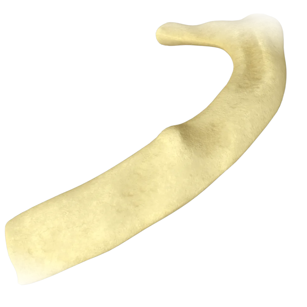
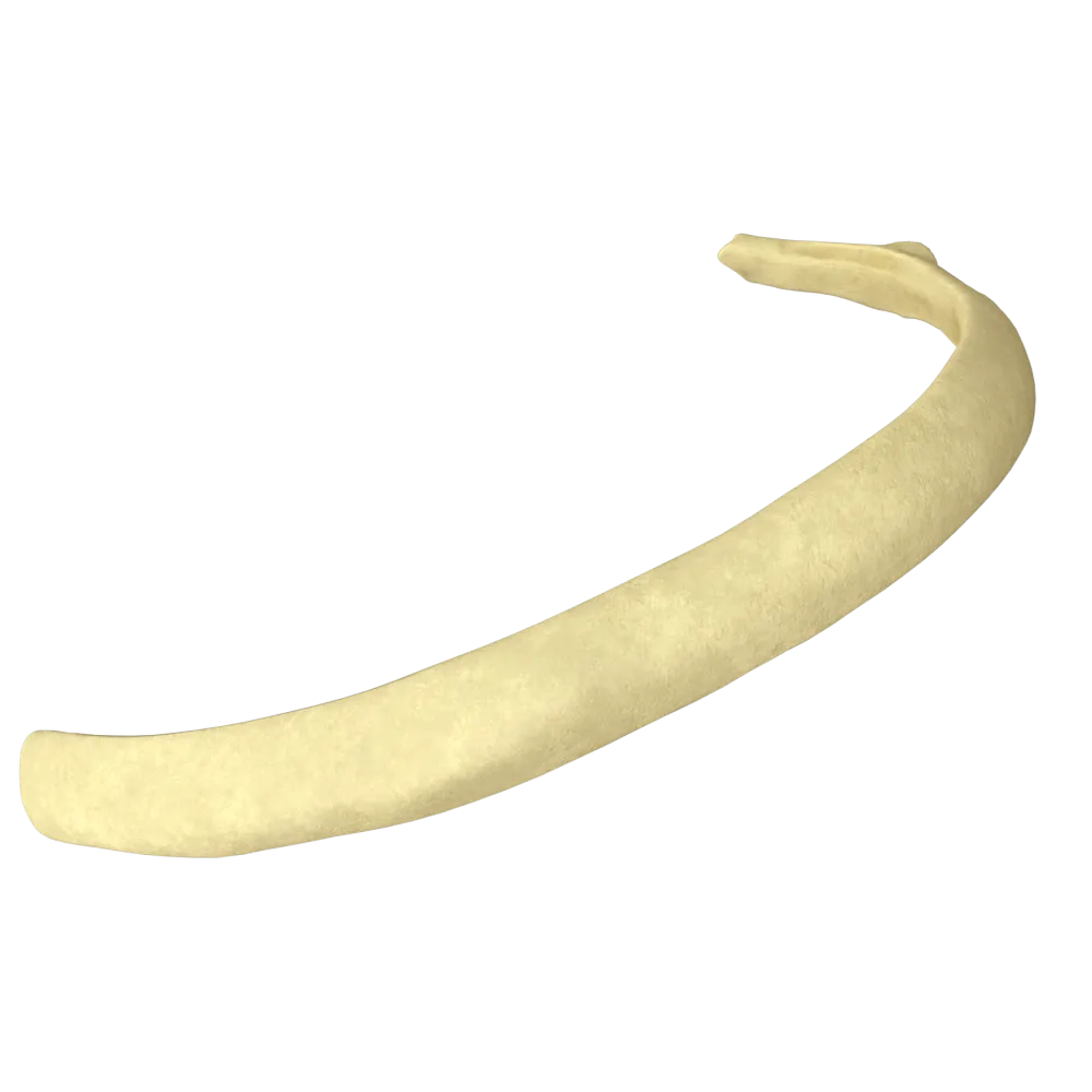
The first rib is the shortest, broadest, almost horizontal, and flat. The second rib is longer, narrower, and less curved than the more inferior ribs, but retains its characteristic curvature.
-
The first rib is curved in three planes; the second is flat
The first rib is the shortest, broadest, almost horizontal, and flat. The second rib is longer, narrower, and less curved than the more inferior ribs, but retains its characteristic curvature.
-
Both ribs have the same shape
The first rib is the shortest, broadest, almost horizontal, and flat. The second rib is longer, narrower, and less curved than the more inferior ribs, but retains its characteristic curvature.
-
The second rib has no angle
The first rib is the shortest, broadest, almost horizontal, and flat. The second rib is longer, narrower, and less curved than the more inferior ribs, but retains its characteristic curvature.
-
I find it difficult to answer
The first rib is the shortest, broadest, almost horizontal, and flat. The second rib is longer, narrower, and less curved than the more inferior ribs, but retains its characteristic curvature.
15. Into which vein do the anterior intercostal veins of the second intercostal space drain?
-
V. azygos
The anterior intercostal veins (vv. intercostales anteriores) accompany the corresponding arteries and drain into the v. thoracica interna (internal thoracic vein).
-
V. thoracica interna
The anterior intercostal veins (vv. intercostales anteriores) accompany the corresponding arteries and drain into the v. thoracica interna (internal thoracic vein).
-
V. subclavia
The anterior intercostal veins (vv. intercostales anteriores) accompany the corresponding arteries and drain into the v. thoracica interna (internal thoracic vein).
-
V. hemiazygos
The anterior intercostal veins (vv. intercostales anteriores) accompany the corresponding arteries and drain into the v. thoracica interna (internal thoracic vein).
-
I find it difficult to answer
The anterior intercostal veins (vv. intercostales anteriores) accompany the corresponding arteries and drain into the v. thoracica interna (internal thoracic vein).
16. Which layer of the pleura is directly adjacent to the internal surface of the second rib?
-
Pleura visceralis
The parietal pleura is adjacent to the internal surfaces of the ribs, specifically its costal part—pleura parietalis, pars costalis. The visceral pleura directly covers the lung.
-
Pleura parietalis (pars costalis)
The parietal pleura is adjacent to the internal surfaces of the ribs, specifically its costal part—pleura parietalis, pars costalis. The visceral pleura directly covers the lung.
-
Pleura mediastinalis
The parietal pleura is adjacent to the internal surfaces of the ribs, specifically its costal part—pleura parietalis, pars costalis. The visceral pleura directly covers the lung.
-
Pleura diaphragmatica
The parietal pleura is adjacent to the internal surfaces of the ribs, specifically its costal part—pleura parietalis, pars costalis. The visceral pleura directly covers the lung.
-
I find it difficult to answer
The parietal pleura is adjacent to the internal surfaces of the ribs, specifically its costal part—pleura parietalis, pars costalis. The visceral pleura directly covers the lung.
17. Which part of the second rib is located between the head and the tubercle?
-
Corpus costae
The collum costae (neck of the rib) is the narrowed part of the rib located between the caput costae (head) and the tuberculum costae (tubercle). It is characteristic of all typical ribs.
-
Collum costae
The collum costae (neck of the rib) is the narrowed part of the rib located between the caput costae (head) and the tuberculum costae (tubercle). It is characteristic of all typical ribs.
-
Angulus costae
The collum costae (neck of the rib) is the narrowed part of the rib located between the caput costae (head) and the tuberculum costae (tubercle). It is characteristic of all typical ribs.
-
Tuberculum costae
The collum costae (neck of the rib) is the narrowed part of the rib located between the caput costae (head) and the tuberculum costae (tubercle). It is characteristic of all typical ribs.
-
I find it difficult to answer
The collum costae (neck of the rib) is the narrowed part of the rib located between the caput costae (head) and the tuberculum costae (tubercle). It is characteristic of all typical ribs.
18. Which spinal cord segment corresponds to the dermatome of the second intercostal nerve?
-
C8
The second intercostal nerve is the anterior ramus of the second thoracic spinal nerve (Th2). The Th2 dermatome corresponds to the skin in the region of the second intercostal space and the medial surface of the arm.
-
Th1
The second intercostal nerve is the anterior ramus of the second thoracic spinal nerve (Th2). The Th2 dermatome corresponds to the skin in the region of the second intercostal space and the medial surface of the arm.
-
Th2
The second intercostal nerve is the anterior ramus of the second thoracic spinal nerve (Th2). The Th2 dermatome corresponds to the skin in the region of the second intercostal space and the medial surface of the arm.
-
Th3
The second intercostal nerve is the anterior ramus of the second thoracic spinal nerve (Th2). The Th2 dermatome corresponds to the skin in the region of the second intercostal space and the medial surface of the arm.
-
I find it difficult to answer
The second intercostal nerve is the anterior ramus of the second thoracic spinal nerve (Th2). The Th2 dermatome corresponds to the skin in the region of the second intercostal space and the medial surface of the arm.
19. What is the superior-to-inferior arrangement of the elements of the neurovascular bundle in the sulcus costae of the second rib?
-
Nerve, artery, vein
In the costal groove (sulcus costae), the neurovascular bundle is arranged from superior to inferior in the following order: vein (v. intercostalis)—artery (a. intercostalis)—nerve (n. intercostalis), designated by the mnemonic VAN.
-
Artery, vein, nerve
In the costal groove (sulcus costae), the neurovascular bundle is arranged from superior to inferior in the following order: vein (v. intercostalis)—artery (a. intercostalis)—nerve (n. intercostalis), designated by the mnemonic VAN.
-
Vein, artery, nerve
In the costal groove (sulcus costae), the neurovascular bundle is arranged from superior to inferior in the following order: vein (v. intercostalis)—artery (a. intercostalis)—nerve (n. intercostalis), designated by the mnemonic VAN.
-
Vein, nerve, artery.
In the costal groove (sulcus costae), the neurovascular bundle is arranged from superior to inferior in the following order: vein (v. intercostalis)—artery (a. intercostalis)—nerve (n. intercostalis), designated by the mnemonic VAN.
-
I find it difficult to answer
In the costal groove (sulcus costae), the neurovascular bundle is arranged from superior to inferior in the following order: vein (v. intercostalis)—artery (a. intercostalis)—nerve (n. intercostalis), designated by the mnemonic VAN.
20. Which ligament reinforces the joint of the head of the second rib from within the joint cavity?
-
Lig Costotransversarium
Lig capitis costae intraarticulare—the intra-articular ligament connecting the crista capitis costae to the intervertebral disc within the cavity of the articulatio capitis costae. It is characteristic of ribs II–IX and divides the joint into two compartments.
-
Lig Capitis costae intraarticulare
Lig capitis costae intraarticulare—the intra-articular ligament connecting the crista capitis costae to the intervertebral disc within the cavity of the articulatio capitis costae. It is characteristic of ribs II–IX and divides the joint into two compartments.
-
Lig sternocostale radiatum
Lig capitis costae intraarticulare—the intra-articular ligament connecting the crista capitis costae to the intervertebral disc within the cavity of the articulatio capitis costae. It is characteristic of ribs II–IX and divides the joint into two compartments.
-
Lig costoclaviculare
Lig capitis costae intraarticulare—the intra-articular ligament connecting the crista capitis costae to the intervertebral disc within the cavity of the articulatio capitis costae. It is characteristic of ribs II–IX and divides the joint into two compartments.
-
I find it difficult to answer
Lig capitis costae intraarticulare—the intra-articular ligament connecting the crista capitis costae to the intervertebral disc within the cavity of the articulatio capitis costae. It is characteristic of ribs II–IX and divides the joint into two compartments.
Retake this quiz?
Your current progress will be reset.
