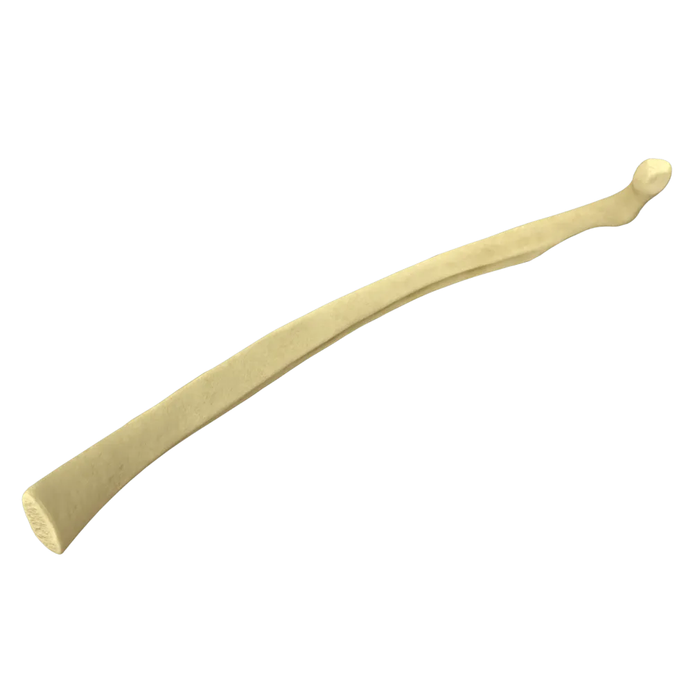
Typical rib anatomy quiz
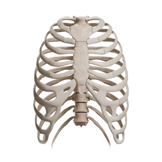
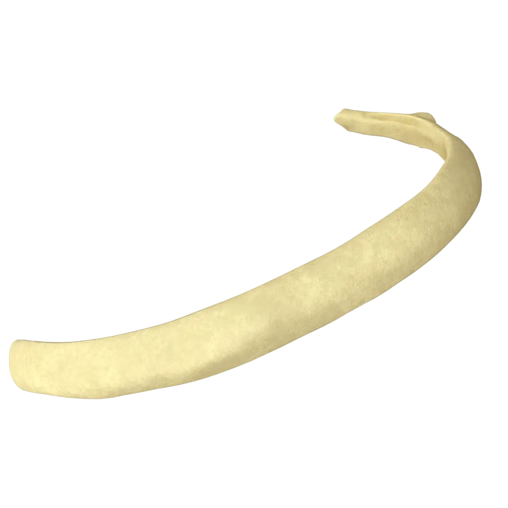
Systemic anatomy quiz on the typical rib: structure, surfaces, angles, grooves, articulations, and functional relationships.
1/20
bold
text
1. Which ribs are classified as typical (true typical) ribs in systemic anatomy?
-
Ribs I, II, and XI
Ribs III–IX, which have all the classical anatomical components—the head, neck, tubercle, and body—are considered typical. Ribs I, II, XI, and XII have atypical structural features.
-
Ribs III–IX
Ribs III–IX, which have all the classical anatomical components—the head, neck, tubercle, and body—are considered typical. Ribs I, II, XI, and XII have atypical structural features.
-
Ribs VIII–X
Ribs III–IX, which have all the classical anatomical components—the head, neck, tubercle, and body—are considered typical. Ribs I, II, XI, and XII have atypical structural features.
-
Ribs I–VII
Ribs III–IX, which have all the classical anatomical components—the head, neck, tubercle, and body—are considered typical. Ribs I, II, XI, and XII have atypical structural features.
-
I find it difficult to answer
Ribs III–IX, which have all the classical anatomical components—the head, neck, tubercle, and body—are considered typical. Ribs I, II, XI, and XII have atypical structural features.
2. Which surface of a typical rib faces the pleural cavity?
-
External (convex)
The internal (concave) surface of the body of the rib faces the pleural cavity. The costal groove (sulcus costae), containing the intercostal vessels and nerve, runs along its inferior border.
-
Internal (concave)
The internal (concave) surface of the body of the rib faces the pleural cavity. The costal groove (sulcus costae), containing the intercostal vessels and nerve, runs along its inferior border.
-
Superior.
The internal (concave) surface of the body of the rib faces the pleural cavity. The costal groove (sulcus costae), containing the intercostal vessels and nerve, runs along its inferior border.
-
Inferior.
The internal (concave) surface of the body of the rib faces the pleural cavity. The costal groove (sulcus costae), containing the intercostal vessels and nerve, runs along its inferior border.
-
I find it difficult to answer
The internal (concave) surface of the body of the rib faces the pleural cavity. The costal groove (sulcus costae), containing the intercostal vessels and nerve, runs along its inferior border.
3. Where is the sulcus costae (costal groove) located on a typical rib?
-
On the external surface of the body along the superior border
The costal groove (sulcus costae) is located on the internal surface of the body of the rib along the inferior border. It contains the v. intercostalis, a. intercostalis, and n. intercostalis (from superior to inferior: vein, artery, nerve).
-
On the internal surface of the body along the inferior border
The costal groove (sulcus costae) is located on the internal surface of the body of the rib along the inferior border. It contains the v. intercostalis, a. intercostalis, and n. intercostalis (from superior to inferior: vein, artery, nerve).
-
On the posterior surface of the neck of the rib
The costal groove (sulcus costae) is located on the internal surface of the body of the rib along the inferior border. It contains the v. intercostalis, a. intercostalis, and n. intercostalis (from superior to inferior: vein, artery, nerve).
-
On the external surface of the body along the inferior border
The costal groove (sulcus costae) is located on the internal surface of the body of the rib along the inferior border. It contains the v. intercostalis, a. intercostalis, and n. intercostalis (from superior to inferior: vein, artery, nerve).
-
I find it difficult to answer
The costal groove (sulcus costae) is located on the internal surface of the body of the rib along the inferior border. It contains the v. intercostalis, a. intercostalis, and n. intercostalis (from superior to inferior: vein, artery, nerve).
4. What are the components of the intercostal neurovascular bundle passing through the sulcus costae of a typical rib (from superior to inferior)?
-
Nerve, artery, vein
In the costal groove, the structures are arranged from superior to inferior: vein (v. intercostalis), artery (a. intercostalis), nerve (n. intercostalis)—the VAN mnemonic.
-
Vein, artery, nerve
In the costal groove, the structures are arranged from superior to inferior: vein (v. intercostalis), artery (a. intercostalis), nerve (n. intercostalis)—the VAN mnemonic.
-
Artery, vein, nerve
In the costal groove, the structures are arranged from superior to inferior: vein (v. intercostalis), artery (a. intercostalis), nerve (n. intercostalis)—the VAN mnemonic.
-
Vein, nerve, artery.
In the costal groove, the structures are arranged from superior to inferior: vein (v. intercostalis), artery (a. intercostalis), nerve (n. intercostalis)—the VAN mnemonic.
-
I find it difficult to answer
In the costal groove, the structures are arranged from superior to inferior: vein (v. intercostalis), artery (a. intercostalis), nerve (n. intercostalis)—the VAN mnemonic.
5. What is the caput costae (head of the rib) of a typical rib?
-
A thickening at the anterior end that articulates with the sternum
The head of a typical rib (caput costae) is located at the posterior end and bears two articular facets separated by a crest (crista capitis costae); each facet forms a demifacet for articulation with the bodies of two adjacent thoracic vertebrae.
-
The posterior end of the rib, with two articular demifacets for articulation with the bodies of adjacent vertebrae
The head of a typical rib (caput costae) is located at the posterior end and bears two articular facets separated by a crest (crista capitis costae); each facet forms a demifacet for articulation with the bodies of two adjacent thoracic vertebrae.
-
A projection on the posterior surface of the neck for muscle attachment
The head of a typical rib (caput costae) is located at the posterior end and bears two articular facets separated by a crest (crista capitis costae); each facet forms a demifacet for articulation with the bodies of two adjacent thoracic vertebrae.
-
A flat facet on the tubercle for articulation with the transverse process of a vertebra
The head of a typical rib (caput costae) is located at the posterior end and bears two articular facets separated by a crest (crista capitis costae); each facet forms a demifacet for articulation with the bodies of two adjacent thoracic vertebrae.
-
I find it difficult to answer
The head of a typical rib (caput costae) is located at the posterior end and bears two articular facets separated by a crest (crista capitis costae); each facet forms a demifacet for articulation with the bodies of two adjacent thoracic vertebrae.
6. What is the joint formed by the tubercle of a typical rib and the transverse process of the corresponding thoracic vertebra called?
-
Joint of the head of the rib
The tubercle of the rib (tuberculum costae) articulates with the costal facet of the transverse process of the thoracic vertebra, forming the articulatio costotransversaria (costotransverse joint).
-
Costotransverse joint
The tubercle of the rib (tuberculum costae) articulates with the costal facet of the transverse process of the thoracic vertebra, forming the articulatio costotransversaria (costotransverse joint).
-
Sternocostal joint
The tubercle of the rib (tuberculum costae) articulates with the costal facet of the transverse process of the thoracic vertebra, forming the articulatio costotransversaria (costotransverse joint).
-
Interchondral joint
The tubercle of the rib (tuberculum costae) articulates with the costal facet of the transverse process of the thoracic vertebra, forming the articulatio costotransversaria (costotransverse joint).
-
I find it difficult to answer
The tubercle of the rib (tuberculum costae) articulates with the costal facet of the transverse process of the thoracic vertebra, forming the articulatio costotransversaria (costotransverse joint).
7. What constitutes the anterior end of a typical rib?
-
An articular facet for articulation with the sternum
The anterior end of a typical rib has a small depression into which the costal cartilage (cartilago costalis) is inserted, connecting the rib to the sternum or to the cartilage above.
-
A cartilaginous part
The anterior end of a typical rib has a small depression into which the costal cartilage (cartilago costalis) is inserted, connecting the rib to the sternum or to the cartilage above.
-
A pointed border without articular surfaces
The anterior end of a typical rib has a small depression into which the costal cartilage (cartilago costalis) is inserted, connecting the rib to the sternum or to the cartilage above.
-
A tubercle with an articular surface
The anterior end of a typical rib has a small depression into which the costal cartilage (cartilago costalis) is inserted, connecting the rib to the sternum or to the cartilage above.
-
I find it difficult to answer
The anterior end of a typical rib has a small depression into which the costal cartilage (cartilago costalis) is inserted, connecting the rib to the sternum or to the cartilage above.
8. Which structure separates the two articular facets on the head of a typical rib?
-
Groove of the head of the rib
The two articular facets of the head of a typical rib are separated by the crest—crista capitis costae—to which the intra-articular ligament (lig. capitis costae intraarticulare) attaches.
-
Crest of the head of the rib
The two articular facets of the head of a typical rib are separated by the crest—crista capitis costae—to which the intra-articular ligament (lig. capitis costae intraarticulare) attaches.
-
Neck of the rib
The two articular facets of the head of a typical rib are separated by the crest—crista capitis costae—to which the intra-articular ligament (lig. capitis costae intraarticulare) attaches.
-
Tubercle of the rib
The two articular facets of the head of a typical rib are separated by the crest—crista capitis costae—to which the intra-articular ligament (lig. capitis costae intraarticulare) attaches.
-
I find it difficult to answer
The two articular facets of the head of a typical rib are separated by the crest—crista capitis costae—to which the intra-articular ligament (lig. capitis costae intraarticulare) attaches.
9. What is the angulus costae (costal angle) of a typical rib?
-
The site where the neck transitions into the body of the rib
The costal angle (angulus costae) is the site of greatest outward and anterior curvature of the body of the rib, located distal (lateral) to the tubercle. The m. iliocostalis attaches at this site.
-
The site of greatest curvature of the body of the rib, located lateral to the tubercle
The costal angle (angulus costae) is the site of greatest outward and anterior curvature of the body of the rib, located distal (lateral) to the tubercle. The m. iliocostalis attaches at this site.
-
The angle between the head and neck of the rib
The costal angle (angulus costae) is the site of greatest outward and anterior curvature of the body of the rib, located distal (lateral) to the tubercle. The m. iliocostalis attaches at this site.
-
The site of attachment of the costal cartilage to the body of the rib
The costal angle (angulus costae) is the site of greatest outward and anterior curvature of the body of the rib, located distal (lateral) to the tubercle. The m. iliocostalis attaches at this site.
-
I find it difficult to answer
The costal angle (angulus costae) is the site of greatest outward and anterior curvature of the body of the rib, located distal (lateral) to the tubercle. The m. iliocostalis attaches at this site.
10. What is the cross-sectional shape of the body of a typical rib?
-
Rounded
The body of a typical rib is flattened dorsoventrally, giving it a characteristic flat cross-sectional shape with distinguishable external and internal surfaces and superior and inferior borders.
-
Square
The body of a typical rib is flattened dorsoventrally, giving it a characteristic flat cross-sectional shape with distinguishable external and internal surfaces and superior and inferior borders.
-
Flattened (flat)
The body of a typical rib is flattened dorsoventrally, giving it a characteristic flat cross-sectional shape with distinguishable external and internal surfaces and superior and inferior borders.
-
Triangular
The body of a typical rib is flattened dorsoventrally, giving it a characteristic flat cross-sectional shape with distinguishable external and internal surfaces and superior and inferior borders.
-
I find it difficult to answer
The body of a typical rib is flattened dorsoventrally, giving it a characteristic flat cross-sectional shape with distinguishable external and internal surfaces and superior and inferior borders.
11. Which of the following structures of a typical rib is the attachment site of the lig. Capitis costae intraarticulare?
-
Tubercle of the rib
The intra-articular ligament of the head of the rib (lig. capitis costae intraarticulare) attaches to the crista capitis costae—the crest separating the two articular facets of the head of the rib.
-
Crest of the head of the rib
The intra-articular ligament of the head of the rib (lig. capitis costae intraarticulare) attaches to the crista capitis costae—the crest separating the two articular facets of the head of the rib.
-
Neck of the rib
The intra-articular ligament of the head of the rib (lig. capitis costae intraarticulare) attaches to the crista capitis costae—the crest separating the two articular facets of the head of the rib.
-
Angle of the rib
The intra-articular ligament of the head of the rib (lig. capitis costae intraarticulare) attaches to the crista capitis costae—the crest separating the two articular facets of the head of the rib.
-
I find it difficult to answer
The intra-articular ligament of the head of the rib (lig. capitis costae intraarticulare) attaches to the crista capitis costae—the crest separating the two articular facets of the head of the rib.
12. What forms the wall of the sulcus costae (costal groove)?
-
The superior border of the body of the rib
The costal groove is formed by the inferior border of the body of the rib, creating a channel for the intercostal neurovascular bundle.
-
The inferior border of the body of the rib
The costal groove is formed by the inferior border of the body of the rib, creating a channel for the intercostal neurovascular bundle.
-
The neck of the rib
The costal groove is formed by the inferior border of the body of the rib, creating a channel for the intercostal neurovascular bundle.
-
The tubercle of the rib
The costal groove is formed by the inferior border of the body of the rib, creating a channel for the intercostal neurovascular bundle.
-
I find it difficult to answer
The costal groove is formed by the inferior border of the body of the rib, creating a channel for the intercostal neurovascular bundle.
13. What is the length of the collum costae (neck of the rib) of a typical rib?
-
The neck is absent in a typical rib
The neck of the rib (collum costae) is a narrowed cylindrical segment approximately 3–4 cm long, located between the head and tubercle of the rib. It is directed posteriorly and laterally.
-
The neck is a narrowed segment approximately 3–4 cm long between the head and tubercle
The neck of the rib (collum costae) is a narrowed cylindrical segment approximately 3–4 cm long, located between the head and tubercle of the rib. It is directed posteriorly and laterally.
-
The neck is equal in length to the body of the rib
The neck of the rib (collum costae) is a narrowed cylindrical segment approximately 3–4 cm long, located between the head and tubercle of the rib. It is directed posteriorly and laterally.
-
The neck of the rib is synonymous with the costal cartilage
The neck of the rib (collum costae) is a narrowed cylindrical segment approximately 3–4 cm long, located between the head and tubercle of the rib. It is directed posteriorly and laterally.
-
I find it difficult to answer
The neck of the rib (collum costae) is a narrowed cylindrical segment approximately 3–4 cm long, located between the head and tubercle of the rib. It is directed posteriorly and laterally.
14. Which type of tissue predominates in the compact and cancellous substances of a typical rib?
-
Hyaline cartilage externally and dense connective tissue internally
As a flat bone, a typical rib consists of external and internal layers of compact bone (laminae), between which cancellous bone (diploe) containing red bone marrow is located.
-
Compact bone externally and cancellous bone internally
As a flat bone, a typical rib consists of external and internal layers of compact bone (laminae), between which cancellous bone (diploe) containing red bone marrow is located.
-
Elastic cartilage externally and compact bone internally
As a flat bone, a typical rib consists of external and internal layers of compact bone (laminae), between which cancellous bone (diploe) containing red bone marrow is located.
-
Compact bone only, without cancellous bone
As a flat bone, a typical rib consists of external and internal layers of compact bone (laminae), between which cancellous bone (diploe) containing red bone marrow is located.
-
I find it difficult to answer
As a flat bone, a typical rib consists of external and internal layers of compact bone (laminae), between which cancellous bone (diploe) containing red bone marrow is located.
15. With the bodies of how many vertebrae does the head of a typical rib (for example, rib V) articulate?
-
Only one—the correspondingly numbered vertebra
The head of a typical rib articulates with two vertebrae: the superior facet with the inferior costal facet of the vertebra above, and the inferior facet with the superior costal facet of the correspondingly numbered vertebra.
-
Two—the inferior demifacet of the vertebra above and the superior demifacet of the corresponding vertebra
The head of a typical rib articulates with two vertebrae: the superior facet with the inferior costal facet of the vertebra above, and the inferior facet with the superior costal facet of the correspondingly numbered vertebra.
-
Three—two adjacent vertebrae and one vertebra below
The head of a typical rib articulates with two vertebrae: the superior facet with the inferior costal facet of the vertebra above, and the inferior facet with the superior costal facet of the correspondingly numbered vertebra.
-
None—the head articulates only with the transverse process
The head of a typical rib articulates with two vertebrae: the superior facet with the inferior costal facet of the vertebra above, and the inferior facet with the superior costal facet of the correspondingly numbered vertebra.
-
I find it difficult to answer
The head of a typical rib articulates with two vertebrae: the superior facet with the inferior costal facet of the vertebra above, and the inferior facet with the superior costal facet of the correspondingly numbered vertebra.
16. Which ligament reinforces the articulatio costotransversaria posteriorly?
-
Lig Capitis costae radiatum
The articulatio costotransversaria is reinforced by several ligaments; posteriorly (laterally), the lig. costotransversarium laterale extends between the tubercle of the rib and the apex of the transverse process.
-
Lig costotransversarium superius
The articulatio costotransversaria is reinforced by several ligaments; posteriorly (laterally), the lig. costotransversarium laterale extends between the tubercle of the rib and the apex of the transverse process.
-
Lig costotransversarium laterale
The articulatio costotransversaria is reinforced by several ligaments; posteriorly (laterally), the lig. costotransversarium laterale extends between the tubercle of the rib and the apex of the transverse process.
-
Lig Capitis costae intraarticulare
The articulatio costotransversaria is reinforced by several ligaments; posteriorly (laterally), the lig. costotransversarium laterale extends between the tubercle of the rib and the apex of the transverse process.
-
I find it difficult to answer
The articulatio costotransversaria is reinforced by several ligaments; posteriorly (laterally), the lig. costotransversarium laterale extends between the tubercle of the rib and the apex of the transverse process.
17. Which movement in the costovertebral joints of a typical rib increases the anteroposterior diameter of the thoracic cage during inspiration?
-
Anterior gliding of the rib without rotation
During inspiration, a typical rib rotates around an axis passing through the neck (through both costovertebral joints). Elevation of the anterior end of the rib increases the anteroposterior diameter of the thoracic cage—the “pump-handle movement.”
-
Rotation of the rib around an axis passing through the neck, with elevation of the body of the rib
During inspiration, a typical rib rotates around an axis passing through the neck (through both costovertebral joints). Elevation of the anterior end of the rib increases the anteroposterior diameter of the thoracic cage—the “pump-handle movement.”
-
Inferior translational displacement of the head along the articular facet
During inspiration, a typical rib rotates around an axis passing through the neck (through both costovertebral joints). Elevation of the anterior end of the rib increases the anteroposterior diameter of the thoracic cage—the “pump-handle movement.”
-
Outward divergence of the rib ends without rotation
During inspiration, a typical rib rotates around an axis passing through the neck (through both costovertebral joints). Elevation of the anterior end of the rib increases the anteroposterior diameter of the thoracic cage—the “pump-handle movement.”
-
I find it difficult to answer
During inspiration, a typical rib rotates around an axis passing through the neck (through both costovertebral joints). Elevation of the anterior end of the rib increases the anteroposterior diameter of the thoracic cage—the “pump-handle movement.”
18. According to the classification of bones by shape, to which category of the skeleton do ribs belong?
-
Long tubular bones
Ribs are classified as flat bones (ossa plana). Despite their curved shape, they consist of two plates of compact bone with a layer of cancellous bone between them—a characteristic feature of flat bones.
-
Flat bones
Ribs are classified as flat bones (ossa plana). Despite their curved shape, they consist of two plates of compact bone with a layer of cancellous bone between them—a characteristic feature of flat bones.
-
Short cancellous bones
Ribs are classified as flat bones (ossa plana). Despite their curved shape, they consist of two plates of compact bone with a layer of cancellous bone between them—a characteristic feature of flat bones.
-
Mixed bones.
Ribs are classified as flat bones (ossa plana). Despite their curved shape, they consist of two plates of compact bone with a layer of cancellous bone between them—a characteristic feature of flat bones.
-
I find it difficult to answer
Ribs are classified as flat bones (ossa plana). Despite their curved shape, they consist of two plates of compact bone with a layer of cancellous bone between them—a characteristic feature of flat bones.
19. Which muscle attaches to the external surface of the angle of a typical rib (angulus costae)?
-
M. serratus posterior inferior
The m. iliocostalis—the lateral tract of the m. erector spinae—attaches to the external surface in the region of the costal angle. It is here, at the angulus costae, that the muscle has the greatest leverage on the rib.
-
M. iliocostalis (part of m. erector spinae)
The m. iliocostalis—the lateral tract of the m. erector spinae—attaches to the external surface in the region of the costal angle. It is here, at the angulus costae, that the muscle has the greatest leverage on the rib.
-
M. scalenus medius
The m. iliocostalis—the lateral tract of the m. erector spinae—attaches to the external surface in the region of the costal angle. It is here, at the angulus costae, that the muscle has the greatest leverage on the rib.
-
External intercostal muscle
The m. iliocostalis—the lateral tract of the m. erector spinae—attaches to the external surface in the region of the costal angle. It is here, at the angulus costae, that the muscle has the greatest leverage on the rib.
-
I find it difficult to answer
The m. iliocostalis—the lateral tract of the m. erector spinae—attaches to the external surface in the region of the costal angle. It is here, at the angulus costae, that the muscle has the greatest leverage on the rib.
20. What is the shape of the articular surface of the tuberculum costae of a typical rib where it articulates with the transverse process of a vertebra?
-
Flat.
In the articulatio costotransversaria, the articular surface of the tubercle of a typical rib is convex, while the corresponding facet of the transverse process of the vertebra is concave. This structure permits rotational movements of the rib around the axis of the neck.
-
Convex (convexity on the rib, concavity on the vertebra)
In the articulatio costotransversaria, the articular surface of the tubercle of a typical rib is convex, while the corresponding facet of the transverse process of the vertebra is concave. This structure permits rotational movements of the rib around the axis of the neck.
-
Concave (concavity on the rib, convexity on the vertebra)
In the articulatio costotransversaria, the articular surface of the tubercle of a typical rib is convex, while the corresponding facet of the transverse process of the vertebra is concave. This structure permits rotational movements of the rib around the axis of the neck.
-
Saddle-shaped.
In the articulatio costotransversaria, the articular surface of the tubercle of a typical rib is convex, while the corresponding facet of the transverse process of the vertebra is concave. This structure permits rotational movements of the rib around the axis of the neck.
-
I find it difficult to answer
In the articulatio costotransversaria, the articular surface of the tubercle of a typical rib is convex, while the corresponding facet of the transverse process of the vertebra is concave. This structure permits rotational movements of the rib around the axis of the neck.
Retake this quiz?
Your current progress will be reset.
