1/20
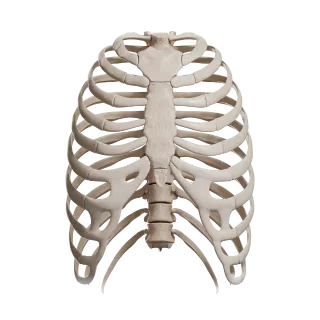
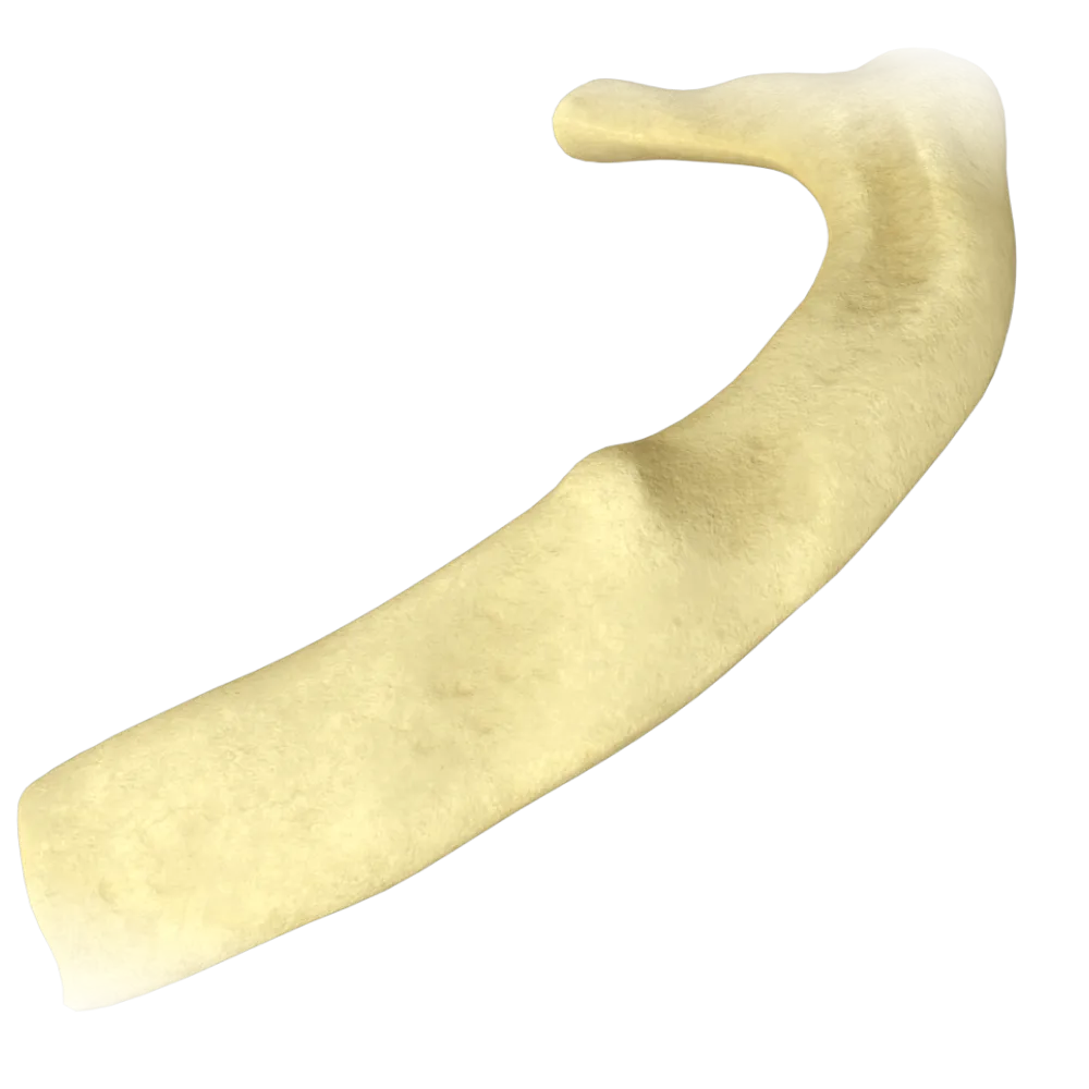
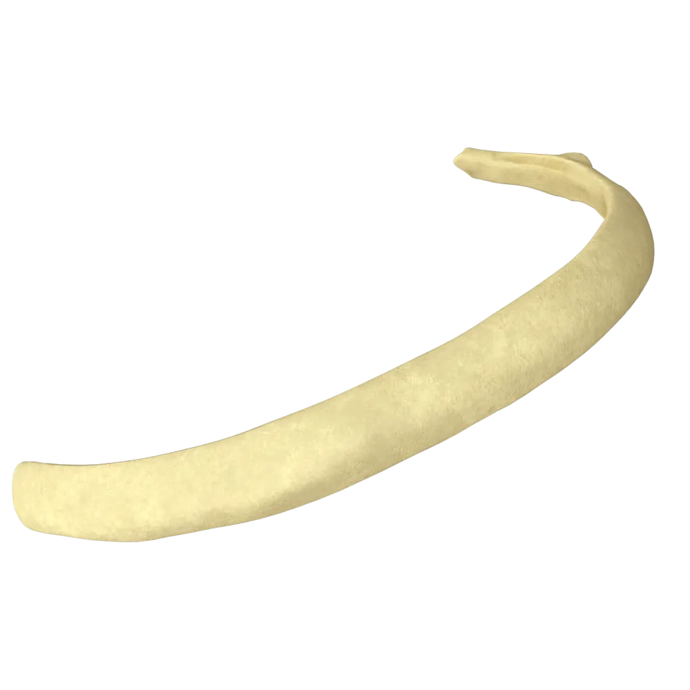
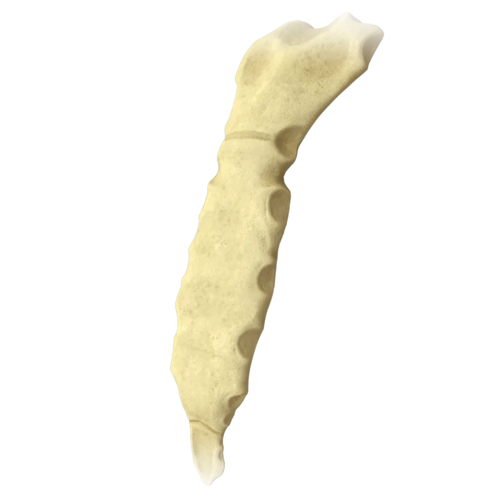
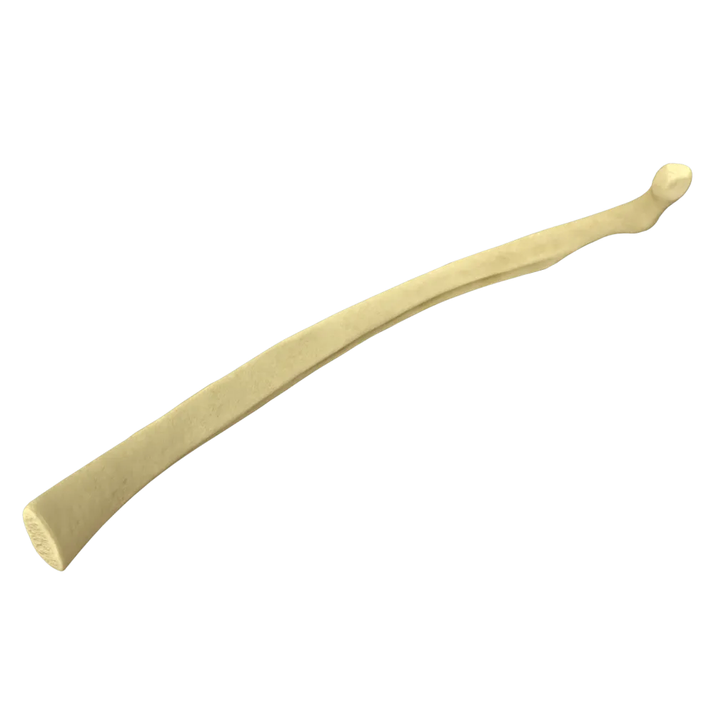
Anatomy quiz of the thoracic skeleton
Quiz on the normal anatomy of the thoracic skeleton: bones, joints, connections and their morphofunctional characteristics.
1. How many pairs of ribs are part of the human thoracic skeleton?
-
10 pairs
The thoracic skeleton includes 12 pairs of ribs.
-
11 pairs
The thoracic skeleton includes 12 pairs of ribs.
-
12 pairs
The thoracic skeleton includes 12 pairs of ribs.
-
13 pairs
The thoracic skeleton includes 12 pairs of ribs.
-
I find it difficult to answer
The thoracic skeleton includes 12 pairs of ribs.
2. What are the ribs called whose cartilages connect directly to the sternum?
-
False ribs (costae spuriae)
True ribs (costae verae) — pairs I–VII — connect directly to the sternum via their cartilages. False ribs connect with the cartilage of the superior rib, while floating ribs terminate freely.
-
Floating ribs (costae fluctuantes)
True ribs (costae verae) — pairs I–VII — connect directly to the sternum via their cartilages. False ribs connect with the cartilage of the superior rib, while floating ribs terminate freely.
-
True ribs (costae verae)
True ribs (costae verae) — pairs I–VII — connect directly to the sternum via their cartilages. False ribs connect with the cartilage of the superior rib, while floating ribs terminate freely.
-
Accessory ribs (costae accessoriae)
True ribs (costae verae) — pairs I–VII — connect directly to the sternum via their cartilages. False ribs connect with the cartilage of the superior rib, while floating ribs terminate freely.
-
I find it difficult to answer
True ribs (costae verae) — pairs I–VII — connect directly to the sternum via their cartilages. False ribs connect with the cartilage of the superior rib, while floating ribs terminate freely.
3. Which ribs are referred to as 'floating ribs' (costae fluctuantes)?
-
VIII–X pairs
Floating ribs — pairs XI and XII — have no cartilaginous connection to either the sternum or the superior ribs; their anterior ends lie freely within the abdominal wall's musculature.
-
VI–VIII pairs
Floating ribs — pairs XI and XII — have no cartilaginous connection to either the sternum or the superior ribs; their anterior ends lie freely within the abdominal wall's musculature.
-
XI–XII pairs
Floating ribs — pairs XI and XII — have no cartilaginous connection to either the sternum or the superior ribs; their anterior ends lie freely within the abdominal wall's musculature.
-
IX–XI pairs
Floating ribs — pairs XI and XII — have no cartilaginous connection to either the sternum or the superior ribs; their anterior ends lie freely within the abdominal wall's musculature.
-
I find it difficult to answer
Floating ribs — pairs XI and XII — have no cartilaginous connection to either the sternum or the superior ribs; their anterior ends lie freely within the abdominal wall's musculature.
4. Which part of the sternum is its superior section and connects with the clavicles?
-
Body of the sternum (corpus sterni)
The manubrium of the sternum (manubrium sterni) is the superior section of the sternum. On its superior edge are located the jugular notch and two clavicular notches for articulating with the clavicles.
-
Xiphoid process (processus xiphoideus)
The manubrium of the sternum (manubrium sterni) is the superior section of the sternum. On its superior edge are located the jugular notch and two clavicular notches for articulating with the clavicles.
-
Manubrium of the sternum (manubrium sterni)
The manubrium of the sternum (manubrium sterni) is the superior section of the sternum. On its superior edge are located the jugular notch and two clavicular notches for articulating with the clavicles.
-
Jugular notch (incisura jugularis)
The manubrium of the sternum (manubrium sterni) is the superior section of the sternum. On its superior edge are located the jugular notch and two clavicular notches for articulating with the clavicles.
-
I find it difficult to answer
The manubrium of the sternum (manubrium sterni) is the superior section of the sternum. On its superior edge are located the jugular notch and two clavicular notches for articulating with the clavicles.
5. What is the name of the angle formed at the junction of the manubrium and the body of the sternum?
-
Angle of Louis (angulus sterni)
Sternal angle (angulus sterni, Angle of Louis) — a forward-protruding angle at the site of the synchondrosis between the manubrium and the body of the sternum. At its level, the cartilage of the second rib joins, serving as an important anatomical landmark.
-
Infrasternal angle (angulus infrasternalis)
Sternal angle (angulus sterni, Angle of Louis) — a forward-protruding angle at the site of the synchondrosis between the manubrium and the body of the sternum. At its level, the cartilage of the second rib joins, serving as an important anatomical landmark.
-
Sharp's angle
Sternal angle (angulus sterni, Angle of Louis) — a forward-protruding angle at the site of the synchondrosis between the manubrium and the body of the sternum. At its level, the cartilage of the second rib joins, serving as an important anatomical landmark.
-
Scarpa's angle (angulus costae)
Sternal angle (angulus sterni, Angle of Louis) — a forward-protruding angle at the site of the synchondrosis between the manubrium and the body of the sternum. At its level, the cartilage of the second rib joins, serving as an important anatomical landmark.
-
I find it difficult to answer
Sternal angle (angulus sterni, Angle of Louis) — a forward-protruding angle at the site of the synchondrosis between the manubrium and the body of the sternum. At its level, the cartilage of the second rib joins, serving as an important anatomical landmark.
6. Which rib's attachment corresponds to the sternal angle (angulus sterni)?
-
I rib
The sternal angle is formed at the boundary between the manubrium and the body of the sternum. At its level, the second rib is attached to the sternum, providing an important point of reference for counting ribs and intercostal spaces.
-
II rib
The sternal angle is formed at the boundary between the manubrium and the body of the sternum. At its level, the second rib is attached to the sternum, providing an important point of reference for counting ribs and intercostal spaces.
-
III rib
The sternal angle is formed at the boundary between the manubrium and the body of the sternum. At its level, the second rib is attached to the sternum, providing an important point of reference for counting ribs and intercostal spaces.
-
IV rib
The sternal angle is formed at the boundary between the manubrium and the body of the sternum. At its level, the second rib is attached to the sternum, providing an important point of reference for counting ribs and intercostal spaces.
-
I find it difficult to answer
The sternal angle is formed at the boundary between the manubrium and the body of the sternum. At its level, the second rib is attached to the sternum, providing an important point of reference for counting ribs and intercostal spaces.
7. Which of the following is an anatomical structure on the posterior surface of the lower edge of a rib?
-
Rib tubercle (tuberculum costae)
The costal groove (sulcus costae) is located on the inner (posterior inferior) surface of the rib's body along its lower edge. Intercostal vessels and a nerve pass through it (from top to bottom: vein, artery, nerve).
-
Costal groove (sulcus costae)
The costal groove (sulcus costae) is located on the inner (posterior inferior) surface of the rib's body along its lower edge. Intercostal vessels and a nerve pass through it (from top to bottom: vein, artery, nerve).
-
Angle of the rib (angulus costae)
The costal groove (sulcus costae) is located on the inner (posterior inferior) surface of the rib's body along its lower edge. Intercostal vessels and a nerve pass through it (from top to bottom: vein, artery, nerve).
-
Neck of the rib (collum costae)
The costal groove (sulcus costae) is located on the inner (posterior inferior) surface of the rib's body along its lower edge. Intercostal vessels and a nerve pass through it (from top to bottom: vein, artery, nerve).
-
I find it difficult to answer
The costal groove (sulcus costae) is located on the inner (posterior inferior) surface of the rib's body along its lower edge. Intercostal vessels and a nerve pass through it (from top to bottom: vein, artery, nerve).
8. What is a costal arch (arcus costalis)?
-
Connection of the cartilages of ribs I–VII with the sternum
The costal arch (arcus costalis) is formed by the cartilages of ribs VII–X, which join together to form an arched lower boundary of the thoracic cage. The cartilage of the seventh rib connects directly to the sternum, while the cartilages of ribs VIII–X connect via connections with the superior cartilages.
-
An arched structure formed by the cartilages of ribs VII–X, creating the lower boundary of the thoracic cage
The costal arch (arcus costalis) is formed by the cartilages of ribs VII–X, which join together to form an arched lower boundary of the thoracic cage. The cartilage of the seventh rib connects directly to the sternum, while the cartilages of ribs VIII–X connect via connections with the superior cartilages.
-
Connection of the anterior ends of ribs XI–XII
The costal arch (arcus costalis) is formed by the cartilages of ribs VII–X, which join together to form an arched lower boundary of the thoracic cage. The cartilage of the seventh rib connects directly to the sternum, while the cartilages of ribs VIII–X connect via connections with the superior cartilages.
-
Connection of the manubrium and the body of the sternum
The costal arch (arcus costalis) is formed by the cartilages of ribs VII–X, which join together to form an arched lower boundary of the thoracic cage. The cartilage of the seventh rib connects directly to the sternum, while the cartilages of ribs VIII–X connect via connections with the superior cartilages.
-
I find it difficult to answer
The costal arch (arcus costalis) is formed by the cartilages of ribs VII–X, which join together to form an arched lower boundary of the thoracic cage. The cartilage of the seventh rib connects directly to the sternum, while the cartilages of ribs VIII–X connect via connections with the superior cartilages.
9. What are "false ribs" in human anatomy?
-
Ribs that are absent in most individuals due to evolutionary changes
False ribs (pairs VIII–X) are connected to the sternum indirectly, joining with the cartilage of the superior rib and forming a costal arch.
-
Ribs (pairs XI–XII) that do not attach to the sternum and end freely within muscles
False ribs (pairs VIII–X) are connected to the sternum indirectly, joining with the cartilage of the superior rib and forming a costal arch.
-
Ribs (pairs VIII–X) that attach not directly to the sternum but to the cartilage of the superior rib
False ribs (pairs VIII–X) are connected to the sternum indirectly, joining with the cartilage of the superior rib and forming a costal arch.
-
Ribs that attach directly to the sternum
False ribs (pairs VIII–X) are connected to the sternum indirectly, joining with the cartilage of the superior rib and forming a costal arch.
-
I find it difficult to answer
False ribs (pairs VIII–X) are connected to the sternum indirectly, joining with the cartilage of the superior rib and forming a costal arch.
10. What is the name of the unpaired indentation located at the center of the upper edge of the manubrium of the sternum?
-
Clavicular notch
The jugular notch (incisura jugularis) is located precisely at the center (midline of the body). It can be easily palpated under the skin at the base of the neck.
-
Costal notch
The jugular notch (incisura jugularis) is located precisely at the center (midline of the body). It can be easily palpated under the skin at the base of the neck.
-
Jugular notch
The jugular notch (incisura jugularis) is located precisely at the center (midline of the body). It can be easily palpated under the skin at the base of the neck.
-
Sublingual notch
The jugular notch (incisura jugularis) is located precisely at the center (midline of the body). It can be easily palpated under the skin at the base of the neck.
-
I find it difficult to answer
The jugular notch (incisura jugularis) is located precisely at the center (midline of the body). It can be easily palpated under the skin at the base of the neck.
11. How many notches are located on the human sternum?
-
18
17 notches: 1 jugular, 2 clavicular, 14 costal.
-
17
17 notches: 1 jugular, 2 clavicular, 14 costal.
-
12
17 notches: 1 jugular, 2 clavicular, 14 costal.
-
10
17 notches: 1 jugular, 2 clavicular, 14 costal.
-
I find it difficult to answer
17 notches: 1 jugular, 2 clavicular, 14 costal.
12. How many costal facets (foveae costales) does the body of a typical thoracic vertebra have?
-
One complete facet on each side
The lateral surfaces of the body of a typical thoracic vertebra have superior and inferior costal demifacets (foveae costales superior et inferior) which, along with the demifacets of adjacent vertebrae, form complete facets for the rib head.
-
Two demifacets (superior and inferior) on each side
The lateral surfaces of the body of a typical thoracic vertebra have superior and inferior costal demifacets (foveae costales superior et inferior) which, along with the demifacets of adjacent vertebrae, form complete facets for the rib head.
-
Three facets on each side
The lateral surfaces of the body of a typical thoracic vertebra have superior and inferior costal demifacets (foveae costales superior et inferior) which, along with the demifacets of adjacent vertebrae, form complete facets for the rib head.
-
Four facets on each side
The lateral surfaces of the body of a typical thoracic vertebra have superior and inferior costal demifacets (foveae costales superior et inferior) which, along with the demifacets of adjacent vertebrae, form complete facets for the rib head.
-
I find it difficult to answer
The lateral surfaces of the body of a typical thoracic vertebra have superior and inferior costal demifacets (foveae costales superior et inferior) which, along with the demifacets of adjacent vertebrae, form complete facets for the rib head.
13. What is the subcostal angle (angulus infrasternalis)?
-
Angle between the manubrium and the body of the sternum
The subcostal angle (angulus infrasternalis) is the angle formed between the right and left costal arches and the base of the xiphoid process. Normally, it constitutes about 70–75°, varying depending on build.
-
Angle between the sternum and the first rib
The subcostal angle (angulus infrasternalis) is the angle formed between the right and left costal arches and the base of the xiphoid process. Normally, it constitutes about 70–75°, varying depending on build.
-
Angle between the cartilages of the right and left costal arches at the base of the xiphoid process
The subcostal angle (angulus infrasternalis) is the angle formed between the right and left costal arches and the base of the xiphoid process. Normally, it constitutes about 70–75°, varying depending on build.
-
Angle between the body and the xiphoid process of the sternum
The subcostal angle (angulus infrasternalis) is the angle formed between the right and left costal arches and the base of the xiphoid process. Normally, it constitutes about 70–75°, varying depending on build.
-
I find it difficult to answer
The subcostal angle (angulus infrasternalis) is the angle formed between the right and left costal arches and the base of the xiphoid process. Normally, it constitutes about 70–75°, varying depending on build.
14. Which of the listed human ribs do not have a rib tubercle (tuberculum costae)?
-
Pairs I and II
Pairs XI and XII ribs (floating ribs) are the least developed: they lack a neck and rib tubercle, and do not form costotransverse joints, attaching only via the head directly to their vertebra's body.
-
Pairs II and XI
Pairs XI and XII ribs (floating ribs) are the least developed: they lack a neck and rib tubercle, and do not form costotransverse joints, attaching only via the head directly to their vertebra's body.
-
Pairs XI and XII
Pairs XI and XII ribs (floating ribs) are the least developed: they lack a neck and rib tubercle, and do not form costotransverse joints, attaching only via the head directly to their vertebra's body.
-
Only pair XII
Pairs XI and XII ribs (floating ribs) are the least developed: they lack a neck and rib tubercle, and do not form costotransverse joints, attaching only via the head directly to their vertebra's body.
-
I find it difficult to answer
Pairs XI and XII ribs (floating ribs) are the least developed: they lack a neck and rib tubercle, and do not form costotransverse joints, attaching only via the head directly to their vertebra's body.
15. Which of the following is part of the body of a rib (corpus costae) and not its head or neck?
-
Crest of the head (crista capitis costae)
The angle of the rib (angulus costae) is the site of greatest curvature of the rib's body, located on the rib's body lateral to the tubercle. It is here that the rib sharply turns forward and downward.
-
Transverse process
The angle of the rib (angulus costae) is the site of greatest curvature of the rib's body, located on the rib's body lateral to the tubercle. It is here that the rib sharply turns forward and downward.
-
Angle of the rib (angulus costae)
The angle of the rib (angulus costae) is the site of greatest curvature of the rib's body, located on the rib's body lateral to the tubercle. It is here that the rib sharply turns forward and downward.
-
Articular surface of the tubercle (facies articularis tuberculi costae)
The angle of the rib (angulus costae) is the site of greatest curvature of the rib's body, located on the rib's body lateral to the tubercle. It is here that the rib sharply turns forward and downward.
-
I find it difficult to answer
The angle of the rib (angulus costae) is the site of greatest curvature of the rib's body, located on the rib's body lateral to the tubercle. It is here that the rib sharply turns forward and downward.
16. In which of the listed human ribs does the location of the rib tubercle coincide with its angle?
-
On the first rib
The angle of the rib (angulus costae) is where the rib bends most sharply forward. In most ribs (pairs II–X), the angle of the rib is located more laterally than the rib tubercle (tuberculum costae). On the first rib, due to its unique shape (it is flattened from top to bottom rather than from sides), the angle coincides with the rib tubercle.
-
On the second rib
The angle of the rib (angulus costae) is where the rib bends most sharply forward. In most ribs (pairs II–X), the angle of the rib is located more laterally than the rib tubercle (tuberculum costae). On the first rib, due to its unique shape (it is flattened from top to bottom rather than from sides), the angle coincides with the rib tubercle.
-
On the seventh rib
The angle of the rib (angulus costae) is where the rib bends most sharply forward. In most ribs (pairs II–X), the angle of the rib is located more laterally than the rib tubercle (tuberculum costae). On the first rib, due to its unique shape (it is flattened from top to bottom rather than from sides), the angle coincides with the rib tubercle.
-
On the twelfth rib
The angle of the rib (angulus costae) is where the rib bends most sharply forward. In most ribs (pairs II–X), the angle of the rib is located more laterally than the rib tubercle (tuberculum costae). On the first rib, due to its unique shape (it is flattened from top to bottom rather than from sides), the angle coincides with the rib tubercle.
-
I find it difficult to answer
The angle of the rib (angulus costae) is where the rib bends most sharply forward. In most ribs (pairs II–X), the angle of the rib is located more laterally than the rib tubercle (tuberculum costae). On the first rib, due to its unique shape (it is flattened from top to bottom rather than from sides), the angle coincides with the rib tubercle.
17. What parts make up a typical rib?
-
Head, body, tubercle
A typical rib consists of a head (caput costae), a neck (collum costae), and a body (corpus costae). The torso near the neck of the rib is marked by a tubercle (tuberculum costae), which is part of the body.
-
Head, neck, body (with tubercle)
A typical rib consists of a head (caput costae), a neck (collum costae), and a body (corpus costae). The torso near the neck of the rib is marked by a tubercle (tuberculum costae), which is part of the body.
-
Base, body, apex
A typical rib consists of a head (caput costae), a neck (collum costae), and a body (corpus costae). The torso near the neck of the rib is marked by a tubercle (tuberculum costae), which is part of the body.
-
Neck, angle, body
A typical rib consists of a head (caput costae), a neck (collum costae), and a body (corpus costae). The torso near the neck of the rib is marked by a tubercle (tuberculum costae), which is part of the body.
-
I find it difficult to answer
A typical rib consists of a head (caput costae), a neck (collum costae), and a body (corpus costae). The torso near the neck of the rib is marked by a tubercle (tuberculum costae), which is part of the body.
18. Which ribs are considered atypical in their structure?
-
Ribs I, II, and XII
Ribs I, XI, and XII are considered atypical. The first rib is flat, wide, and features a scalene tubercle and grooves for the subclavian vessels. Ribs XI and XII lack articular tubercles.
-
Ribs I, II, XI, and XII
Ribs I, XI, and XII are considered atypical. The first rib is flat, wide, and features a scalene tubercle and grooves for the subclavian vessels. Ribs XI and XII lack articular tubercles.
-
Ribs I, XI, and XII
Ribs I, XI, and XII are considered atypical. The first rib is flat, wide, and features a scalene tubercle and grooves for the subclavian vessels. Ribs XI and XII lack articular tubercles.
-
Ribs II, XI, and XII
Ribs I, XI, and XII are considered atypical. The first rib is flat, wide, and features a scalene tubercle and grooves for the subclavian vessels. Ribs XI and XII lack articular tubercles.
-
I find it difficult to answer
Ribs I, XI, and XII are considered atypical. The first rib is flat, wide, and features a scalene tubercle and grooves for the subclavian vessels. Ribs XI and XII lack articular tubercles.
19. What is the shape of the superior thoracic aperture (apertura thoracis superior)?
-
Round
The superior thoracic aperture (apertura thoracis superior) is kidney-shaped. It is bordered posteriorly by the first thoracic vertebra, laterally by the first ribs, and anteriorly by the superior border of the manubrium of the sternum.
-
Triangular
The superior thoracic aperture (apertura thoracis superior) is kidney-shaped. It is bordered posteriorly by the first thoracic vertebra, laterally by the first ribs, and anteriorly by the superior border of the manubrium of the sternum.
-
Kidney-shaped
The superior thoracic aperture (apertura thoracis superior) is kidney-shaped. It is bordered posteriorly by the first thoracic vertebra, laterally by the first ribs, and anteriorly by the superior border of the manubrium of the sternum.
-
Square
The superior thoracic aperture (apertura thoracis superior) is kidney-shaped. It is bordered posteriorly by the first thoracic vertebra, laterally by the first ribs, and anteriorly by the superior border of the manubrium of the sternum.
-
I find it difficult to answer
The superior thoracic aperture (apertura thoracis superior) is kidney-shaped. It is bordered posteriorly by the first thoracic vertebra, laterally by the first ribs, and anteriorly by the superior border of the manubrium of the sternum.
20. With what part does the rib connect to the sternum?
-
Head of the rib
The anterior (sternal) end of the bony part of the rib transitions into the costal cartilage (cartilago costalis), through which the rib attaches to the costal notches of the sternum (directly in pairs I-VII and indirectly in pairs VIII-X). The cartilaginous connection provides flexibility and mobility to the thoracic cage during respiration.
-
Costal cartilage
The anterior (sternal) end of the bony part of the rib transitions into the costal cartilage (cartilago costalis), through which the rib attaches to the costal notches of the sternum (directly in pairs I-VII and indirectly in pairs VIII-X). The cartilaginous connection provides flexibility and mobility to the thoracic cage during respiration.
-
Tubercle of the rib
The anterior (sternal) end of the bony part of the rib transitions into the costal cartilage (cartilago costalis), through which the rib attaches to the costal notches of the sternum (directly in pairs I-VII and indirectly in pairs VIII-X). The cartilaginous connection provides flexibility and mobility to the thoracic cage during respiration.
-
Angle of the rib
The anterior (sternal) end of the bony part of the rib transitions into the costal cartilage (cartilago costalis), through which the rib attaches to the costal notches of the sternum (directly in pairs I-VII and indirectly in pairs VIII-X). The cartilaginous connection provides flexibility and mobility to the thoracic cage during respiration.
-
I find it difficult to answer
The anterior (sternal) end of the bony part of the rib transitions into the costal cartilage (cartilago costalis), through which the rib attaches to the costal notches of the sternum (directly in pairs I-VII and indirectly in pairs VIII-X). The cartilaginous connection provides flexibility and mobility to the thoracic cage during respiration.
Retake this quiz?
Your current progress will be reset.
Thoracic skeleton
Typical rib
0/20

Cookie Consent
We use cookies to enhance your browsing experience, analyze site traffic, and deliver content. Please choose whether you accept all cookies or wish to reject non-essential tracking.
Cookie Preferences
Manage your cookie preferences below:
Essential cookies enable basic functions and are necessary for the proper function of the website.
Name
Description
Duration
Geolocation Config
This cookie is used to store the consent settings based on the visitor's location.
30 days
Cookie Preferences
This cookie is used to store the user's cookie consent preferences.
30 days
Google reCAPTCHA helps protect websites from spam and abuse by verifying user interactions through challenges.
Name
Description
Duration
_GRECAPTCHA
Google reCAPTCHA sets a necessary cookie (_GRECAPTCHA) when executed for the purpose of providing its risk analysis.
179 days
Statistics cookies collect information anonymously. This information helps us understand how visitors use our website.
Google Analytics is a powerful tool that tracks and analyzes website traffic for informed marketing decisions.
Service URL: policies.google.com (opens in a new window)
Name
Description
Duration
__utma
ID used to identify users and sessions
2 years after last activity
__utmt
Used to monitor number of Google Analytics server requests
10 minutes
__utmb
Used to distinguish new sessions and visits. This cookie is set when the GA.js javascript library is loaded and there is no existing __utmb cookie. The cookie is updated every time data is sent to the Google Analytics server.
30 minutes after last activity
__utmc
Used only with old Urchin versions of Google Analytics and not with GA.js. Was used to distinguish between new sessions and visits at the end of a session.
End of session (browser)
__utmz
Contains information about the traffic source or campaign that directed user to the website. The cookie is set when the GA.js javascript is loaded and updated when data is sent to the Google Anaytics server
6 months after last activity
__utmv
Contains custom information set by the web developer via the _setCustomVar method in Google Analytics. This cookie is updated every time new data is sent to the Google Analytics server.
2 years after last activity
__utmx
Used to determine whether a user is included in an A / B or Multivariate test.
18 months
_ga
ID used to identify users
2 years
_gali
Used by Google Analytics to determine which links on a page are being clicked
30 seconds
_ga_
ID used to identify users
2 years
_gid
ID used to identify users for 24 hours after last activity
24 hours
_gat
Used to monitor number of Google Analytics server requests when using Google Tag Manager
1 minute
_gac_
Contains information related to marketing campaigns of the user. These are shared with Google AdWords / Google Ads when the Google Ads and Google Analytics accounts are linked together.
90 days
Clarity is a web analytics service that tracks and reports website traffic.
Service URL: clarity.microsoft.com (opens in a new window)
Name
Description
Duration
_clsk
Connects multiple page views by a user into a single Clarity session recording.
1 day
_clck
Stores a unique user identifier to analyze visitor behavior through heatmaps and session recordings.
1 year
ANONCHK
Indicates whether MUID is transferred to ANID, a cookie used for advertising. Clarity doesn't use ANID and so this is always set to 0.
Session
CLID
Identifies the first-time Clarity saw this user on any site using Clarity.
12 months
_clsk
Connects multiple page views by a user into a single Clarity session recording.
12 months
_clck
Persists the Clarity User ID and preferences, unique to that site is attributed to the same user ID.
12 months