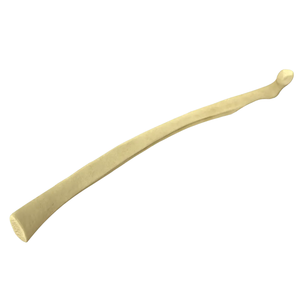
First rib anatomy quiz
Quiz on the systemic anatomy of the first rib: structure, grooves, tubercles, muscle attachments, vessels, and nerves.
1/20
bold
text
1. To which anatomical classification of ribs does the first rib belong?
-
False rib
The first rib is classified as a true rib (costae verae) because it connects directly to the sternum through its own costal cartilage. Ribs I–VII are considered true ribs.
-
True rib
The first rib is classified as a true rib (costae verae) because it connects directly to the sternum through its own costal cartilage. Ribs I–VII are considered true ribs.
-
Floating rib
The first rib is classified as a true rib (costae verae) because it connects directly to the sternum through its own costal cartilage. Ribs I–VII are considered true ribs.
-
Accessory rib
The first rib is classified as a true rib (costae verae) because it connects directly to the sternum through its own costal cartilage. Ribs I–VII are considered true ribs.
-
I find it difficult to answer
The first rib is classified as a true rib (costae verae) because it connects directly to the sternum through its own costal cartilage. Ribs I–VII are considered true ribs.
2. Which surface of the first rib faces superiorly (cranially)?
-
External surface
Unlike most ribs, the first rib lies almost in the horizontal plane; therefore, it has superior (facies superior) and inferior (facies inferior) surfaces rather than external and internal surfaces.
-
Internal surface
Unlike most ribs, the first rib lies almost in the horizontal plane; therefore, it has superior (facies superior) and inferior (facies inferior) surfaces rather than external and internal surfaces.
-
Superior surface
Unlike most ribs, the first rib lies almost in the horizontal plane; therefore, it has superior (facies superior) and inferior (facies inferior) surfaces rather than external and internal surfaces.
-
Inferior surface
Unlike most ribs, the first rib lies almost in the horizontal plane; therefore, it has superior (facies superior) and inferior (facies inferior) surfaces rather than external and internal surfaces.
-
I find it difficult to answer
Unlike most ribs, the first rib lies almost in the horizontal plane; therefore, it has superior (facies superior) and inferior (facies inferior) surfaces rather than external and internal surfaces.
3. Where is the groove for the subclavian artery located on the superior surface of the first rib?
-
Anterior to the scalene tubercle
The sulcus arteriae subclaviae is located poster to the tuberculum musculi scaleni anterioris—the attachment site of the anterior scalene muscle. The subclavian vein passes anterior to the tubercle.
-
Poster to the scalene tubercle
The sulcus arteriae subclaviae is located poster to the tuberculum musculi scaleni anterioris—the attachment site of the anterior scalene muscle. The subclavian vein passes anterior to the tubercle.
-
Anterior to the head of the rib
The sulcus arteriae subclaviae is located poster to the tuberculum musculi scaleni anterioris—the attachment site of the anterior scalene muscle. The subclavian vein passes anterior to the tubercle.
-
On the internal border of the rib
The sulcus arteriae subclaviae is located poster to the tuberculum musculi scaleni anterioris—the attachment site of the anterior scalene muscle. The subclavian vein passes anterior to the tubercle.
-
I find it difficult to answer
The sulcus arteriae subclaviae is located poster to the tuberculum musculi scaleni anterioris—the attachment site of the anterior scalene muscle. The subclavian vein passes anterior to the tubercle.
4. What is the tubercle on the superior surface of the first rib to which the anterior scalene muscle attaches called?
-
Tubercle of rib
The scalene tubercle (tuberculum musculi scaleni anterioris, or Lisfranc’s tubercle) is a characteristic landmark on the superior surface of the first rib that separates the grooves for the subclavian vein and artery.
-
Anterior scalene tubercle
The scalene tubercle (tuberculum musculi scaleni anterioris, or Lisfranc’s tubercle) is a characteristic landmark on the superior surface of the first rib that separates the grooves for the subclavian vein and artery.
-
Middle scalene tubercle
The scalene tubercle (tuberculum musculi scaleni anterioris, or Lisfranc’s tubercle) is a characteristic landmark on the superior surface of the first rib that separates the grooves for the subclavian vein and artery.
-
Tubercle of the subclavian artery
The scalene tubercle (tuberculum musculi scaleni anterioris, or Lisfranc’s tubercle) is a characteristic landmark on the superior surface of the first rib that separates the grooves for the subclavian vein and artery.
-
I find it difficult to answer
The scalene tubercle (tuberculum musculi scaleni anterioris, or Lisfranc’s tubercle) is a characteristic landmark on the superior surface of the first rib that separates the grooves for the subclavian vein and artery.
5. Which muscle attaches to Lisfranc’s tubercle of the first rib?
-
Scalenus medius muscle
The anterior scalene muscle (m. scalenus anterior) attaches to the tuberculum musculi scaleni anterioris. The middle scalene muscle attaches to the first rib poster to the groove for the subclavian artery.
-
Scalenus posterior muscle
The anterior scalene muscle (m. scalenus anterior) attaches to the tuberculum musculi scaleni anterioris. The middle scalene muscle attaches to the first rib poster to the groove for the subclavian artery.
-
Scalenus anterior muscle
The anterior scalene muscle (m. scalenus anterior) attaches to the tuberculum musculi scaleni anterioris. The middle scalene muscle attaches to the first rib poster to the groove for the subclavian artery.
-
Pectoralis minor muscle
The anterior scalene muscle (m. scalenus anterior) attaches to the tuberculum musculi scaleni anterioris. The middle scalene muscle attaches to the first rib poster to the groove for the subclavian artery.
-
I find it difficult to answer
The anterior scalene muscle (m. scalenus anterior) attaches to the tuberculum musculi scaleni anterioris. The middle scalene muscle attaches to the first rib poster to the groove for the subclavian artery.
6. Where does the subclavian vein pass relative to the scalene tubercle on the first rib?
-
Poster to the tubercle
The subclavian vein (v. subclavia) passes over the first rib anteverted to the scalene tubercle, whereas the subclavian artery passes poster to it. The groove for the vein is located anteromedial to the tubercle.
-
Anterior to the tubercle
The subclavian vein (v. subclavia) passes over the first rib anteverted to the scalene tubercle, whereas the subclavian artery passes poster to it. The groove for the vein is located anteromedial to the tubercle.
-
Medial to the head of the rib
The subclavian vein (v. subclavia) passes over the first rib anteverted to the scalene tubercle, whereas the subclavian artery passes poster to it. The groove for the vein is located anteromedial to the tubercle.
-
In the groove poster to the tubercle
The subclavian vein (v. subclavia) passes over the first rib anteverted to the scalene tubercle, whereas the subclavian artery passes poster to it. The groove for the vein is located anteromedial to the tubercle.
-
I find it difficult to answer
The subclavian vein (v. subclavia) passes over the first rib anteverted to the scalene tubercle, whereas the subclavian artery passes poster to it. The groove for the vein is located anteromedial to the tubercle.
7. The first rib has pronounced curvature. How is its shape described?
-
Straight, without curves
The first rib is the shortest, broadest, and most curved of all the ribs. It is flat and broad, with a single curve in the horizontal plane, which distinguishes it from the long, narrow ribs below it.
-
S-shaped, with two curves
The first rib is the shortest, broadest, and most curved of all the ribs. It is flat and broad, with a single curve in the horizontal plane, which distinguishes it from the long, narrow ribs below it.
-
Flat and broad, with a single curve in the horizontal plane
The first rib is the shortest, broadest, and most curved of all the ribs. It is flat and broad, with a single curve in the horizontal plane, which distinguishes it from the long, narrow ribs below it.
-
Cylindrical and tubular
The first rib is the shortest, broadest, and most curved of all the ribs. It is flat and broad, with a single curve in the horizontal plane, which distinguishes it from the long, narrow ribs below it.
-
I find it difficult to answer
The first rib is the shortest, broadest, and most curved of all the ribs. It is flat and broad, with a single curve in the horizontal plane, which distinguishes it from the long, narrow ribs below it.
8. With which vertebra does the head of the first rib articulate?
-
Only with C7
The head of the first rib (caput costae I) comprises one articular surface and articulates only with the body of one vertebra—Th1 (the first thoracic vertebra). This distinguishes the first rib from most ribs, whose heads articulate with two adjacent vertebrae.
-
Only with Th1
The head of the first rib (caput costae I) comprises one articular surface and articulates only with the body of one vertebra—Th1 (the first thoracic vertebra). This distinguishes the first rib from most ribs, whose heads articulate with two adjacent vertebrae.
-
With C7 and Th1
The head of the first rib (caput costae I) comprises one articular surface and articulates only with the body of one vertebra—Th1 (the first thoracic vertebra). This distinguishes the first rib from most ribs, whose heads articulate with two adjacent vertebrae.
-
With Th1 and Th2
The head of the first rib (caput costae I) comprises one articular surface and articulates only with the body of one vertebra—Th1 (the first thoracic vertebra). This distinguishes the first rib from most ribs, whose heads articulate with two adjacent vertebrae.
-
I find it difficult to answer
The head of the first rib (caput costae I) comprises one articular surface and articulates only with the body of one vertebra—Th1 (the first thoracic vertebra). This distinguishes the first rib from most ribs, whose heads articulate with two adjacent vertebrae.
9. How many articular surfaces are present on the head of the first rib?
-
One.
The head of the first rib bears one articular surface (facies articularis capitis costae) because it articulates with only one vertebra—Th1. The heads of ribs II–X comprise two surfaces separated by a crest.
-
Two.
The head of the first rib bears one articular surface (facies articularis capitis costae) because it articulates with only one vertebra—Th1. The heads of ribs II–X comprise two surfaces separated by a crest.
-
Three.
The head of the first rib bears one articular surface (facies articularis capitis costae) because it articulates with only one vertebra—Th1. The heads of ribs II–X comprise two surfaces separated by a crest.
-
The head of the first rib has no articular surfaces
The head of the first rib bears one articular surface (facies articularis capitis costae) because it articulates with only one vertebra—Th1. The heads of ribs II–X comprise two surfaces separated by a crest.
-
I find it difficult to answer
The head of the first rib bears one articular surface (facies articularis capitis costae) because it articulates with only one vertebra—Th1. The heads of ribs II–X comprise two surfaces separated by a crest.
10. Where is the tubercle of the rib (tuberculum costae) located on the first rib?
-
On the superior surface, near the internal border
The tuberculum costae is located on the posterior (external) surface of the rib at the junction of the neck and body. It bears an articular surface for articulation with the transverse process of the corresponding vertebra (costotransverse joint).
-
On the external surface of the neck, at the junction of the neck and body
The tuberculum costae is located on the posterior (external) surface of the rib at the junction of the neck and body. It bears an articular surface for articulation with the transverse process of the corresponding vertebra (costotransverse joint).
-
On the inferior surface of the body, near the external border
The tuberculum costae is located on the posterior (external) surface of the rib at the junction of the neck and body. It bears an articular surface for articulation with the transverse process of the corresponding vertebra (costotransverse joint).
-
On the internal surface of the body, in the middle
The tuberculum costae is located on the posterior (external) surface of the rib at the junction of the neck and body. It bears an articular surface for articulation with the transverse process of the corresponding vertebra (costotransverse joint).
-
I find it difficult to answer
The tuberculum costae is located on the posterior (external) surface of the rib at the junction of the neck and body. It bears an articular surface for articulation with the transverse process of the corresponding vertebra (costotransverse joint).
11. Which muscle attaches to the first rib poster to the groove for the subclavian artery?
-
Scalenus anterior muscle
The middle scalene muscle (m. scalenus medius) attaches to the superior surface of the first rib poster to the sulcus arteriae subclaviae—posterior to the subclavian artery. The anterior scalene muscle attaches to the tuberculum musculi scaleni anterioris.
-
Pectoralis minor muscle
The middle scalene muscle (m. scalenus medius) attaches to the superior surface of the first rib poster to the sulcus arteriae subclaviae—posterior to the subclavian artery. The anterior scalene muscle attaches to the tuberculum musculi scaleni anterioris.
-
Scalenus medius muscle
The middle scalene muscle (m. scalenus medius) attaches to the superior surface of the first rib poster to the sulcus arteriae subclaviae—posterior to the subclavian artery. The anterior scalene muscle attaches to the tuberculum musculi scaleni anterioris.
-
Subclavius muscle
The middle scalene muscle (m. scalenus medius) attaches to the superior surface of the first rib poster to the sulcus arteriae subclaviae—posterior to the subclavian artery. The anterior scalene muscle attaches to the tuberculum musculi scaleni anterioris.
-
I find it difficult to answer
The middle scalene muscle (m. scalenus medius) attaches to the superior surface of the first rib poster to the sulcus arteriae subclaviae—posterior to the subclavian artery. The anterior scalene muscle attaches to the tuberculum musculi scaleni anterioris.
12. To which structure of the first rib does the subclavius muscle (m. subclavius) attach?
-
To the head of the rib
The subclavius muscle (m. subclavius) originates from the cartilage of the first rib and the inferior surface of the osseous part of the first rib near the sternal end. It depresses the clavicle and stabilizes the sternoclavicular joint.
-
To the tubercle of the rib
The subclavius muscle (m. subclavius) originates from the cartilage of the first rib and the inferior surface of the osseous part of the first rib near the sternal end. It depresses the clavicle and stabilizes the sternoclavicular joint.
-
To the cartilage of the first rib and the inferior surface of the body of the first rib near the sternal end
The subclavius muscle (m. subclavius) originates from the cartilage of the first rib and the inferior surface of the osseous part of the first rib near the sternal end. It depresses the clavicle and stabilizes the sternoclavicular joint.
-
To the neck of the rib
The subclavius muscle (m. subclavius) originates from the cartilage of the first rib and the inferior surface of the osseous part of the first rib near the sternal end. It depresses the clavicle and stabilizes the sternoclavicular joint.
-
I find it difficult to answer
The subclavius muscle (m. subclavius) originates from the cartilage of the first rib and the inferior surface of the osseous part of the first rib near the sternal end. It depresses the clavicle and stabilizes the sternoclavicular joint.
13. What is the articulation between the first rib and the sternum called?
-
Diarthrosis (synovial joint)
The first rib is connected to the manubrium of the sternum by a synchondrosis (synchondrosis costosternalis I)—a cartilaginous joint without a joint cavity. Unlike ribs II–VII, movement here is minimal.
-
Synchondrosis (cartilaginous joint)
The first rib is connected to the manubrium of the sternum by a synchondrosis (synchondrosis costosternalis I)—a cartilaginous joint without a joint cavity. Unlike ribs II–VII, movement here is minimal.
-
Syndesmosis (fibrous joint)
The first rib is connected to the manubrium of the sternum by a synchondrosis (synchondrosis costosternalis I)—a cartilaginous joint without a joint cavity. Unlike ribs II–VII, movement here is minimal.
-
Synostosis (osseous union)
The first rib is connected to the manubrium of the sternum by a synchondrosis (synchondrosis costosternalis I)—a cartilaginous joint without a joint cavity. Unlike ribs II–VII, movement here is minimal.
-
I find it difficult to answer
The first rib is connected to the manubrium of the sternum by a synchondrosis (synchondrosis costosternalis I)—a cartilaginous joint without a joint cavity. Unlike ribs II–VII, movement here is minimal.
14. Which nerve structure passes directly over the superior surface of the first rib?
-
Ulnar nerve
The trunks of the brachial plexus (trunci plexus brachialis) pass between the anterior and middle scalene muscles and over the superior surface of the first rib poster to the subclavian artery, curving around the rib on their course to the axillary region.
-
Axillary nerve
The trunks of the brachial plexus (trunci plexus brachialis) pass between the anterior and middle scalene muscles and over the superior surface of the first rib poster to the subclavian artery, curving around the rib on their course to the axillary region.
-
Trunks of the brachial plexus
The trunks of the brachial plexus (trunci plexus brachialis) pass between the anterior and middle scalene muscles and over the superior surface of the first rib poster to the subclavian artery, curving around the rib on their course to the axillary region.
-
Radial nerve
The trunks of the brachial plexus (trunci plexus brachialis) pass between the anterior and middle scalene muscles and over the superior surface of the first rib poster to the subclavian artery, curving around the rib on their course to the axillary region.
-
I find it difficult to answer
The trunks of the brachial plexus (trunci plexus brachialis) pass between the anterior and middle scalene muscles and over the superior surface of the first rib poster to the subclavian artery, curving around the rib on their course to the axillary region.
15. Which part of the first rib is called the “neck” (collum costae)?
-
The flattened part between the costal cartilage and the tubercle
The neck of the rib (collum costae) is the narrowed part located between the head (caput costae) and the tubercle (tuberculum costae). The neck of the first rib is short and well defined.
-
The narrowed part between the head and tubercle of the rib
The neck of the rib (collum costae) is the narrowed part located between the head (caput costae) and the tubercle (tuberculum costae). The neck of the first rib is short and well defined.
-
The broad part of the body of the rib
The neck of the rib (collum costae) is the narrowed part located between the head (caput costae) and the tubercle (tuberculum costae). The neck of the first rib is short and well defined.
-
The junction of the rib and costal cartilage
The neck of the rib (collum costae) is the narrowed part located between the head (caput costae) and the tubercle (tuberculum costae). The neck of the first rib is short and well defined.
-
I find it difficult to answer
The neck of the rib (collum costae) is the narrowed part located between the head (caput costae) and the tubercle (tuberculum costae). The neck of the first rib is short and well defined.
16. Which vessel produces a groove (sulcus) on the superior surface of the first rib poster to the scalene tubercle?
-
Subclavian vein
The subclavian artery (a. subclavia) produces a groove (sulcus arteriae subclaviae) on the superior surface of the first rib poster to the tuberculum musculi scaleni anterioris. The groove for the subclavian vein is located anteverted to this tubercle.
-
Subclavian artery
The subclavian artery (a. subclavia) produces a groove (sulcus arteriae subclaviae) on the superior surface of the first rib poster to the tuberculum musculi scaleni anterioris. The groove for the subclavian vein is located anteverted to this tubercle.
-
Common carotid artery
The subclavian artery (a. subclavia) produces a groove (sulcus arteriae subclaviae) on the superior surface of the first rib poster to the tuberculum musculi scaleni anterioris. The groove for the subclavian vein is located anteverted to this tubercle.
-
Vertebral artery
The subclavian artery (a. subclavia) produces a groove (sulcus arteriae subclaviae) on the superior surface of the first rib poster to the tuberculum musculi scaleni anterioris. The groove for the subclavian vein is located anteverted to this tubercle.
-
I find it difficult to answer
The subclavian artery (a. subclavia) produces a groove (sulcus arteriae subclaviae) on the superior surface of the first rib poster to the tuberculum musculi scaleni anterioris. The groove for the subclavian vein is located anteverted to this tubercle.
17. Which anatomical opening (aperture) does the first rib contribute to forming?
-
Inferior thoracic aperture
Together with the manubrium of the sternum and the first thoracic vertebra (Th1), the first rib bounds the superior thoracic aperture (apertura thoracis superior), through which the great vessels, nerves, and trachea pass.
-
Superior thoracic aperture
Together with the manubrium of the sternum and the first thoracic vertebra (Th1), the first rib bounds the superior thoracic aperture (apertura thoracis superior), through which the great vessels, nerves, and trachea pass.
-
First intercostal space
Together with the manubrium of the sternum and the first thoracic vertebra (Th1), the first rib bounds the superior thoracic aperture (apertura thoracis superior), through which the great vessels, nerves, and trachea pass.
-
Aperture of the pulmonary trunk
Together with the manubrium of the sternum and the first thoracic vertebra (Th1), the first rib bounds the superior thoracic aperture (apertura thoracis superior), through which the great vessels, nerves, and trachea pass.
-
I find it difficult to answer
Together with the manubrium of the sternum and the first thoracic vertebra (Th1), the first rib bounds the superior thoracic aperture (apertura thoracis superior), through which the great vessels, nerves, and trachea pass.
18. The angle of the rib (angulus costae) in the first rib:
-
Is sharply defined and located near the head
In the first rib, the angle of the rib (angulus costae) practically coincides with the tubercle (tuberculum costae) and is not separately defined. This distinguishes the first rib from the ribs below it, in which the angle is progressively displaced farther poster to the tubercle.
-
Coincides with the tubercle of the rib and is practically not separately defined
In the first rib, the angle of the rib (angulus costae) practically coincides with the tubercle (tuberculum costae) and is not separately defined. This distinguishes the first rib from the ribs below it, in which the angle is progressively displaced farther poster to the tubercle.
-
Is located in the middle of the body of the rib
In the first rib, the angle of the rib (angulus costae) practically coincides with the tubercle (tuberculum costae) and is not separately defined. This distinguishes the first rib from the ribs below it, in which the angle is progressively displaced farther poster to the tubercle.
-
Is absent in the first rib
In the first rib, the angle of the rib (angulus costae) practically coincides with the tubercle (tuberculum costae) and is not separately defined. This distinguishes the first rib from the ribs below it, in which the angle is progressively displaced farther poster to the tubercle.
-
I find it difficult to answer
In the first rib, the angle of the rib (angulus costae) practically coincides with the tubercle (tuberculum costae) and is not separately defined. This distinguishes the first rib from the ribs below it, in which the angle is progressively displaced farther poster to the tubercle.
19. How does the groove for the subclavian vein on the first rib compare with the groove for the subclavian artery?
-
The groove for the vein is located posteriorly and is deeper
The groove for the subclavian vein (sulcus venae subclaviae) lies anteverted to the tuberculum musculi scaleni anterioris and is shallower than the sulcus arteriae subclaviae, which lies poster to the tubercle and is more pronounced.
-
The groove for the vein is located anteriorly and is shallower
The groove for the subclavian vein (sulcus venae subclaviae) lies anteverted to the tuberculum musculi scaleni anterioris and is shallower than the sulcus arteriae subclaviae, which lies poster to the tubercle and is more pronounced.
-
The grooves for the vein and artery are indistinguishable
The groove for the subclavian vein (sulcus venae subclaviae) lies anteverted to the tuberculum musculi scaleni anterioris and is shallower than the sulcus arteriae subclaviae, which lies poster to the tubercle and is more pronounced.
-
The groove for the vein is absent from the first rib
The groove for the subclavian vein (sulcus venae subclaviae) lies anteverted to the tuberculum musculi scaleni anterioris and is shallower than the sulcus arteriae subclaviae, which lies poster to the tubercle and is more pronounced.
-
I find it difficult to answer
The groove for the subclavian vein (sulcus venae subclaviae) lies anteverted to the tuberculum musculi scaleni anterioris and is shallower than the sulcus arteriae subclaviae, which lies poster to the tubercle and is more pronounced.
20. The costotransverse joint of the first rib is formed by the tubercle of the rib and which process of the vertebra?
-
Spinous process of Th1
The costotransverse joint (articulatio costotransversaria) of the first rib is formed by the articular surface of the tubercle of the first rib and the costal facet of the transverse process of the first thoracic vertebra (Th1). This is a true synovial joint.
-
Transverse process of Th1
The costotransverse joint (articulatio costotransversaria) of the first rib is formed by the articular surface of the tubercle of the first rib and the costal facet of the transverse process of the first thoracic vertebra (Th1). This is a true synovial joint.
-
Articular process of Th1
The costotransverse joint (articulatio costotransversaria) of the first rib is formed by the articular surface of the tubercle of the first rib and the costal facet of the transverse process of the first thoracic vertebra (Th1). This is a true synovial joint.
-
Transverse process of C7
The costotransverse joint (articulatio costotransversaria) of the first rib is formed by the articular surface of the tubercle of the first rib and the costal facet of the transverse process of the first thoracic vertebra (Th1). This is a true synovial joint.
-
I find it difficult to answer
The costotransverse joint (articulatio costotransversaria) of the first rib is formed by the articular surface of the tubercle of the first rib and the costal facet of the transverse process of the first thoracic vertebra (Th1). This is a true synovial joint.
Retake this quiz?
Your current progress will be reset.
