
Sphenoid bone anatomy quiz
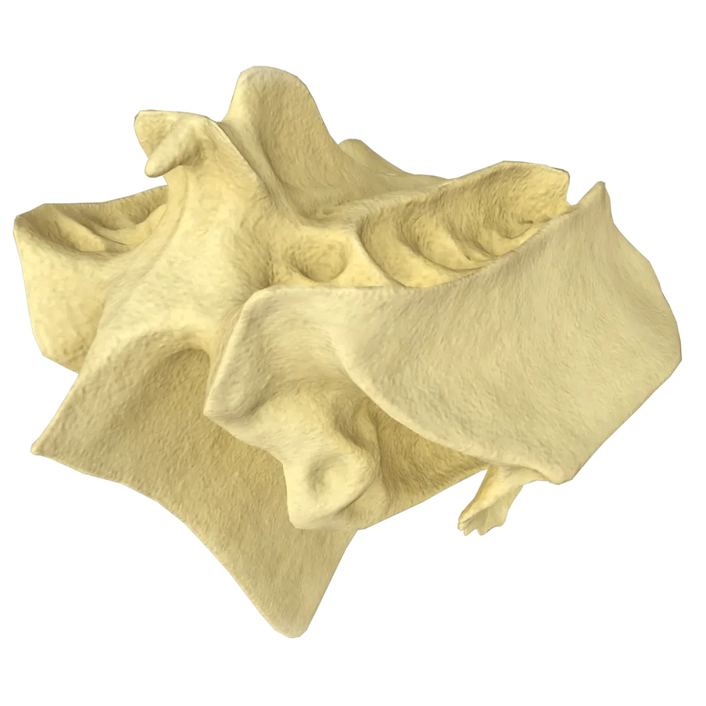
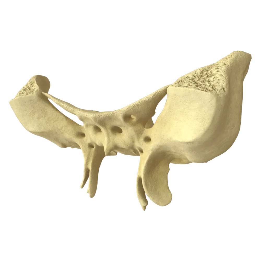
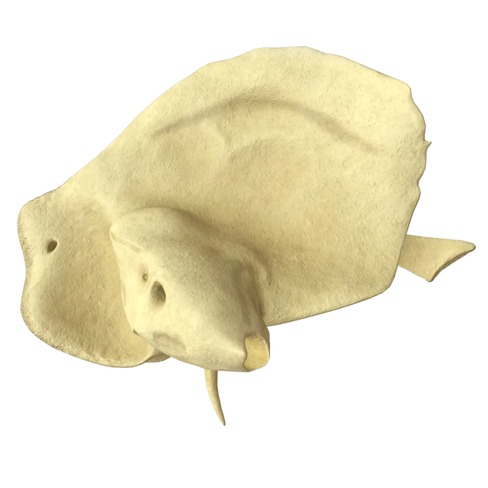
The quiz covers the structure of the sphenoid bone: its parts, processes, foramina, canals, articular surfaces, and anatomical landmarks.
1/20
bold
text
1. The sphenoid bone consists of the following main parts:
-
A body, two greater wings, two lesser wings, and two pterygoid processes
The sphenoid bone (os sphenoidale) consists of a body (corpus), two greater wings (alae majores), two lesser wings (alae minores), and two pterygoid processes (processus pterygoidei). This is the complete list of its parts according to Terminologia Anatomica.
-
A body, two greater wings, and two lesser wings
The sphenoid bone (os sphenoidale) consists of a body (corpus), two greater wings (alae majores), two lesser wings (alae minores), and two pterygoid processes (processus pterygoidei). This is the complete list of its parts according to Terminologia Anatomica.
-
A body, two pterygoid processes, and two zygomatic processes
The sphenoid bone (os sphenoidale) consists of a body (corpus), two greater wings (alae majores), two lesser wings (alae minores), and two pterygoid processes (processus pterygoidei). This is the complete list of its parts according to Terminologia Anatomica.
-
A body, two lesser wings, and two pterygoid processes
The sphenoid bone (os sphenoidale) consists of a body (corpus), two greater wings (alae majores), two lesser wings (alae minores), and two pterygoid processes (processus pterygoidei). This is the complete list of its parts according to Terminologia Anatomica.
-
I find it difficult to answer
The sphenoid bone (os sphenoidale) consists of a body (corpus), two greater wings (alae majores), two lesser wings (alae minores), and two pterygoid processes (processus pterygoidei). This is the complete list of its parts according to Terminologia Anatomica.
2. Which structure is located on the superior surface of the body of the sphenoid bone and contains the pituitary gland?
-
Hypophyseal fossa (fossa hypophysialis)
The sella turcica (sella turcica) is located on the superior surface of the body of the sphenoid bone; its central part is occupied by the hypophyseal fossa (fossa hypophysialis), a depression for the pituitary gland. The optic chiasm is located anterior to the sella but is not a bony structure.
-
Optic chiasm (chiasma opticum)
The sella turcica (sella turcica) is located on the superior surface of the body of the sphenoid bone; its central part is occupied by the hypophyseal fossa (fossa hypophysialis), a depression for the pituitary gland. The optic chiasm is located anterior to the sella but is not a bony structure.
-
Meckel's cave (cavum trigeminale)
The sella turcica (sella turcica) is located on the superior surface of the body of the sphenoid bone; its central part is occupied by the hypophyseal fossa (fossa hypophysialis), a depression for the pituitary gland. The optic chiasm is located anterior to the sella but is not a bony structure.
-
Foramen lacerum (foramen lacerum)
The sella turcica (sella turcica) is located on the superior surface of the body of the sphenoid bone; its central part is occupied by the hypophyseal fossa (fossa hypophysialis), a depression for the pituitary gland. The optic chiasm is located anterior to the sella but is not a bony structure.
-
I find it difficult to answer
The sella turcica (sella turcica) is located on the superior surface of the body of the sphenoid bone; its central part is occupied by the hypophyseal fossa (fossa hypophysialis), a depression for the pituitary gland. The optic chiasm is located anterior to the sella but is not a bony structure.
3. The sella turcica (sella turcica) of the sphenoid bone is bounded posteriorly by the following structure:
-
Tuberculum sellae (tuberculum sellae)
The sella turcica is bounded posteriorly by the dorsum sellae (dorsum sellae), a plate that rises above the hypophyseal fossa. Anterior to the hypophyseal fossa is the tuberculum sellae (tuberculum sellae), and further anteriorly is the prechiasmatic sulcus (sulcus prechiasmatis).
-
Sphenoidal crest (crista sphenoidalis)
The sella turcica is bounded posteriorly by the dorsum sellae (dorsum sellae), a plate that rises above the hypophyseal fossa. Anterior to the hypophyseal fossa is the tuberculum sellae (tuberculum sellae), and further anteriorly is the prechiasmatic sulcus (sulcus prechiasmatis).
-
Dorsum sellae (dorsum sellae)
The sella turcica is bounded posteriorly by the dorsum sellae (dorsum sellae), a plate that rises above the hypophyseal fossa. Anterior to the hypophyseal fossa is the tuberculum sellae (tuberculum sellae), and further anteriorly is the prechiasmatic sulcus (sulcus prechiasmatis).
-
Prechiasmatic sulcus (sulcus prechiasmatis)
The sella turcica is bounded posteriorly by the dorsum sellae (dorsum sellae), a plate that rises above the hypophyseal fossa. Anterior to the hypophyseal fossa is the tuberculum sellae (tuberculum sellae), and further anteriorly is the prechiasmatic sulcus (sulcus prechiasmatis).
-
I find it difficult to answer
The sella turcica is bounded posteriorly by the dorsum sellae (dorsum sellae), a plate that rises above the hypophyseal fossa. Anterior to the hypophyseal fossa is the tuberculum sellae (tuberculum sellae), and further anteriorly is the prechiasmatic sulcus (sulcus prechiasmatis).
4. The optic canal (canalis opticus) is formed by:
-
The greater wing of the sphenoid bone
The optic canal (canalis opticus) is formed at the root of the lesser wing (ala minor) of the sphenoid bone. The optic nerve (n. opticus) and ophthalmic artery (a. ophthalmica) pass through it.
-
The lesser wing of the sphenoid bone at its root
The optic canal (canalis opticus) is formed at the root of the lesser wing (ala minor) of the sphenoid bone. The optic nerve (n. opticus) and ophthalmic artery (a. ophthalmica) pass through it.
-
The body of the sphenoid bone together with the ethmoid bone
The optic canal (canalis opticus) is formed at the root of the lesser wing (ala minor) of the sphenoid bone. The optic nerve (n. opticus) and ophthalmic artery (a. ophthalmica) pass through it.
-
The pterygoid process of the sphenoid bone
The optic canal (canalis opticus) is formed at the root of the lesser wing (ala minor) of the sphenoid bone. The optic nerve (n. opticus) and ophthalmic artery (a. ophthalmica) pass through it.
-
I find it difficult to answer
The optic canal (canalis opticus) is formed at the root of the lesser wing (ala minor) of the sphenoid bone. The optic nerve (n. opticus) and ophthalmic artery (a. ophthalmica) pass through it.
5. The following nerves pass through the superior orbital fissure (fissura orbitalis superior), EXCEPT:
-
Oculomotor nerve (n. oculomotorius, CN III)
CN III, CN IV, CN VI, and CN V1 (ophthalmic nerve), as well as the ophthalmic vein, pass through the superior orbital fissure. The optic nerve (CN II) passes through the optic canal (canalis opticus), not through the fissure.
-
Trochlear nerve (n. trochlearis, CN IV)
CN III, CN IV, CN VI, and CN V1 (ophthalmic nerve), as well as the ophthalmic vein, pass through the superior orbital fissure. The optic nerve (CN II) passes through the optic canal (canalis opticus), not through the fissure.
-
Ophthalmic nerve (n. ophthalmicus, CN V1)
CN III, CN IV, CN VI, and CN V1 (ophthalmic nerve), as well as the ophthalmic vein, pass through the superior orbital fissure. The optic nerve (CN II) passes through the optic canal (canalis opticus), not through the fissure.
-
Optic nerve (n. opticus, CN II)
CN III, CN IV, CN VI, and CN V1 (ophthalmic nerve), as well as the ophthalmic vein, pass through the superior orbital fissure. The optic nerve (CN II) passes through the optic canal (canalis opticus), not through the fissure.
-
I find it difficult to answer
CN III, CN IV, CN VI, and CN V1 (ophthalmic nerve), as well as the ophthalmic vein, pass through the superior orbital fissure. The optic nerve (CN II) passes through the optic canal (canalis opticus), not through the fissure.
6. Which foramen of the sphenoid bone is the largest of the three foramina in its greater wing?
-
Foramen rotundum
Among the foramina of the greater wing (foramen rotundum, foramen ovale, foramen spinosum), the foramen ovale (foramen ovale) has the largest diameter; the mandibular nerve (n. mandibularis, CN V3) passes through it.
-
Foramen ovale
Among the foramina of the greater wing (foramen rotundum, foramen ovale, foramen spinosum), the foramen ovale (foramen ovale) has the largest diameter; the mandibular nerve (n. mandibularis, CN V3) passes through it.
-
Foramen spinosum
Among the foramina of the greater wing (foramen rotundum, foramen ovale, foramen spinosum), the foramen ovale (foramen ovale) has the largest diameter; the mandibular nerve (n. mandibularis, CN V3) passes through it.
-
Foramen lacerum (foramen lacerum)
Among the foramina of the greater wing (foramen rotundum, foramen ovale, foramen spinosum), the foramen ovale (foramen ovale) has the largest diameter; the mandibular nerve (n. mandibularis, CN V3) passes through it.
-
I find it difficult to answer
Among the foramina of the greater wing (foramen rotundum, foramen ovale, foramen spinosum), the foramen ovale (foramen ovale) has the largest diameter; the mandibular nerve (n. mandibularis, CN V3) passes through it.
7. The following passes through the foramen rotundum (foramen rotundum) of the sphenoid bone:
-
Mandibular nerve (n. mandibularis, CN V3)
The foramen rotundum (foramen rotundum) is located in the greater wing of the sphenoid bone and provides a route for the maxillary nerve (n. maxillaris, CN V2) to exit the skull into the pterygopalatine fossa (fossa pterygopalatina).
-
Middle meningeal artery (a. meningea media)
The foramen rotundum (foramen rotundum) is located in the greater wing of the sphenoid bone and provides a route for the maxillary nerve (n. maxillaris, CN V2) to exit the skull into the pterygopalatine fossa (fossa pterygopalatina).
-
Maxillary nerve (n. maxillaris, CN V2)
The foramen rotundum (foramen rotundum) is located in the greater wing of the sphenoid bone and provides a route for the maxillary nerve (n. maxillaris, CN V2) to exit the skull into the pterygopalatine fossa (fossa pterygopalatina).
-
Ophthalmic nerve (n. ophthalmicus, CN V1)
The foramen rotundum (foramen rotundum) is located in the greater wing of the sphenoid bone and provides a route for the maxillary nerve (n. maxillaris, CN V2) to exit the skull into the pterygopalatine fossa (fossa pterygopalatina).
-
I find it difficult to answer
The foramen rotundum (foramen rotundum) is located in the greater wing of the sphenoid bone and provides a route for the maxillary nerve (n. maxillaris, CN V2) to exit the skull into the pterygopalatine fossa (fossa pterygopalatina).
8. The following passes through the foramen spinosum (foramen spinosum) of the sphenoid bone:
-
Middle meningeal artery (a. meningea media) and the meningeal branch of the mandibular nerve
Through the foramen spinosum (foramen spinosum), the middle meningeal artery (a. meningea media) — a branch of the a. maxillaris — enters the cranial cavity, while the meningeal branch of the mandibular nerve (ramus meningeus n. mandibularis) exits.
-
Mandibular nerve (n. mandibularis)
Through the foramen spinosum (foramen spinosum), the middle meningeal artery (a. meningea media) — a branch of the a. maxillaris — enters the cranial cavity, while the meningeal branch of the mandibular nerve (ramus meningeus n. mandibularis) exits.
-
Maxillary nerve (n. maxillaris)
Through the foramen spinosum (foramen spinosum), the middle meningeal artery (a. meningea media) — a branch of the a. maxillaris — enters the cranial cavity, while the meningeal branch of the mandibular nerve (ramus meningeus n. mandibularis) exits.
-
Lesser petrosal nerve (n. petrosus minor)
Through the foramen spinosum (foramen spinosum), the middle meningeal artery (a. meningea media) — a branch of the a. maxillaris — enters the cranial cavity, while the meningeal branch of the mandibular nerve (ramus meningeus n. mandibularis) exits.
-
I find it difficult to answer
Through the foramen spinosum (foramen spinosum), the middle meningeal artery (a. meningea media) — a branch of the a. maxillaris — enters the cranial cavity, while the meningeal branch of the mandibular nerve (ramus meningeus n. mandibularis) exits.
9. The pterygoid canal (canalis pterygoideus) of the sphenoid bone connects:
-
The middle cranial fossa with the infratemporal fossa
The pterygoid canal (canalis pterygoideus, Vidian canal) passes through the base of the pterygoid process and connects the foramen lacerum (foramen lacerum) anteriorly with the pterygopalatine fossa (fossa pterygopalatina). The Vidian nerve (n. canalis pterygoidei) passes through it.
-
The foramen lacerum with the pterygopalatine fossa
The pterygoid canal (canalis pterygoideus, Vidian canal) passes through the base of the pterygoid process and connects the foramen lacerum (foramen lacerum) anteriorly with the pterygopalatine fossa (fossa pterygopalatina). The Vidian nerve (n. canalis pterygoidei) passes through it.
-
The orbit with the pterygopalatine fossa
The pterygoid canal (canalis pterygoideus, Vidian canal) passes through the base of the pterygoid process and connects the foramen lacerum (foramen lacerum) anteriorly with the pterygopalatine fossa (fossa pterygopalatina). The Vidian nerve (n. canalis pterygoidei) passes through it.
-
The nasal cavity with the pterygopalatine fossa
The pterygoid canal (canalis pterygoideus, Vidian canal) passes through the base of the pterygoid process and connects the foramen lacerum (foramen lacerum) anteriorly with the pterygopalatine fossa (fossa pterygopalatina). The Vidian nerve (n. canalis pterygoidei) passes through it.
-
I find it difficult to answer
The pterygoid canal (canalis pterygoideus, Vidian canal) passes through the base of the pterygoid process and connects the foramen lacerum (foramen lacerum) anteriorly with the pterygopalatine fossa (fossa pterygopalatina). The Vidian nerve (n. canalis pterygoidei) passes through it.
10. The medial plate of the pterygoid process (lamina medialis processus pterygoidei) terminates inferiorly in the following structure:
-
Pterygoid hamulus (hamulus pterygoideus)
The medial plate of the pterygoid process terminates inferiorly in the pterygoid hamulus (hamulus pterygoideus), around which the tendon of the tensor veli palatini muscle (m. tensor veli palatini) passes.
-
Pyramidal process
The medial plate of the pterygoid process terminates inferiorly in the pterygoid hamulus (hamulus pterygoideus), around which the tendon of the tensor veli palatini muscle (m. tensor veli palatini) passes.
-
Pterygoid fossa (fossa pterygoidea)
The medial plate of the pterygoid process terminates inferiorly in the pterygoid hamulus (hamulus pterygoideus), around which the tendon of the tensor veli palatini muscle (m. tensor veli palatini) passes.
-
Scaphoid fossa (fossa scaphoidea)
The medial plate of the pterygoid process terminates inferiorly in the pterygoid hamulus (hamulus pterygoideus), around which the tendon of the tensor veli palatini muscle (m. tensor veli palatini) passes.
-
I find it difficult to answer
The medial plate of the pterygoid process terminates inferiorly in the pterygoid hamulus (hamulus pterygoideus), around which the tendon of the tensor veli palatini muscle (m. tensor veli palatini) passes.
11. The scaphoid fossa (fossa scaphoidea) of the sphenoid bone is located:
-
On the lateral plate of the pterygoid process, at its apex
The scaphoid fossa (fossa scaphoidea) is located on the anterior aspect of the base of the medial plate of the pterygoid process. The medial portion of the tensor veli palatini muscle originates from it.
-
At the base of the medial plate of the pterygoid process, posteriorly
The scaphoid fossa (fossa scaphoidea) is located on the anterior aspect of the base of the medial plate of the pterygoid process. The medial portion of the tensor veli palatini muscle originates from it.
-
On the greater wing, medial to the foramen spinosum
The scaphoid fossa (fossa scaphoidea) is located on the anterior aspect of the base of the medial plate of the pterygoid process. The medial portion of the tensor veli palatini muscle originates from it.
-
At the base of the medial plate of the pterygoid process, anteriorly
The scaphoid fossa (fossa scaphoidea) is located on the anterior aspect of the base of the medial plate of the pterygoid process. The medial portion of the tensor veli palatini muscle originates from it.
-
I find it difficult to answer
The scaphoid fossa (fossa scaphoidea) is located on the anterior aspect of the base of the medial plate of the pterygoid process. The medial portion of the tensor veli palatini muscle originates from it.
12. The superior orbital fissure (fissura orbitalis superior) is formed between:
-
The lesser wing of the sphenoid bone and the frontal bone
The superior orbital fissure (fissura orbitalis superior) is bounded superiorly by the lesser wing (ala minor) and inferiorly by the greater wing (ala major) of the sphenoid bone. This slit-like space leads from the middle cranial fossa into the orbit.
-
Greater wing of the sphenoid bone and the frontal bone
The superior orbital fissure (fissura orbitalis superior) is bounded superiorly by the lesser wing (ala minor) and inferiorly by the greater wing (ala major) of the sphenoid bone. This slit-like space leads from the middle cranial fossa into the orbit.
-
The lesser and greater wings of the sphenoid bone
The superior orbital fissure (fissura orbitalis superior) is bounded superiorly by the lesser wing (ala minor) and inferiorly by the greater wing (ala major) of the sphenoid bone. This slit-like space leads from the middle cranial fossa into the orbit.
-
The greater wing of the sphenoid bone and the zygomatic bone
The superior orbital fissure (fissura orbitalis superior) is bounded superiorly by the lesser wing (ala minor) and inferiorly by the greater wing (ala major) of the sphenoid bone. This slit-like space leads from the middle cranial fossa into the orbit.
-
I find it difficult to answer
The superior orbital fissure (fissura orbitalis superior) is bounded superiorly by the lesser wing (ala minor) and inferiorly by the greater wing (ala major) of the sphenoid bone. This slit-like space leads from the middle cranial fossa into the orbit.
13. The inferior orbital fissure (fissura orbitalis inferior) is formed between the greater wing of the sphenoid bone and:
-
The maxillary and palatine bones
The inferior orbital fissure (fissura orbitalis inferior) is located between the greater wing of the sphenoid bone superiorly and the maxilla (os maxillare) and palatine bone (os palatinum) inferiorly. The maxillary nerve passes through it.
-
The frontal and zygomatic bones
The inferior orbital fissure (fissura orbitalis inferior) is located between the greater wing of the sphenoid bone superiorly and the maxilla (os maxillare) and palatine bone (os palatinum) inferiorly. The maxillary nerve passes through it.
-
The zygomatic and ethmoid bones
The inferior orbital fissure (fissura orbitalis inferior) is located between the greater wing of the sphenoid bone superiorly and the maxilla (os maxillare) and palatine bone (os palatinum) inferiorly. The maxillary nerve passes through it.
-
The lesser wing of the sphenoid bone and the lacrimal bone
The inferior orbital fissure (fissura orbitalis inferior) is located between the greater wing of the sphenoid bone superiorly and the maxilla (os maxillare) and palatine bone (os palatinum) inferiorly. The maxillary nerve passes through it.
-
I find it difficult to answer
The inferior orbital fissure (fissura orbitalis inferior) is located between the greater wing of the sphenoid bone superiorly and the maxilla (os maxillare) and palatine bone (os palatinum) inferiorly. The maxillary nerve passes through it.
14. The sphenoidal sinus (sinus sphenoidalis) is located in the:
-
Lesser wing of the sphenoid bone
The sphenoidal sinus (sinus sphenoidalis) is a paired air-filled cavity located within the body (corpus) of the sphenoid bone. It opens through an aperture on the anterior surface of the body into the sphenoethmoidal recess (recessus sphenoethmoidalis).
-
Greater wing of the sphenoid bone
The sphenoidal sinus (sinus sphenoidalis) is a paired air-filled cavity located within the body (corpus) of the sphenoid bone. It opens through an aperture on the anterior surface of the body into the sphenoethmoidal recess (recessus sphenoethmoidalis).
-
Body of the sphenoid bone
The sphenoidal sinus (sinus sphenoidalis) is a paired air-filled cavity located within the body (corpus) of the sphenoid bone. It opens through an aperture on the anterior surface of the body into the sphenoethmoidal recess (recessus sphenoethmoidalis).
-
Pterygoid process of the sphenoid bone
The sphenoidal sinus (sinus sphenoidalis) is a paired air-filled cavity located within the body (corpus) of the sphenoid bone. It opens through an aperture on the anterior surface of the body into the sphenoethmoidal recess (recessus sphenoethmoidalis).
-
I find it difficult to answer
The sphenoidal sinus (sinus sphenoidalis) is a paired air-filled cavity located within the body (corpus) of the sphenoid bone. It opens through an aperture on the anterior surface of the body into the sphenoethmoidal recess (recessus sphenoethmoidalis).
15. The sphenopalatine foramen (foramen sphenopalatinum) is formed by the sphenoid and palatine bones and leads from the pterygopalatine fossa into the:
-
Nasal cavity
The sphenopalatine foramen (foramen sphenopalatinum) connects the pterygopalatine fossa (fossa pterygopalatina) with the nasal cavity (cavitas nasi). The nasal branches of the maxillary nerve and the sphenopalatine artery pass through it.
-
Oral cavity
The sphenopalatine foramen (foramen sphenopalatinum) connects the pterygopalatine fossa (fossa pterygopalatina) with the nasal cavity (cavitas nasi). The nasal branches of the maxillary nerve and the sphenopalatine artery pass through it.
-
Orbit
The sphenopalatine foramen (foramen sphenopalatinum) connects the pterygopalatine fossa (fossa pterygopalatina) with the nasal cavity (cavitas nasi). The nasal branches of the maxillary nerve and the sphenopalatine artery pass through it.
-
Infratemporal fossa
The sphenopalatine foramen (foramen sphenopalatinum) connects the pterygopalatine fossa (fossa pterygopalatina) with the nasal cavity (cavitas nasi). The nasal branches of the maxillary nerve and the sphenopalatine artery pass through it.
-
I find it difficult to answer
The sphenopalatine foramen (foramen sphenopalatinum) connects the pterygopalatine fossa (fossa pterygopalatina) with the nasal cavity (cavitas nasi). The nasal branches of the maxillary nerve and the sphenopalatine artery pass through it.
16. The following is located on the anterior surface of the body of the sphenoid bone:
-
Sphenoidal crest (crista sphenoidalis) and sphenoidal rostrum (rostrum sphenoidale)
On the anterior surface of the body of the sphenoid bone is the vertical sphenoidal crest (crista sphenoidalis), which continues inferiorly as the sphenoidal rostrum (rostrum sphenoidale), which enters the groove of the vomer. The apertures of the sphenoidal sinus also open here.
-
Prechiasmatic sulcus (sulcus prechiasmatis)
On the anterior surface of the body of the sphenoid bone is the vertical sphenoidal crest (crista sphenoidalis), which continues inferiorly as the sphenoidal rostrum (rostrum sphenoidale), which enters the groove of the vomer. The apertures of the sphenoidal sinus also open here.
-
Dorsum sellae (dorsum sellae)
On the anterior surface of the body of the sphenoid bone is the vertical sphenoidal crest (crista sphenoidalis), which continues inferiorly as the sphenoidal rostrum (rostrum sphenoidale), which enters the groove of the vomer. The apertures of the sphenoidal sinus also open here.
-
Hypophyseal fossa (fossa hypophysialis)
On the anterior surface of the body of the sphenoid bone is the vertical sphenoidal crest (crista sphenoidalis), which continues inferiorly as the sphenoidal rostrum (rostrum sphenoidale), which enters the groove of the vomer. The apertures of the sphenoidal sinus also open here.
-
I find it difficult to answer
On the anterior surface of the body of the sphenoid bone is the vertical sphenoidal crest (crista sphenoidalis), which continues inferiorly as the sphenoidal rostrum (rostrum sphenoidale), which enters the groove of the vomer. The apertures of the sphenoidal sinus also open here.
17. Which suture is formed between the sphenoid and frontal bones on the external cranial base?
-
Sphenoparietal suture (sutura sphenoparietalis)
The sphenofrontal suture (sutura sphenofrontalis) is formed where the lesser wing and part of the greater wing of the sphenoid bone articulate with the frontal bone. It is visible on the internal cranial base and on the lateral wall of the orbit.
-
Sphenofrontal suture (sutura sphenofrontalis)
The sphenofrontal suture (sutura sphenofrontalis) is formed where the lesser wing and part of the greater wing of the sphenoid bone articulate with the frontal bone. It is visible on the internal cranial base and on the lateral wall of the orbit.
-
Coronal suture (sutura coronalis)
The sphenofrontal suture (sutura sphenofrontalis) is formed where the lesser wing and part of the greater wing of the sphenoid bone articulate with the frontal bone. It is visible on the internal cranial base and on the lateral wall of the orbit.
-
Sphenozygomatic suture (sutura sphenozygomatica)
The sphenofrontal suture (sutura sphenofrontalis) is formed where the lesser wing and part of the greater wing of the sphenoid bone articulate with the frontal bone. It is visible on the internal cranial base and on the lateral wall of the orbit.
-
I find it difficult to answer
The sphenofrontal suture (sutura sphenofrontalis) is formed where the lesser wing and part of the greater wing of the sphenoid bone articulate with the frontal bone. It is visible on the internal cranial base and on the lateral wall of the orbit.
18. The greater wing of the sphenoid bone has a cerebral surface (facies cerebralis). Which impressions on this surface correspond to the cerebral gyri?
-
Digital impressions
On the cerebral surface of the greater wing (facies cerebralis alae majoris), impressiones digitatae are visible—digitiform impressions corresponding to the gyri of the frontal and temporal lobes. Juga cerebralia are elevations between the impressions; sulci venosi are grooves for the venous sinuses.
-
Cerebral ridges
On the cerebral surface of the greater wing (facies cerebralis alae majoris), impressiones digitatae are visible—digitiform impressions corresponding to the gyri of the frontal and temporal lobes. Juga cerebralia are elevations between the impressions; sulci venosi are grooves for the venous sinuses.
-
Venous grooves
On the cerebral surface of the greater wing (facies cerebralis alae majoris), impressiones digitatae are visible—digitiform impressions corresponding to the gyri of the frontal and temporal lobes. Juga cerebralia are elevations between the impressions; sulci venosi are grooves for the venous sinuses.
-
Nerve foramina
On the cerebral surface of the greater wing (facies cerebralis alae majoris), impressiones digitatae are visible—digitiform impressions corresponding to the gyri of the frontal and temporal lobes. Juga cerebralia are elevations between the impressions; sulci venosi are grooves for the venous sinuses.
-
I find it difficult to answer
On the cerebral surface of the greater wing (facies cerebralis alae majoris), impressiones digitatae are visible—digitiform impressions corresponding to the gyri of the frontal and temporal lobes. Juga cerebralia are elevations between the impressions; sulci venosi are grooves for the venous sinuses.
19. The synchondrosis between the body of the sphenoid bone and the body of the occipital bone is called:
-
Spheno-occipital synchondrosis (synchondrosis sphenooccipitalis)
The spheno-occipital synchondrosis (synchondrosis sphenooccipitalis) is a primary cartilaginous joint between the posterior surface of the body of the sphenoid bone and the basilar part of the occipital bone. It is an important growth center of the cranial base and undergoes synostosis at approximately 18–25 years of age.
-
Sphenopetrosal synchondrosis (synchondrosis sphenopetrosa)
The spheno-occipital synchondrosis (synchondrosis sphenooccipitalis) is a primary cartilaginous joint between the posterior surface of the body of the sphenoid bone and the basilar part of the occipital bone. It is an important growth center of the cranial base and undergoes synostosis at approximately 18–25 years of age.
-
Sphenoethmoidal synchondrosis (synchondrosis sphenoethmoidalis)
The spheno-occipital synchondrosis (synchondrosis sphenooccipitalis) is a primary cartilaginous joint between the posterior surface of the body of the sphenoid bone and the basilar part of the occipital bone. It is an important growth center of the cranial base and undergoes synostosis at approximately 18–25 years of age.
-
Occipitomastoid suture (sutura occipitomastoidea)
The spheno-occipital synchondrosis (synchondrosis sphenooccipitalis) is a primary cartilaginous joint between the posterior surface of the body of the sphenoid bone and the basilar part of the occipital bone. It is an important growth center of the cranial base and undergoes synostosis at approximately 18–25 years of age.
-
I find it difficult to answer
The spheno-occipital synchondrosis (synchondrosis sphenooccipitalis) is a primary cartilaginous joint between the posterior surface of the body of the sphenoid bone and the basilar part of the occipital bone. It is an important growth center of the cranial base and undergoes synostosis at approximately 18–25 years of age.
20. The pterygoid fossa (fossa pterygoidea) is bounded by the medial and lateral plates of the pterygoid process. Which muscle originates in this fossa?
-
Lateral pterygoid muscle (m. pterygoideus lateralis)
The deep head of the medial pterygoid muscle (m. pterygoideus medialis) originates in the pterygoid fossa (fossa pterygoidea), which is formed between the medial and lateral plates of the pterygoid process. The lateral pterygoid muscle originates from the external surface of the lateral plate.
-
Medial pterygoid muscle (m. pterygoideus medialis)
The deep head of the medial pterygoid muscle (m. pterygoideus medialis) originates in the pterygoid fossa (fossa pterygoidea), which is formed between the medial and lateral plates of the pterygoid process. The lateral pterygoid muscle originates from the external surface of the lateral plate.
-
Masseter muscle (m. masseter)
The deep head of the medial pterygoid muscle (m. pterygoideus medialis) originates in the pterygoid fossa (fossa pterygoidea), which is formed between the medial and lateral plates of the pterygoid process. The lateral pterygoid muscle originates from the external surface of the lateral plate.
-
Tensor veli palatini muscle (m. tensor veli palatini)
The deep head of the medial pterygoid muscle (m. pterygoideus medialis) originates in the pterygoid fossa (fossa pterygoidea), which is formed between the medial and lateral plates of the pterygoid process. The lateral pterygoid muscle originates from the external surface of the lateral plate.
-
I find it difficult to answer
The deep head of the medial pterygoid muscle (m. pterygoideus medialis) originates in the pterygoid fossa (fossa pterygoidea), which is formed between the medial and lateral plates of the pterygoid process. The lateral pterygoid muscle originates from the external surface of the lateral plate.
Retake this quiz?
Your current progress will be reset.
