1/20

Neurocranium Anatomy Quiz
Quiz on the anatomy of neurocranium bones: structure, sutures, foramina, canals, and topographical relationships.
1. Which bone forms most of the cranial vault in the parietal region?
-
Frontal bone
Parietal bone is a paired bone, forming the main area of the cranial vault in the parietal region. The frontal bone is located anteriorly, occipital bone is located posteriorly, and the temporal squama – laterally.
-
Parietal bone
Parietal bone is a paired bone, forming the main area of the cranial vault in the parietal region. The frontal bone is located anteriorly, occipital bone is located posteriorly, and the temporal squama – laterally.
-
Occipital bone
Parietal bone is a paired bone, forming the main area of the cranial vault in the parietal region. The frontal bone is located anteriorly, occipital bone is located posteriorly, and the temporal squama – laterally.
-
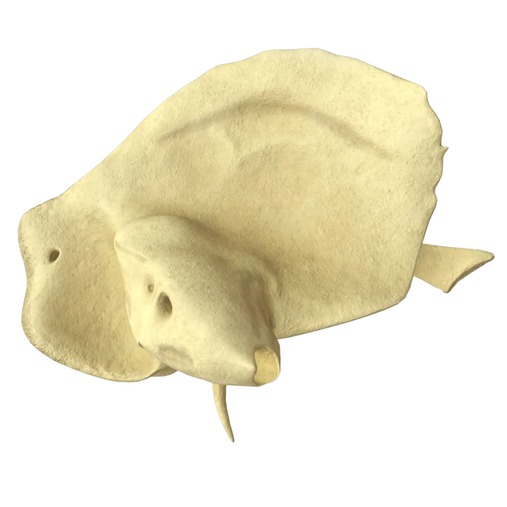
Temporal bone squama
Parietal bone is a paired bone, forming the main area of the cranial vault in the parietal region. The frontal bone is located anteriorly, occipital bone is located posteriorly, and the temporal squama – laterally.
-
I find it difficult to answer
Parietal bone is a paired bone, forming the main area of the cranial vault in the parietal region. The frontal bone is located anteriorly, occipital bone is located posteriorly, and the temporal squama – laterally.
2. Which suture connects the frontal and parietal bones?
-
Lambdoid suture
The coronal suture passes in the frontal plane between the frontal and two parietal bones.
-
Squamous suture
The coronal suture passes in the frontal plane between the frontal and two parietal bones.
-
Coronal suture
The coronal suture passes in the frontal plane between the frontal and two parietal bones.
-
Sagittal suture
The coronal suture passes in the frontal plane between the frontal and two parietal bones.
-
I find it difficult to answer
The coronal suture passes in the frontal plane between the frontal and two parietal bones.
3. Through which foramen of the occipital bone does the vertebral artery pass?
-
Foramen magnum
The vertebral artery enters the cranial cavity through the foramen magnum
-
Jugular foramen
The vertebral artery enters the cranial cavity through the foramen magnum
-
Hypoglossal canal
The vertebral artery enters the cranial cavity through the foramen magnum
-
Condylar canal
The vertebral artery enters the cranial cavity through the foramen magnum
-
I find it difficult to answer
The vertebral artery enters the cranial cavity through the foramen magnum
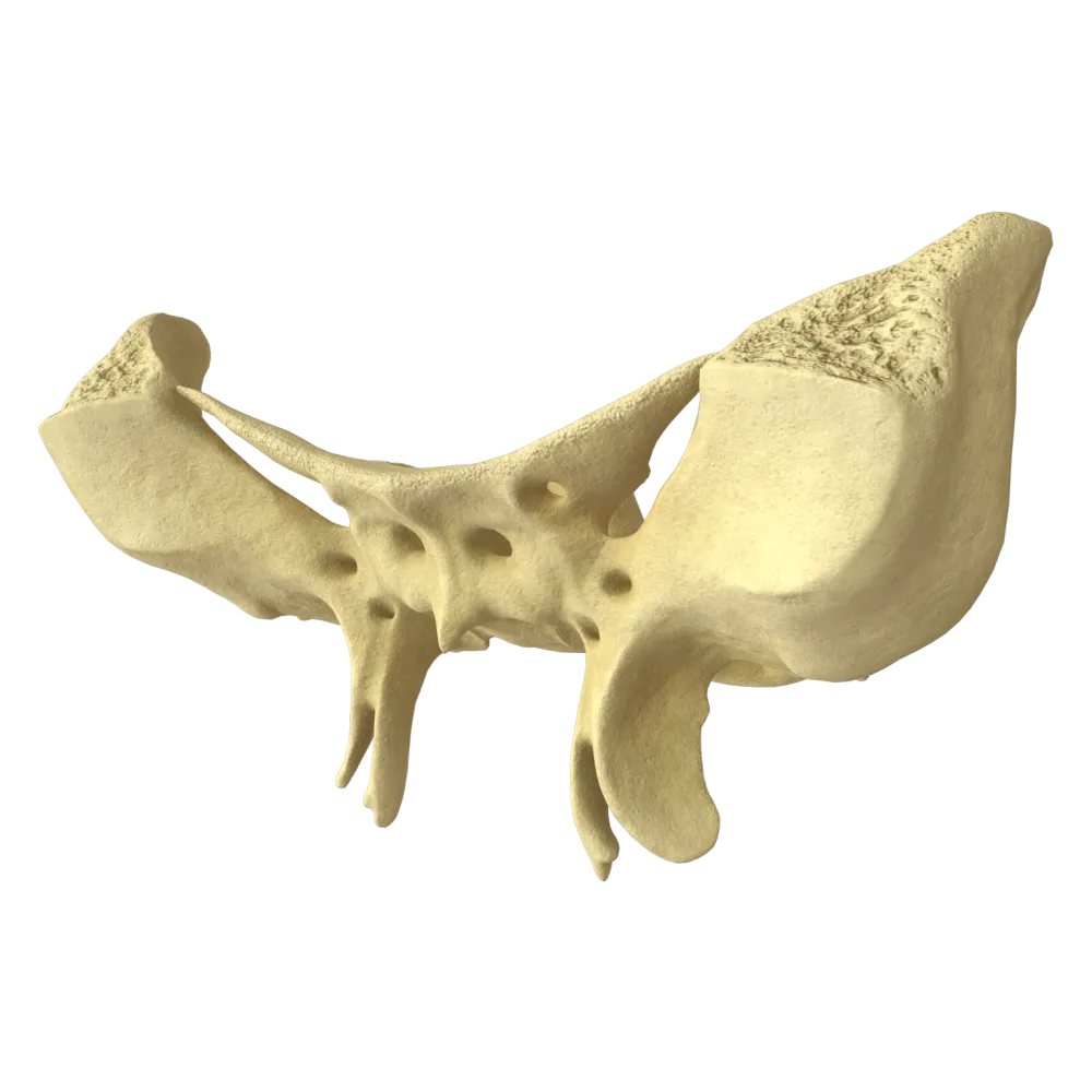
4. Which of the listed elements is part of the sphenoid bone?
-
Pyramid
The sphenoid bone comprises the body, greater wings, lesser wings, and pterygoid processes. The pyramid is a part of the temporal bone; crista galli is part of the ethmoid bone; the zygomatic process is a process of the frontal and temporal bones.
-
Crista galli
The sphenoid bone comprises the body, greater wings, lesser wings, and pterygoid processes. The pyramid is a part of the temporal bone; crista galli is part of the ethmoid bone; the zygomatic process is a process of the frontal and temporal bones.
-
Lesser wings
The sphenoid bone comprises the body, greater wings, lesser wings, and pterygoid processes. The pyramid is a part of the temporal bone; crista galli is part of the ethmoid bone; the zygomatic process is a process of the frontal and temporal bones.
-
Zygomatic process
The sphenoid bone comprises the body, greater wings, lesser wings, and pterygoid processes. The pyramid is a part of the temporal bone; crista galli is part of the ethmoid bone; the zygomatic process is a process of the frontal and temporal bones.
-
I find it difficult to answer
The sphenoid bone comprises the body, greater wings, lesser wings, and pterygoid processes. The pyramid is a part of the temporal bone; crista galli is part of the ethmoid bone; the zygomatic process is a process of the frontal and temporal bones.
5. Where is the sella turcica located?
-
On the squama of the occipital bone
The sella turcica is a depression on the upper surface of the body of the sphenoid bone that houses the pituitary gland. The basilar part of the occipital bone forms the clivus, and the ethmoid bone contains the cribriform plate and crista galli.
-
On the body of the sphenoid bone
The sella turcica is a depression on the upper surface of the body of the sphenoid bone that houses the pituitary gland. The basilar part of the occipital bone forms the clivus, and the ethmoid bone contains the cribriform plate and crista galli.
-
On the body of the ethmoid bone
The sella turcica is a depression on the upper surface of the body of the sphenoid bone that houses the pituitary gland. The basilar part of the occipital bone forms the clivus, and the ethmoid bone contains the cribriform plate and crista galli.
-
On the basilar part of the occipital bone
The sella turcica is a depression on the upper surface of the body of the sphenoid bone that houses the pituitary gland. The basilar part of the occipital bone forms the clivus, and the ethmoid bone contains the cribriform plate and crista galli.
-
I find it difficult to answer
The sella turcica is a depression on the upper surface of the body of the sphenoid bone that houses the pituitary gland. The basilar part of the occipital bone forms the clivus, and the ethmoid bone contains the cribriform plate and crista galli.
6. Through what canal of the sphenoid bone does the internal carotid artery pass?
-
Foramen rotundum
The internal carotid artery enters the cranial base through the carotid canal located in the pyramid of the temporal bone.
-
Carotid canal
The internal carotid artery enters the cranial base through the carotid canal located in the pyramid of the temporal bone.
-
Foramen spinosum
The internal carotid artery enters the cranial base through the carotid canal located in the pyramid of the temporal bone.
-
Optic canal
The internal carotid artery enters the cranial base through the carotid canal located in the pyramid of the temporal bone.
-
I find it difficult to answer
The internal carotid artery enters the cranial base through the carotid canal located in the pyramid of the temporal bone.
7. Which suture joins the two parietal bones together?
-
Coronal suture
The sagittal suture runs in the sagittal plane along the median line between the two parietal bones.
-
Lambdoid suture
The sagittal suture runs in the sagittal plane along the median line between the two parietal bones.
-
Sagittal suture
The sagittal suture runs in the sagittal plane along the median line between the two parietal bones.
-
Squamous suture
The sagittal suture runs in the sagittal plane along the median line between the two parietal bones.
-
I find it difficult to answer
The sagittal suture runs in the sagittal plane along the median line between the two parietal bones.
8. Which structure delimits the hypophyseal fossa anteriorly?
-
Dorsum sellae
The tuberculum sellae forms the anterior boundary of the hypophyseal fossa in the sella turcica of the sphenoid bone. The dorsum sellae delimits it posteriorly.
-
Tuberculum sellae
The tuberculum sellae forms the anterior boundary of the hypophyseal fossa in the sella turcica of the sphenoid bone. The dorsum sellae delimits it posteriorly.
-
Optic chiasm = Optic chiasma
The tuberculum sellae forms the anterior boundary of the hypophyseal fossa in the sella turcica of the sphenoid bone. The dorsum sellae delimits it posteriorly.
-
Diaphragma sellae
The tuberculum sellae forms the anterior boundary of the hypophyseal fossa in the sella turcica of the sphenoid bone. The dorsum sellae delimits it posteriorly.
-
I find it difficult to answer
The tuberculum sellae forms the anterior boundary of the hypophyseal fossa in the sella turcica of the sphenoid bone. The dorsum sellae delimits it posteriorly.
9. Which bone forms the crista galli?
-
Sphenoid bone
The crista galli is a projection of the ethmoid bone directed upwards into the anterior cranial fossa. The falx cerebri is attached to it. The frontal, sphenoid, and parietal bones do not form the crista galli.
-
Frontal bone
The crista galli is a projection of the ethmoid bone directed upwards into the anterior cranial fossa. The falx cerebri is attached to it. The frontal, sphenoid, and parietal bones do not form the crista galli.
-
Ethmoid bone
The crista galli is a projection of the ethmoid bone directed upwards into the anterior cranial fossa. The falx cerebri is attached to it. The frontal, sphenoid, and parietal bones do not form the crista galli.
-
Parietal bone
The crista galli is a projection of the ethmoid bone directed upwards into the anterior cranial fossa. The falx cerebri is attached to it. The frontal, sphenoid, and parietal bones do not form the crista galli.
-
I find it difficult to answer
The crista galli is a projection of the ethmoid bone directed upwards into the anterior cranial fossa. The falx cerebri is attached to it. The frontal, sphenoid, and parietal bones do not form the crista galli.
10. Through which opening of the sphenoid bone does the mandibular nerve (V3) pass?
-
Foramen rotundum
The mandibular nerve (V3) exits the cranial cavity through the foramen ovale.
-
Foramen spinosum
The mandibular nerve (V3) exits the cranial cavity through the foramen ovale.
-
Foramen ovale
The mandibular nerve (V3) exits the cranial cavity through the foramen ovale.
-
Foramen lacerum
The mandibular nerve (V3) exits the cranial cavity through the foramen ovale.
-
I find it difficult to answer
The mandibular nerve (V3) exits the cranial cavity through the foramen ovale.
11. Which part of the occipital bone forms the clivus involving the body of the sphenoid bone?
-
Squamous part of the occipital bone
The clivus is formed by the basilar part of the occipital bone and the body of the sphenoid bone. The occipital squama forms the posterior wall of the cranium; the lateral parts bear the condyles and jugular processes.
-
Lateral parts of the occipital bone
The clivus is formed by the basilar part of the occipital bone and the body of the sphenoid bone. The occipital squama forms the posterior wall of the cranium; the lateral parts bear the condyles and jugular processes.
-
Basilar part of the occipital bone
The clivus is formed by the basilar part of the occipital bone and the body of the sphenoid bone. The occipital squama forms the posterior wall of the cranium; the lateral parts bear the condyles and jugular processes.
-
Occipital condyles
The clivus is formed by the basilar part of the occipital bone and the body of the sphenoid bone. The occipital squama forms the posterior wall of the cranium; the lateral parts bear the condyles and jugular processes.
-
I find it difficult to answer
The clivus is formed by the basilar part of the occipital bone and the body of the sphenoid bone. The occipital squama forms the posterior wall of the cranium; the lateral parts bear the condyles and jugular processes.
12. In which cranial fossa is the foramen magnum located?
-
Anterior cranial fossa
The foramen magnum is located in the posterior cranial fossa, formed predominantly by the occipital bone and partially by the pyramids of the temporal bones.
-
Middle cranial fossa
The foramen magnum is located in the posterior cranial fossa, formed predominantly by the occipital bone and partially by the pyramids of the temporal bones.
-
Posterior cranial fossa
The foramen magnum is located in the posterior cranial fossa, formed predominantly by the occipital bone and partially by the pyramids of the temporal bones.
-
At the boundary of the middle and posterior fossae
The foramen magnum is located in the posterior cranial fossa, formed predominantly by the occipital bone and partially by the pyramids of the temporal bones.
-
I find it difficult to answer
The foramen magnum is located in the posterior cranial fossa, formed predominantly by the occipital bone and partially by the pyramids of the temporal bones.
13. Which bone contributes most significantly to the formation of the floor of the middle cranial fossa?
-
Frontal bone
The floor of the middle cranial fossa is mainly formed by the greater wings of the sphenoid bone and the anterior surfaces of the temporal bone pyramids. The frontal bone forms the anterior fossa, the occipital forms the posterior fossa, while the parietal does not contribute to the formation of the cranial fossae.
-
Parietal bone
The floor of the middle cranial fossa is mainly formed by the greater wings of the sphenoid bone and the anterior surfaces of the temporal bone pyramids. The frontal bone forms the anterior fossa, the occipital forms the posterior fossa, while the parietal does not contribute to the formation of the cranial fossae.
-
Sphenoid and temporal bones
The floor of the middle cranial fossa is mainly formed by the greater wings of the sphenoid bone and the anterior surfaces of the temporal bone pyramids. The frontal bone forms the anterior fossa, the occipital forms the posterior fossa, while the parietal does not contribute to the formation of the cranial fossae.
-
Occipital bone
The floor of the middle cranial fossa is mainly formed by the greater wings of the sphenoid bone and the anterior surfaces of the temporal bone pyramids. The frontal bone forms the anterior fossa, the occipital forms the posterior fossa, while the parietal does not contribute to the formation of the cranial fossae.
-
I find it difficult to answer
The floor of the middle cranial fossa is mainly formed by the greater wings of the sphenoid bone and the anterior surfaces of the temporal bone pyramids. The frontal bone forms the anterior fossa, the occipital forms the posterior fossa, while the parietal does not contribute to the formation of the cranial fossae.
14. Where is the groove of the superior sagittal sinus located?
-
On the internal surface of the occipital bone
The groove of the superior sagittal sinus runs along the median line on the internal surface of the frontal, parietal, and occipital bones, corresponding to the course of the sagittal suture. The corresponding dural venous sinus is located within it.
-
Along the sagittal suture on the internal surface of the cranial vault
The groove of the superior sagittal sinus runs along the median line on the internal surface of the frontal, parietal, and occipital bones, corresponding to the course of the sagittal suture. The corresponding dural venous sinus is located within it.
-
On the internal surface of the sphenoid bone
The groove of the superior sagittal sinus runs along the median line on the internal surface of the frontal, parietal, and occipital bones, corresponding to the course of the sagittal suture. The corresponding dural venous sinus is located within it.
-
On the pyramid of the temporal bone
The groove of the superior sagittal sinus runs along the median line on the internal surface of the frontal, parietal, and occipital bones, corresponding to the course of the sagittal suture. The corresponding dural venous sinus is located within it.
-
I find it difficult to answer
The groove of the superior sagittal sinus runs along the median line on the internal surface of the frontal, parietal, and occipital bones, corresponding to the course of the sagittal suture. The corresponding dural venous sinus is located within it.
15. How many parts make up the occipital bone in an adult?
-
Two parts
The occipital bone consists of four parts: squamous part, two lateral parts, and a basilar part. In children, these parts are separated by the cartilage and fuse by the age of 3–6.
-
Three parts
The occipital bone consists of four parts: squamous part, two lateral parts, and a basilar part. In children, these parts are separated by the cartilage and fuse by the age of 3–6.
-
Four parts
The occipital bone consists of four parts: squamous part, two lateral parts, and a basilar part. In children, these parts are separated by the cartilage and fuse by the age of 3–6.
-
Five parts
The occipital bone consists of four parts: squamous part, two lateral parts, and a basilar part. In children, these parts are separated by the cartilage and fuse by the age of 3–6.
-
I find it difficult to answer
The occipital bone consists of four parts: squamous part, two lateral parts, and a basilar part. In children, these parts are separated by the cartilage and fuse by the age of 3–6.
16. What structure is located in the groove on the superior surface of the temporal bone pyramid and corresponds to the greater petrosal nerve?
-
Groove for inferior petrosal sinus
On the anterior surface of the temporal bone pyramid there is a groove for the greater petrosal nerve, housing the greater petrosal nerve — a branch of the facial nerve. The groove for the sigmoid sinus is located on the posterior surface of the pyramid.
-
Groove for greater petrosal nerve
On the anterior surface of the temporal bone pyramid there is a groove for the greater petrosal nerve, housing the greater petrosal nerve — a branch of the facial nerve. The groove for the sigmoid sinus is located on the posterior surface of the pyramid.
-
Groove for sigmoid sinus
On the anterior surface of the temporal bone pyramid there is a groove for the greater petrosal nerve, housing the greater petrosal nerve — a branch of the facial nerve. The groove for the sigmoid sinus is located on the posterior surface of the pyramid.
-
Groove for middle meningeal artery
On the anterior surface of the temporal bone pyramid there is a groove for the greater petrosal nerve, housing the greater petrosal nerve — a branch of the facial nerve. The groove for the sigmoid sinus is located on the posterior surface of the pyramid.
-
I find it difficult to answer
On the anterior surface of the temporal bone pyramid there is a groove for the greater petrosal nerve, housing the greater petrosal nerve — a branch of the facial nerve. The groove for the sigmoid sinus is located on the posterior surface of the pyramid.
17. What forms the optic canal?
-
Lesser wing of the sphenoid bone
The optic canal is bounded above and laterally by the lesser wing of the sphenoid bone and medially by the root of the lesser wing (at the body of the sphenoid bone). The optic nerve (II) and ophthalmic artery pass through it.
-
Greater wing of the sphenoid bone and the frontal bone
The optic canal is bounded above and laterally by the lesser wing of the sphenoid bone and medially by the root of the lesser wing (at the body of the sphenoid bone). The optic nerve (II) and ophthalmic artery pass through it.
-
Lesser wing of the sphenoid bone and the root of the lesser wing
The optic canal is bounded above and laterally by the lesser wing of the sphenoid bone and medially by the root of the lesser wing (at the body of the sphenoid bone). The optic nerve (II) and ophthalmic artery pass through it.
-
Body of the sphenoid bone and the ethmoid bone
The optic canal is bounded above and laterally by the lesser wing of the sphenoid bone and medially by the root of the lesser wing (at the body of the sphenoid bone). The optic nerve (II) and ophthalmic artery pass through it.
-
I find it difficult to answer
The optic canal is bounded above and laterally by the lesser wing of the sphenoid bone and medially by the root of the lesser wing (at the body of the sphenoid bone). The optic nerve (II) and ophthalmic artery pass through it.
18. Which of the fontanelles in a newborn is located at the intersection of the sagittal and lambdoid sutures?
-
Anterior (frontal) fontanelle
The posterior (occipital) fontanelle is located at the convergence of the sagittal and lambdoid sutures (lambda point). It ossifies by the age of 2–3 months.
-
Posterior (occipital) fontanelle
The posterior (occipital) fontanelle is located at the convergence of the sagittal and lambdoid sutures (lambda point). It ossifies by the age of 2–3 months.
-
Sphenoidal fontanelle
The posterior (occipital) fontanelle is located at the convergence of the sagittal and lambdoid sutures (lambda point). It ossifies by the age of 2–3 months.
-
Mastoid fontanelle
The posterior (occipital) fontanelle is located at the convergence of the sagittal and lambdoid sutures (lambda point). It ossifies by the age of 2–3 months.
-
I find it difficult to answer
The posterior (occipital) fontanelle is located at the convergence of the sagittal and lambdoid sutures (lambda point). It ossifies by the age of 2–3 months.
19. Which of the listed openings allows communication between the inner ear and the posterior cranial fossa?
-
Jugular foramen
The internal acoustic opening is located on the posterior surface of the petrous part of the temporal bone and opens into the posterior cranial fossa. Through it passes the facial nerve (VII), the vestibulocochlear nerve (VIII) and the labyrinthine artery. The stylomastoid foramen is the site where the VII nerve exits on the external surface of the skull base.
-
Stylomastoid foramen
The internal acoustic opening is located on the posterior surface of the petrous part of the temporal bone and opens into the posterior cranial fossa. Through it passes the facial nerve (VII), the vestibulocochlear nerve (VIII) and the labyrinthine artery. The stylomastoid foramen is the site where the VII nerve exits on the external surface of the skull base.
-
Internal acoustic opening
The internal acoustic opening is located on the posterior surface of the petrous part of the temporal bone and opens into the posterior cranial fossa. Through it passes the facial nerve (VII), the vestibulocochlear nerve (VIII) and the labyrinthine artery. The stylomastoid foramen is the site where the VII nerve exits on the external surface of the skull base.
-
Bony external acoustic opening
The internal acoustic opening is located on the posterior surface of the petrous part of the temporal bone and opens into the posterior cranial fossa. Through it passes the facial nerve (VII), the vestibulocochlear nerve (VIII) and the labyrinthine artery. The stylomastoid foramen is the site where the VII nerve exits on the external surface of the skull base.
-
I find it difficult to answer
The internal acoustic opening is located on the posterior surface of the petrous part of the temporal bone and opens into the posterior cranial fossa. Through it passes the facial nerve (VII), the vestibulocochlear nerve (VIII) and the labyrinthine artery. The stylomastoid foramen is the site where the VII nerve exits on the external surface of the skull base.
20. Which bone is involved in forming the anterior fontanelle of a newborn?
-
Only the frontal and parietal bones
The anterior (large) fontanelle is bordered by the two halves of the frontal bone in front and the two parietal bones behind. It is located at the bregma, at the intersection of the coronal and sagittal sutures, and closes by 1.5–2 years of age. The sphenoid and occipital bones are not involved in its formation.
-
Frontal, parietal, and sphenoid bones
The anterior (large) fontanelle is bordered by the two halves of the frontal bone in front and the two parietal bones behind. It is located at the bregma, at the intersection of the coronal and sagittal sutures, and closes by 1.5–2 years of age. The sphenoid and occipital bones are not involved in its formation.
-
Frontal, parietal, occipital, and sphenoid bones
The anterior (large) fontanelle is bordered by the two halves of the frontal bone in front and the two parietal bones behind. It is located at the bregma, at the intersection of the coronal and sagittal sutures, and closes by 1.5–2 years of age. The sphenoid and occipital bones are not involved in its formation.
-
Frontal and occipital bones
The anterior (large) fontanelle is bordered by the two halves of the frontal bone in front and the two parietal bones behind. It is located at the bregma, at the intersection of the coronal and sagittal sutures, and closes by 1.5–2 years of age. The sphenoid and occipital bones are not involved in its formation.
-
I find it difficult to answer
The anterior (large) fontanelle is bordered by the two halves of the frontal bone in front and the two parietal bones behind. It is located at the bregma, at the intersection of the coronal and sagittal sutures, and closes by 1.5–2 years of age. The sphenoid and occipital bones are not involved in its formation.
Retake this quiz?
Your current progress will be reset.
Neurocranium bones
Temporal bone
0/20

Cookie Consent
We use cookies to enhance your browsing experience, analyze site traffic, and deliver content. Please choose whether you accept all cookies or wish to reject non-essential tracking.
Cookie Preferences
Manage your cookie preferences below:
Essential cookies enable basic functions and are necessary for the proper function of the website.
Name
Description
Duration
Geolocation Config
This cookie is used to store the consent settings based on the visitor's location.
30 days
Cookie Preferences
This cookie is used to store the user's cookie consent preferences.
30 days
Google reCAPTCHA helps protect websites from spam and abuse by verifying user interactions through challenges.
Name
Description
Duration
_GRECAPTCHA
Google reCAPTCHA sets a necessary cookie (_GRECAPTCHA) when executed for the purpose of providing its risk analysis.
179 days
Statistics cookies collect information anonymously. This information helps us understand how visitors use our website.
Google Analytics is a powerful tool that tracks and analyzes website traffic for informed marketing decisions.
Service URL: policies.google.com (opens in a new window)
Name
Description
Duration
__utma
ID used to identify users and sessions
2 years after last activity
__utmt
Used to monitor number of Google Analytics server requests
10 minutes
__utmb
Used to distinguish new sessions and visits. This cookie is set when the GA.js javascript library is loaded and there is no existing __utmb cookie. The cookie is updated every time data is sent to the Google Analytics server.
30 minutes after last activity
__utmc
Used only with old Urchin versions of Google Analytics and not with GA.js. Was used to distinguish between new sessions and visits at the end of a session.
End of session (browser)
__utmz
Contains information about the traffic source or campaign that directed user to the website. The cookie is set when the GA.js javascript is loaded and updated when data is sent to the Google Anaytics server
6 months after last activity
__utmv
Contains custom information set by the web developer via the _setCustomVar method in Google Analytics. This cookie is updated every time new data is sent to the Google Analytics server.
2 years after last activity
__utmx
Used to determine whether a user is included in an A / B or Multivariate test.
18 months
_ga
ID used to identify users
2 years
_gali
Used by Google Analytics to determine which links on a page are being clicked
30 seconds
_ga_
ID used to identify users
2 years
_gid
ID used to identify users for 24 hours after last activity
24 hours
_gat
Used to monitor number of Google Analytics server requests when using Google Tag Manager
1 minute
_gac_
Contains information related to marketing campaigns of the user. These are shared with Google AdWords / Google Ads when the Google Ads and Google Analytics accounts are linked together.
90 days
Clarity is a web analytics service that tracks and reports website traffic.
Service URL: clarity.microsoft.com (opens in a new window)
Name
Description
Duration
_clsk
Connects multiple page views by a user into a single Clarity session recording.
1 day
_clck
Stores a unique user identifier to analyze visitor behavior through heatmaps and session recordings.
1 year
ANONCHK
Indicates whether MUID is transferred to ANID, a cookie used for advertising. Clarity doesn't use ANID and so this is always set to 0.
Session
CLID
Identifies the first-time Clarity saw this user on any site using Clarity.
12 months
_clsk
Connects multiple page views by a user into a single Clarity session recording.
12 months
_clck
Persists the Clarity User ID and preferences, unique to that site is attributed to the same user ID.
12 months