
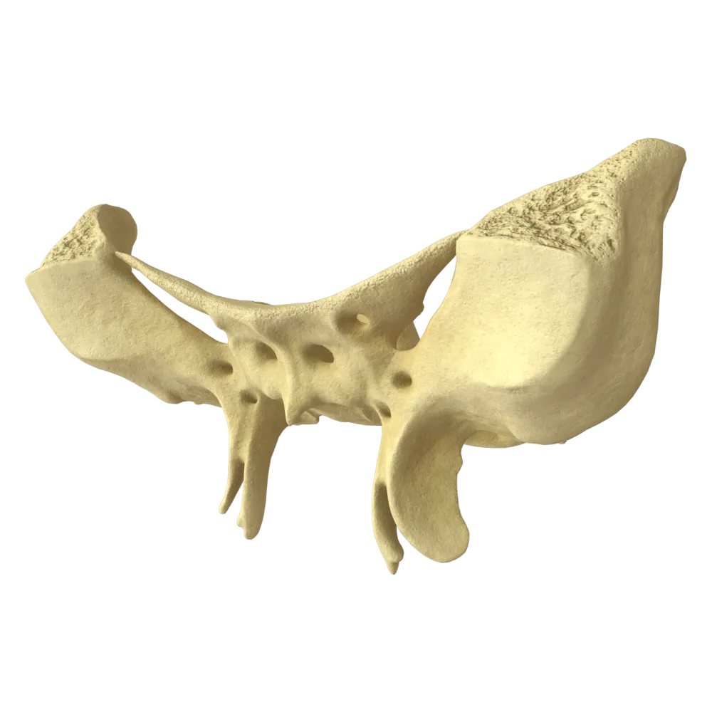
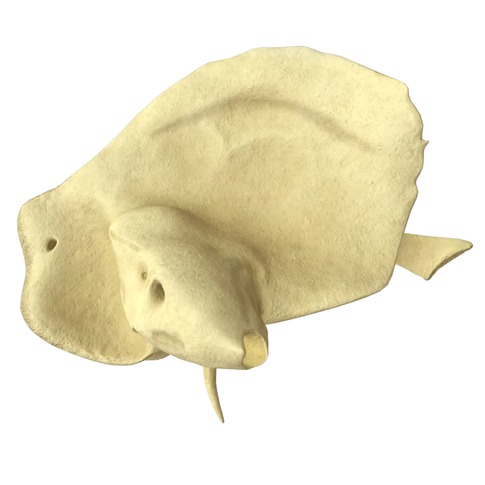
Occipital bone anatomy quiz
Normal anatomy quiz of the occipital bone: structure, foramina, canals, articular surfaces, and topographic relationships.
1/20
bold
text
1. Which part of the occipital bone contributes to the formation of the foramen magnum laterally?
-
Squamous part of the occipital bone
The foramen magnum (foramen magnum) is bounded anteriorly by the basilar part, laterally by the lateral (condylar) parts, and posteriorly by the squamous part of the occipital bone.
-
Lateral parts of the occipital bone
The foramen magnum (foramen magnum) is bounded anteriorly by the basilar part, laterally by the lateral (condylar) parts, and posteriorly by the squamous part of the occipital bone.
-
Basilar part of the occipital bone
The foramen magnum (foramen magnum) is bounded anteriorly by the basilar part, laterally by the lateral (condylar) parts, and posteriorly by the squamous part of the occipital bone.
-
Pharyngeal tubercle
The foramen magnum (foramen magnum) is bounded anteriorly by the basilar part, laterally by the lateral (condylar) parts, and posteriorly by the squamous part of the occipital bone.
-
I find it difficult to answer
The foramen magnum (foramen magnum) is bounded anteriorly by the basilar part, laterally by the lateral (condylar) parts, and posteriorly by the squamous part of the occipital bone.
2. Where is the pharyngeal tubercle (tuberculum pharyngeum) located on the occipital bone?
-
On the external surface of the squamous part, along the median line
The tuberculum pharyngeum is located on the inferior (external) surface of the basilar part of the occipital bone along the median line.
-
On the inferior surface of the basilar part, along the median line
The tuberculum pharyngeum is located on the inferior (external) surface of the basilar part of the occipital bone along the median line.
-
On the condylar part, medially to the occipital condyle
The tuberculum pharyngeum is located on the inferior (external) surface of the basilar part of the occipital bone along the median line.
-
On the internal surface of the squamous part, near the cruciform eminence
The tuberculum pharyngeum is located on the inferior (external) surface of the basilar part of the occipital bone along the median line.
-
I find it difficult to answer
The tuberculum pharyngeum is located on the inferior (external) surface of the basilar part of the occipital bone along the median line.
3. Which canal passes through the base of the occipital condyles?
-
Hypoglossal canal
The canalis nervi hypoglossi (hypoglossal canal, cranial nerve XII) passes through the base of the occipital condyles.
-
Carotid canal
The canalis nervi hypoglossi (hypoglossal canal, cranial nerve XII) passes through the base of the occipital condyles.
-
Facial canal
The canalis nervi hypoglossi (hypoglossal canal, cranial nerve XII) passes through the base of the occipital condyles.
-
Cochlear canal
The canalis nervi hypoglossi (hypoglossal canal, cranial nerve XII) passes through the base of the occipital condyles.
-
I find it difficult to answer
The canalis nervi hypoglossi (hypoglossal canal, cranial nerve XII) passes through the base of the occipital condyles.
4. What forms the cruciform eminence (eminentia cruciformis) on the internal surface of the squamous part of the occipital bone?
-
The intersection of the internal occipital crest and the groove for the superior sagittal sinus with the grooves for the transverse sinuses
The eminentia cruciformis is formed by the internal occipital protuberance (protuberantia occipitalis interna), from which structures extend in four directions: superiorly, the groove for the superior sagittal sinus; inferiorly, the internal occipital crest; and to the right and left, the grooves for the transverse sinuses.
-
The intersection of the external occipital crest with the lines of the nuchal surface
The eminentia cruciformis is formed by the internal occipital protuberance (protuberantia occipitalis interna), from which structures extend in four directions: superiorly, the groove for the superior sagittal sinus; inferiorly, the internal occipital crest; and to the right and left, the grooves for the transverse sinuses.
-
The confluence of the grooves for the sigmoid sinuses with the jugular notches
The eminentia cruciformis is formed by the internal occipital protuberance (protuberantia occipitalis interna), from which structures extend in four directions: superiorly, the groove for the superior sagittal sinus; inferiorly, the internal occipital crest; and to the right and left, the grooves for the transverse sinuses.
-
The intersection of the groove for the superior petrosal sinus with the groove for the transverse sinus
The eminentia cruciformis is formed by the internal occipital protuberance (protuberantia occipitalis interna), from which structures extend in four directions: superiorly, the groove for the superior sagittal sinus; inferiorly, the internal occipital crest; and to the right and left, the grooves for the transverse sinuses.
-
I find it difficult to answer
The eminentia cruciformis is formed by the internal occipital protuberance (protuberantia occipitalis interna), from which structures extend in four directions: superiorly, the groove for the superior sagittal sinus; inferiorly, the internal occipital crest; and to the right and left, the grooves for the transverse sinuses.
5. Which groove is located on the internal surface of the squamous part of the occipital bone, runs horizontally, and contains the corresponding venous sinus?
-
Groove for inferior petrosal sinus
The sulcus sinus transversi (groove for the transverse sinus) is located on the internal surface of the squamous part of the occipital bone and extends laterally toward the angles of the occipital bone, where it continues as the groove for the sigmoid sinus.
-
Groove for transverse sinus
The sulcus sinus transversi (groove for the transverse sinus) is located on the internal surface of the squamous part of the occipital bone and extends laterally toward the angles of the occipital bone, where it continues as the groove for the sigmoid sinus.
-
Groove for sigmoid sinus
The sulcus sinus transversi (groove for the transverse sinus) is located on the internal surface of the squamous part of the occipital bone and extends laterally toward the angles of the occipital bone, where it continues as the groove for the sigmoid sinus.
-
Groove for the superior petrosal sinus
The sulcus sinus transversi (groove for the transverse sinus) is located on the internal surface of the squamous part of the occipital bone and extends laterally toward the angles of the occipital bone, where it continues as the groove for the sigmoid sinus.
-
I find it difficult to answer
The sulcus sinus transversi (groove for the transverse sinus) is located on the internal surface of the squamous part of the occipital bone and extends laterally toward the angles of the occipital bone, where it continues as the groove for the sigmoid sinus.
6. On which part of the occipital bone is the groove for the sigmoid sinus (sulcus sinus sigmoidei) located?
-
On the basilar part
The groove for the sigmoid sinus (sulcus sinus sigmoidei) of the occipital bone is located on the internal (cerebral) surface of its lateral part (pars lateralis), curving around the jugular process. At this site, the paired sigmoid sinus courses toward the jugular foramen, where it continues as the internal jugular vein.
-
On the squamous part of the occipital bone superior to the cruciform eminence
The groove for the sigmoid sinus (sulcus sinus sigmoidei) of the occipital bone is located on the internal (cerebral) surface of its lateral part (pars lateralis), curving around the jugular process. At this site, the paired sigmoid sinus courses toward the jugular foramen, where it continues as the internal jugular vein.
-
On the lateral (side) part
The groove for the sigmoid sinus (sulcus sinus sigmoidei) of the occipital bone is located on the internal (cerebral) surface of its lateral part (pars lateralis), curving around the jugular process. At this site, the paired sigmoid sinus courses toward the jugular foramen, where it continues as the internal jugular vein.
-
There is no such groove
The groove for the sigmoid sinus (sulcus sinus sigmoidei) of the occipital bone is located on the internal (cerebral) surface of its lateral part (pars lateralis), curving around the jugular process. At this site, the paired sigmoid sinus courses toward the jugular foramen, where it continues as the internal jugular vein.
-
I find it difficult to answer
The groove for the sigmoid sinus (sulcus sinus sigmoidei) of the occipital bone is located on the internal (cerebral) surface of its lateral part (pars lateralis), curving around the jugular process. At this site, the paired sigmoid sinus courses toward the jugular foramen, where it continues as the internal jugular vein.
7. What is the structure of the occipital bone that contributes to the formation of the atlanto-occipital joint called?
-
Jugular process
The occipital condyle (condylus occipitalis) is an ellipsoidal structure on the inferior surface of the lateral part of the occipital bone that articulates with the superior articular facet of the atlas in the art. atlantooccipitalis.
-
Occipital condyle
The occipital condyle (condylus occipitalis) is an ellipsoidal structure on the inferior surface of the lateral part of the occipital bone that articulates with the superior articular facet of the atlas in the art. atlantooccipitalis.
-
Basilar part
The occipital condyle (condylus occipitalis) is an ellipsoidal structure on the inferior surface of the lateral part of the occipital bone that articulates with the superior articular facet of the atlas in the art. atlantooccipitalis.
-
Internal occipital protuberance
The occipital condyle (condylus occipitalis) is an ellipsoidal structure on the inferior surface of the lateral part of the occipital bone that articulates with the superior articular facet of the atlas in the art. atlantooccipitalis.
-
I find it difficult to answer
The occipital condyle (condylus occipitalis) is an ellipsoidal structure on the inferior surface of the lateral part of the occipital bone that articulates with the superior articular facet of the atlas in the art. atlantooccipitalis.
8. What passes through the jugular foramen (foramen jugulare), which is formed by the occipital and temporal bones?
-
Internal carotid artery, cranial nerve XII
The following exit through the foramen jugulare: the internal jugular vein (v. jugularis interna) through the posterior part, as well as cranial nerves IX (n. glossopharyngeus), X (n. vagus), and XI (n. accessorius) through the anterior (petrosal) part of the foramen.
-
Internal jugular vein and cranial nerves IX, X, and XI
The following exit through the foramen jugulare: the internal jugular vein (v. jugularis interna) through the posterior part, as well as cranial nerves IX (n. glossopharyngeus), X (n. vagus), and XI (n. accessorius) through the anterior (petrosal) part of the foramen.
-
Vertebral artery and cranial nerve XI
The following exit through the foramen jugulare: the internal jugular vein (v. jugularis interna) through the posterior part, as well as cranial nerves IX (n. glossopharyngeus), X (n. vagus), and XI (n. accessorius) through the anterior (petrosal) part of the foramen.
-
External carotid artery and cranial nerve IX
The following exit through the foramen jugulare: the internal jugular vein (v. jugularis interna) through the posterior part, as well as cranial nerves IX (n. glossopharyngeus), X (n. vagus), and XI (n. accessorius) through the anterior (petrosal) part of the foramen.
-
I find it difficult to answer
The following exit through the foramen jugulare: the internal jugular vein (v. jugularis interna) through the posterior part, as well as cranial nerves IX (n. glossopharyngeus), X (n. vagus), and XI (n. accessorius) through the anterior (petrosal) part of the foramen.
9. By which suture does the occipital bone articulate with the parietal bones?
-
Spheno-occipital suture
The occipital bone articulates with both parietal bones by means of the sutura lambdoidea—the lambdoid suture, named for its resemblance to the Greek letter λ.
-
Coronal suture
The occipital bone articulates with both parietal bones by means of the sutura lambdoidea—the lambdoid suture, named for its resemblance to the Greek letter λ.
-
Lambdoid suture
The occipital bone articulates with both parietal bones by means of the sutura lambdoidea—the lambdoid suture, named for its resemblance to the Greek letter λ.
-
Sagittal suture
The occipital bone articulates with both parietal bones by means of the sutura lambdoidea—the lambdoid suture, named for its resemblance to the Greek letter λ.
-
I find it difficult to answer
The occipital bone articulates with both parietal bones by means of the sutura lambdoidea—the lambdoid suture, named for its resemblance to the Greek letter λ.
10. Where is the external occipital protuberance (protuberantia occipitalis externa) located?
-
On the inferior surface of the basilar part
The protuberantia occipitalis externa is a readily palpable bony projection on the external (posterior) surface of the squamous part of the occipital bone along the median line; the external occipital crest and superior nuchal lines extend from it.
-
On the internal surface of the squamous part along the median line
The protuberantia occipitalis externa is a readily palpable bony projection on the external (posterior) surface of the squamous part of the occipital bone along the median line; the external occipital crest and superior nuchal lines extend from it.
-
On the external surface of the squamous part along the median line
The protuberantia occipitalis externa is a readily palpable bony projection on the external (posterior) surface of the squamous part of the occipital bone along the median line; the external occipital crest and superior nuchal lines extend from it.
-
On the lateral part, superior to the occipital condyle
The protuberantia occipitalis externa is a readily palpable bony projection on the external (posterior) surface of the squamous part of the occipital bone along the median line; the external occipital crest and superior nuchal lines extend from it.
-
I find it difficult to answer
The protuberantia occipitalis externa is a readily palpable bony projection on the external (posterior) surface of the squamous part of the occipital bone along the median line; the external occipital crest and superior nuchal lines extend from it.
11. What are the transverse arcuate lines on the external surface of the squamous part of the occipital bone to which the muscles and fasciae of the neck attach called?
-
Temporal lines
On the external surface of the squamous part of the occipital bone, paired nuchal lines (lineae nuchales) extend laterally from the external occipital protuberance (protuberantia occipitalis externa).
-
Suboccipital lines
On the external surface of the squamous part of the occipital bone, paired nuchal lines (lineae nuchales) extend laterally from the external occipital protuberance (protuberantia occipitalis externa).
-
Trapezoid lines
On the external surface of the squamous part of the occipital bone, paired nuchal lines (lineae nuchales) extend laterally from the external occipital protuberance (protuberantia occipitalis externa).
-
Nuchal lines
On the external surface of the squamous part of the occipital bone, paired nuchal lines (lineae nuchales) extend laterally from the external occipital protuberance (protuberantia occipitalis externa).
-
I find it difficult to answer
On the external surface of the squamous part of the occipital bone, paired nuchal lines (lineae nuchales) extend laterally from the external occipital protuberance (protuberantia occipitalis externa).
12. Through which foramen of the occipital bone does the spinal cord pass as it transitions into the medulla oblongata?
-
Hypoglossal canal
Foramen magnum (foramen magnum) is the largest foramen of the skull, through which the cranial cavity communicates with the vertebral canal; here, the medulla oblongata transitions into the spinal
-
Jugular foramen
Foramen magnum (foramen magnum) is the largest foramen of the skull, through which the cranial cavity communicates with the vertebral canal; here, the medulla oblongata transitions into the spinal
-
Foramen magnum
Foramen magnum (foramen magnum) is the largest foramen of the skull, through which the cranial cavity communicates with the vertebral canal; here, the medulla oblongata transitions into the spinal
-
Condylar canal
Foramen magnum (foramen magnum) is the largest foramen of the skull, through which the cranial cavity communicates with the vertebral canal; here, the medulla oblongata transitions into the spinal
-
I find it difficult to answer
Foramen magnum (foramen magnum) is the largest foramen of the skull, through which the cranial cavity communicates with the vertebral canal; here, the medulla oblongata transitions into the spinal
13. What is the process of the occipital bone located laterally to the jugular notch called?
-
Basilar process
The processus jugularis is a process of the lateral part of the occipital bone located lateral to the jugular notch (incisura jugularis); together with the jugular process of the temporal bone, it bounds the jugular foramen.
-
Jugular process
The processus jugularis is a process of the lateral part of the occipital bone located lateral to the jugular notch (incisura jugularis); together with the jugular process of the temporal bone, it bounds the jugular foramen.
-
Condylar process
The processus jugularis is a process of the lateral part of the occipital bone located lateral to the jugular notch (incisura jugularis); together with the jugular process of the temporal bone, it bounds the jugular foramen.
-
Styloid process
The processus jugularis is a process of the lateral part of the occipital bone located lateral to the jugular notch (incisura jugularis); together with the jugular process of the temporal bone, it bounds the jugular foramen.
-
I find it difficult to answer
The processus jugularis is a process of the lateral part of the occipital bone located lateral to the jugular notch (incisura jugularis); together with the jugular process of the temporal bone, it bounds the jugular foramen.
14. Which groove on the basilar part of the occipital bone contains the inferior petrosal sinus?
-
Sulcus sinus transversi
The sulcus sinus petrosi inferioris is located along the lateral margin of the basilar part of the occipital bone and contains the inferior petrosal sinus (sinus petrosus inferior); this groove bounds the jugular foramen anteriorly.
-
Sulcus sinus sigmoidei
The sulcus sinus petrosi inferioris is located along the lateral margin of the basilar part of the occipital bone and contains the inferior petrosal sinus (sinus petrosus inferior); this groove bounds the jugular foramen anteriorly.
-
Sulcus sinus petrosi inferioris
The sulcus sinus petrosi inferioris is located along the lateral margin of the basilar part of the occipital bone and contains the inferior petrosal sinus (sinus petrosus inferior); this groove bounds the jugular foramen anteriorly.
-
Sulcus sinus sagittalis superioris
The sulcus sinus petrosi inferioris is located along the lateral margin of the basilar part of the occipital bone and contains the inferior petrosal sinus (sinus petrosus inferior); this groove bounds the jugular foramen anteriorly.
-
I find it difficult to answer
The sulcus sinus petrosi inferioris is located along the lateral margin of the basilar part of the occipital bone and contains the inferior petrosal sinus (sinus petrosus inferior); this groove bounds the jugular foramen anteriorly.
15. How many parts are recognized in the occipital bone of an adult?
-
Two parts
Four parts are recognized in the occipital bone: the squamous part (squama occipitalis), the basilar part (pars basilaris), and two lateral (condylar) parts (partes laterales—the right and left), which surround the foramen magnum.
-
Three parts
Four parts are recognized in the occipital bone: the squamous part (squama occipitalis), the basilar part (pars basilaris), and two lateral (condylar) parts (partes laterales—the right and left), which surround the foramen magnum.
-
Four parts
Four parts are recognized in the occipital bone: the squamous part (squama occipitalis), the basilar part (pars basilaris), and two lateral (condylar) parts (partes laterales—the right and left), which surround the foramen magnum.
-
Five parts
Four parts are recognized in the occipital bone: the squamous part (squama occipitalis), the basilar part (pars basilaris), and two lateral (condylar) parts (partes laterales—the right and left), which surround the foramen magnum.
-
I find it difficult to answer
Four parts are recognized in the occipital bone: the squamous part (squama occipitalis), the basilar part (pars basilaris), and two lateral (condylar) parts (partes laterales—the right and left), which surround the foramen magnum.
16. Where is the condylar fossa (fossa condylaris) of the occipital bone located, and what may be found within it?
-
Anterior to the occipital condyle; it contains the pharyngeal tonsil
The fossa condylaris is located immediately posterior to the occipital condyle; the canalis condylaris frequently opens in its floor and transmits an emissary vein connecting the sigmoid sinus with the external veins.
-
Posterior to the occipital condyle; the condylar canal (canalis condylaris) opens into it
The fossa condylaris is located immediately posterior to the occipital condyle; the canalis condylaris frequently opens in its floor and transmits an emissary vein connecting the sigmoid sinus with the external veins.
-
Medial to the occipital condyle; the hypoglossal canal begins within it
The fossa condylaris is located immediately posterior to the occipital condyle; the canalis condylaris frequently opens in its floor and transmits an emissary vein connecting the sigmoid sinus with the external veins.
-
On the internal surface of the squamous part; the groove for the transverse sinus passes through it
The fossa condylaris is located immediately posterior to the occipital condyle; the canalis condylaris frequently opens in its floor and transmits an emissary vein connecting the sigmoid sinus with the external veins.
-
I find it difficult to answer
The fossa condylaris is located immediately posterior to the occipital condyle; the canalis condylaris frequently opens in its floor and transmits an emissary vein connecting the sigmoid sinus with the external veins.
17. How many pairs of nuchal lines are recognized on the external surface of the squamous part of the occipital bone?
-
1 pair
On the external surface of the squamous part of the occipital bone, there are 3 pairs of arcuate nuchal lines (serving as attachment sites for the muscles and fasciae of the neck).
-
2 pairs
On the external surface of the squamous part of the occipital bone, there are 3 pairs of arcuate nuchal lines (serving as attachment sites for the muscles and fasciae of the neck).
-
3 pairs
On the external surface of the squamous part of the occipital bone, there are 3 pairs of arcuate nuchal lines (serving as attachment sites for the muscles and fasciae of the neck).
-
4 pairs
On the external surface of the squamous part of the occipital bone, there are 3 pairs of arcuate nuchal lines (serving as attachment sites for the muscles and fasciae of the neck).
-
I find it difficult to answer
On the external surface of the squamous part of the occipital bone, there are 3 pairs of arcuate nuchal lines (serving as attachment sites for the muscles and fasciae of the neck).
18. Through which structure of the occipital bone does the vertebral artery (a. vertebralis) enter the cranial cavity?
-
Through the hypoglossal canal
After passing through the foramina of the transverse processes of the cervical vertebrae, the vertebral artery enters the cranial cavity through the foramen magnum (foramen magnum), lying lateral to the medulla oblongata.
-
Through the jugular foramen
After passing through the foramina of the transverse processes of the cervical vertebrae, the vertebral artery enters the cranial cavity through the foramen magnum (foramen magnum), lying lateral to the medulla oblongata.
-
Through the large occipital foramen
After passing through the foramina of the transverse processes of the cervical vertebrae, the vertebral artery enters the cranial cavity through the foramen magnum (foramen magnum), lying lateral to the medulla oblongata.
-
Through the condylar canal
After passing through the foramina of the transverse processes of the cervical vertebrae, the vertebral artery enters the cranial cavity through the foramen magnum (foramen magnum), lying lateral to the medulla oblongata.
-
I find it difficult to answer
After passing through the foramina of the transverse processes of the cervical vertebrae, the vertebral artery enters the cranial cavity through the foramen magnum (foramen magnum), lying lateral to the medulla oblongata.
19. Which eminence on the internal surface of the occipital bone corresponds to the site of the confluence of the venous sinuses (confluens sinuum)?
-
Pharyngeal tubercle
The protuberantia occipitalis interna is the central point of the cruciform eminence on the internal surface of the squamous part; the confluens sinuum (confluence of sinuses) is located precisely here.
-
Internal occipital protuberance (protuberantia occipitalis interna)
The protuberantia occipitalis interna is the central point of the cruciform eminence on the internal surface of the squamous part; the confluens sinuum (confluence of sinuses) is located precisely here.
-
Cruciform eminence (eminentia cruciformis)
The protuberantia occipitalis interna is the central point of the cruciform eminence on the internal surface of the squamous part; the confluens sinuum (confluence of sinuses) is located precisely here.
-
Jugular process
The protuberantia occipitalis interna is the central point of the cruciform eminence on the internal surface of the squamous part; the confluens sinuum (confluence of sinuses) is located precisely here.
-
I find it difficult to answer
The protuberantia occipitalis interna is the central point of the cruciform eminence on the internal surface of the squamous part; the confluens sinuum (confluence of sinuses) is located precisely here.
20. Does the occipital bone contribute to the formation of the clivus (clivus) of the cranial base, and if so, by which part?
-
No, the clivus is formed entirely and exclusively by the body of the sphenoid bone
The clivus (clivus) is an inclined bony surface on the internal surface of the cranial base. It is formed by fusion of the basilar part of the occipital bone with the body of the sphenoid bone.
-
Yes, the clivus is formed by the basilar part of the occipital bone together with the body of the sphenoid bone
The clivus (clivus) is an inclined bony surface on the internal surface of the cranial base. It is formed by fusion of the basilar part of the occipital bone with the body of the sphenoid bone.
-
Yes, the clivus is formed by the lateral (side) parts of the occipital bone
The clivus (clivus) is an inclined bony surface on the internal surface of the cranial base. It is formed by fusion of the basilar part of the occipital bone with the body of the sphenoid bone.
-
No, the clivus is located on the internal surface of the squamous part of the occipital bone
The clivus (clivus) is an inclined bony surface on the internal surface of the cranial base. It is formed by fusion of the basilar part of the occipital bone with the body of the sphenoid bone.
-
I find it difficult to answer
The clivus (clivus) is an inclined bony surface on the internal surface of the cranial base. It is formed by fusion of the basilar part of the occipital bone with the body of the sphenoid bone.
Retake this quiz?
Your current progress will be reset.
