
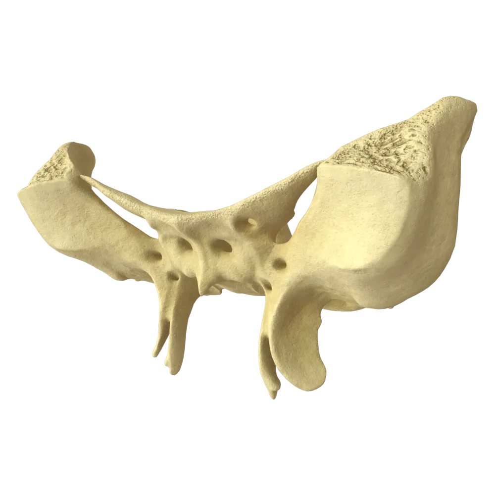
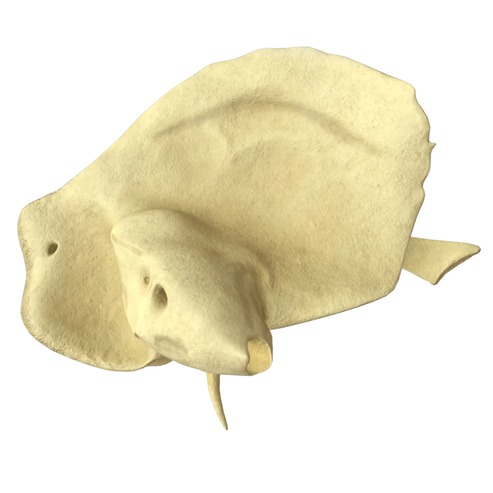
Frontal bone anatomy quiz
Systematic anatomy quiz on the frontal bone: structure, parts, foramina, sutures, processes, and functional relationships.
1/20
bold
text
1. Which part of the frontal bone forms the superior wall of the orbit?
-
Nasal part
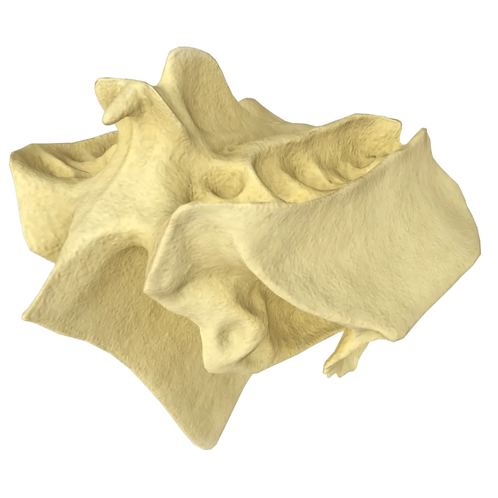
The orbital part (pars orbitalis) of the frontal bone is a horizontal plate that forms the superior wall of the orbit (orbital roof).
-
Squamous part of the frontal bone
The orbital part (pars orbitalis) of the frontal bone is a horizontal plate that forms the superior wall of the orbit (orbital roof).
-
Orbital part
The orbital part (pars orbitalis) of the frontal bone is a horizontal plate that forms the superior wall of the orbit (orbital roof).
-
Zygomatic process
The orbital part (pars orbitalis) of the frontal bone is a horizontal plate that forms the superior wall of the orbit (orbital roof).
-
I find it difficult to answer
The orbital part (pars orbitalis) of the frontal bone is a horizontal plate that forms the superior wall of the orbit (orbital roof).
2. What is the depression on the orbital part of the frontal bone that accommodates the lacrimal gland called?
-
Trochlear fovea
The lacrimal gland fossa (fossa glandulae lacrimalis) is located in the lateral part of the orbital surface of the frontal bone and accommodates the lacrimal gland.
-
Fossa for lacrimal gland
The lacrimal gland fossa (fossa glandulae lacrimalis) is located in the lateral part of the orbital surface of the frontal bone and accommodates the lacrimal gland.
-
Ethmoidal notch
The lacrimal gland fossa (fossa glandulae lacrimalis) is located in the lateral part of the orbital surface of the frontal bone and accommodates the lacrimal gland.
-
Supraorbital notch
The lacrimal gland fossa (fossa glandulae lacrimalis) is located in the lateral part of the orbital surface of the frontal bone and accommodates the lacrimal gland.
-
I find it difficult to answer
The lacrimal gland fossa (fossa glandulae lacrimalis) is located in the lateral part of the orbital surface of the frontal bone and accommodates the lacrimal gland.
3. Where is the trochlear fovea (fovea trochlearis) located on the frontal bone?
-
On the lateral part of the orbital surface
The trochlear fovea (fovea trochlearis) is located on the medial part of the orbital surface of the frontal bone.
-
On the nasal part of the frontal bone
The trochlear fovea (fovea trochlearis) is located on the medial part of the orbital surface of the frontal bone.
-
On the medial part of the orbital surface
The trochlear fovea (fovea trochlearis) is located on the medial part of the orbital surface of the frontal bone.
-
On the internal surface of the squamous part
The trochlear fovea (fovea trochlearis) is located on the medial part of the orbital surface of the frontal bone.
-
I find it difficult to answer
The trochlear fovea (fovea trochlearis) is located on the medial part of the orbital surface of the frontal bone.
4. Which suture connects the frontal bone with the parietal bones?
-
Squamous suture
The coronal suture (sutura coronalis) connects the frontal bone with both parietal bones. The sagittal suture connects the parietal bones to each other, the lambdoid suture connects the parietal bones with the occipital bone, and the squamous suture connects the parietal bones with the temporal bones.
-
Coronal suture
The coronal suture (sutura coronalis) connects the frontal bone with both parietal bones. The sagittal suture connects the parietal bones to each other, the lambdoid suture connects the parietal bones with the occipital bone, and the squamous suture connects the parietal bones with the temporal bones.
-
Sagittal suture
The coronal suture (sutura coronalis) connects the frontal bone with both parietal bones. The sagittal suture connects the parietal bones to each other, the lambdoid suture connects the parietal bones with the occipital bone, and the squamous suture connects the parietal bones with the temporal bones.
-
Lambdoid suture
The coronal suture (sutura coronalis) connects the frontal bone with both parietal bones. The sagittal suture connects the parietal bones to each other, the lambdoid suture connects the parietal bones with the occipital bone, and the squamous suture connects the parietal bones with the temporal bones.
-
I find it difficult to answer
The coronal suture (sutura coronalis) connects the frontal bone with both parietal bones. The sagittal suture connects the parietal bones to each other, the lambdoid suture connects the parietal bones with the occipital bone, and the squamous suture connects the parietal bones with the temporal bones.
5. What is the foramen or notch on the supraorbital margin of the frontal bone through which the supraorbital neurovascular bundle passes called?
-
Supratrochlear foramen
The supraorbital foramen or notch (foramen/incisura supraorbitalis) is located on the supraorbital margin of the frontal bone and transmits the supraorbital nerve (a branch of the frontal nerve from V1) and the supraorbital vessels.
-
Supraorbital foramen (notch)
The supraorbital foramen or notch (foramen/incisura supraorbitalis) is located on the supraorbital margin of the frontal bone and transmits the supraorbital nerve (a branch of the frontal nerve from V1) and the supraorbital vessels.
-
Ethmoidal foramen
The supraorbital foramen or notch (foramen/incisura supraorbitalis) is located on the supraorbital margin of the frontal bone and transmits the supraorbital nerve (a branch of the frontal nerve from V1) and the supraorbital vessels.
-
Frontal foramen (notch)
The supraorbital foramen or notch (foramen/incisura supraorbitalis) is located on the supraorbital margin of the frontal bone and transmits the supraorbital nerve (a branch of the frontal nerve from V1) and the supraorbital vessels.
-
I find it difficult to answer
The supraorbital foramen or notch (foramen/incisura supraorbitalis) is located on the supraorbital margin of the frontal bone and transmits the supraorbital nerve (a branch of the frontal nerve from V1) and the supraorbital vessels.
6. What is the ethmoidal notch (incisura ethmoidalis) of the frontal bone?
-
A groove on the external surface of the squamous part for the passage of the meningeal arteries
The ethmoidal notch (incisura ethmoidalis) is the space between the two orbital parts of the frontal bone.
-
The space between the two orbital parts
The ethmoidal notch (incisura ethmoidalis) is the space between the two orbital parts of the frontal bone.
-
A notch on the supraorbital margin for the passage of a nerve
The ethmoidal notch (incisura ethmoidalis) is the space between the two orbital parts of the frontal bone.
-
A foramen in the nasal part of the frontal bone
The ethmoidal notch (incisura ethmoidalis) is the space between the two orbital parts of the frontal bone.
-
I find it difficult to answer
The ethmoidal notch (incisura ethmoidalis) is the space between the two orbital parts of the frontal bone.
7. Which bone is situated in the ethmoidal notch?
-
Sphenoid bone = Sphenoid = Sphenoidal bone
The ethmoid bone (os ethmoidale) articulates with the frontal bone through the ethmoidal notch (incisura ethmoidalis).
-
Nasal bones
The ethmoid bone (os ethmoidale) articulates with the frontal bone through the ethmoidal notch (incisura ethmoidalis).
-
Ethmoid bone = Ethmoid = Ethmoidal bone
The ethmoid bone (os ethmoidale) articulates with the frontal bone through the ethmoidal notch (incisura ethmoidalis).
-
Lacrimal bone
The ethmoid bone (os ethmoidale) articulates with the frontal bone through the ethmoidal notch (incisura ethmoidalis).
-
I find it difficult to answer
The ethmoid bone (os ethmoidale) articulates with the frontal bone through the ethmoidal notch (incisura ethmoidalis).
8. What is the crest on the internal surface of the squamous part of the frontal bone to which the falx cerebri attaches called?
-
Frontal crest
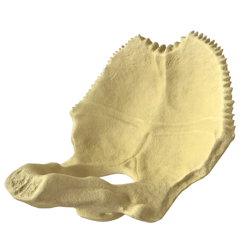
The frontal crest (crista frontalis) is a median crest on the internal (cerebral) surface of the squamous part of the frontal bone. The falx cerebri is attached to it.
-
Crista galli
The frontal crest (crista frontalis) is a median crest on the internal (cerebral) surface of the squamous part of the frontal bone. The falx cerebri is attached to it.
-
Internal occipital crest
The frontal crest (crista frontalis) is a median crest on the internal (cerebral) surface of the squamous part of the frontal bone. The falx cerebri is attached to it.
-
Ethmoidal crest
The frontal crest (crista frontalis) is a median crest on the internal (cerebral) surface of the squamous part of the frontal bone. The falx cerebri is attached to it.
-
I find it difficult to answer
The frontal crest (crista frontalis) is a median crest on the internal (cerebral) surface of the squamous part of the frontal bone. The falx cerebri is attached to it.
9. Where does the groove for the superior sagittal sinus begin on the internal surface of the frontal bone?
-
At the frontal crest and foramen caecum
The groove for the superior sagittal sinus (sulcus sinus sagittalis superioris) begins at the foramen caecum (foramen caecum) near the frontal crest on the internal surface of the squamous part of the frontal bone, then continues onto the parietal bones and the occipital bone.
-
At the level of the coronal suture
The groove for the superior sagittal sinus (sulcus sinus sagittalis superioris) begins at the foramen caecum (foramen caecum) near the frontal crest on the internal surface of the squamous part of the frontal bone, then continues onto the parietal bones and the occipital bone.
-
In the region of the supraorbital notch
The groove for the superior sagittal sinus (sulcus sinus sagittalis superioris) begins at the foramen caecum (foramen caecum) near the frontal crest on the internal surface of the squamous part of the frontal bone, then continues onto the parietal bones and the occipital bone.
-
At the level of the orbital parts
The groove for the superior sagittal sinus (sulcus sinus sagittalis superioris) begins at the foramen caecum (foramen caecum) near the frontal crest on the internal surface of the squamous part of the frontal bone, then continues onto the parietal bones and the occipital bone.
-
I find it difficult to answer
The groove for the superior sagittal sinus (sulcus sinus sagittalis superioris) begins at the foramen caecum (foramen caecum) near the frontal crest on the internal surface of the squamous part of the frontal bone, then continues onto the parietal bones and the occipital bone.
10. What is the foramen at the base of the frontal crest called?
-
Supraorbital foramen
The foramen caecum (foramen caecum) is located at the base of the frontal crest.
-
Blind foramen
The foramen caecum (foramen caecum) is located at the base of the frontal crest.
-
Ethmoidal foramen
The foramen caecum (foramen caecum) is located at the base of the frontal crest.
-
Styloid foramen
The foramen caecum (foramen caecum) is located at the base of the frontal crest.
-
I find it difficult to answer
The foramen caecum (foramen caecum) is located at the base of the frontal crest.
11. How many anatomical notches (or foramina) are located in the medial part of each supraorbital margin of the frontal bone?
-
1
On each supraorbital margin (margo supraorbitalis) of the frontal bone, closer to the nasal part, there are two notches (which may sometimes be converted into complete foramina by bony bridges): the supraorbital notch / foramen (incisura / foramen supraorbitale) and the frontal notch / foramen (incisura / foramen frontale).
-
2
On each supraorbital margin (margo supraorbitalis) of the frontal bone, closer to the nasal part, there are two notches (which may sometimes be converted into complete foramina by bony bridges): the supraorbital notch / foramen (incisura / foramen supraorbitale) and the frontal notch / foramen (incisura / foramen frontale).
-
3
On each supraorbital margin (margo supraorbitalis) of the frontal bone, closer to the nasal part, there are two notches (which may sometimes be converted into complete foramina by bony bridges): the supraorbital notch / foramen (incisura / foramen supraorbitale) and the frontal notch / foramen (incisura / foramen frontale).
-
4
On each supraorbital margin (margo supraorbitalis) of the frontal bone, closer to the nasal part, there are two notches (which may sometimes be converted into complete foramina by bony bridges): the supraorbital notch / foramen (incisura / foramen supraorbitale) and the frontal notch / foramen (incisura / foramen frontale).
-
I find it difficult to answer
On each supraorbital margin (margo supraorbitalis) of the frontal bone, closer to the nasal part, there are two notches (which may sometimes be converted into complete foramina by bony bridges): the supraorbital notch / foramen (incisura / foramen supraorbitale) and the frontal notch / foramen (incisura / foramen frontale).
12. Which bones articulate with the frontal bone in the region of the glabella (glabella)?
-
Nasal bones and maxillae
In the inferior part of the squamous part of the frontal bone—in the region of the glabella—the frontal bone articulates with the nasal bones (through the frontonasal suture) and with the frontal processes of the maxillae. These articulations form the region of the root of the nose.
-
Parietal bones and sphenoid bone
In the inferior part of the squamous part of the frontal bone—in the region of the glabella—the frontal bone articulates with the nasal bones (through the frontonasal suture) and with the frontal processes of the maxillae. These articulations form the region of the root of the nose.
-
Lacrimal bones and palatine bones
In the inferior part of the squamous part of the frontal bone—in the region of the glabella—the frontal bone articulates with the nasal bones (through the frontonasal suture) and with the frontal processes of the maxillae. These articulations form the region of the root of the nose.
-
Zygomatic bones and lacrimal bones
In the inferior part of the squamous part of the frontal bone—in the region of the glabella—the frontal bone articulates with the nasal bones (through the frontonasal suture) and with the frontal processes of the maxillae. These articulations form the region of the root of the nose.
-
I find it difficult to answer
In the inferior part of the squamous part of the frontal bone—in the region of the glabella—the frontal bone articulates with the nasal bones (through the frontonasal suture) and with the frontal processes of the maxillae. These articulations form the region of the root of the nose.
13. What is the paired elevation on the external surface of the squamous part of the frontal bone corresponding to the center of ossification called?
-
Supraorbital margin
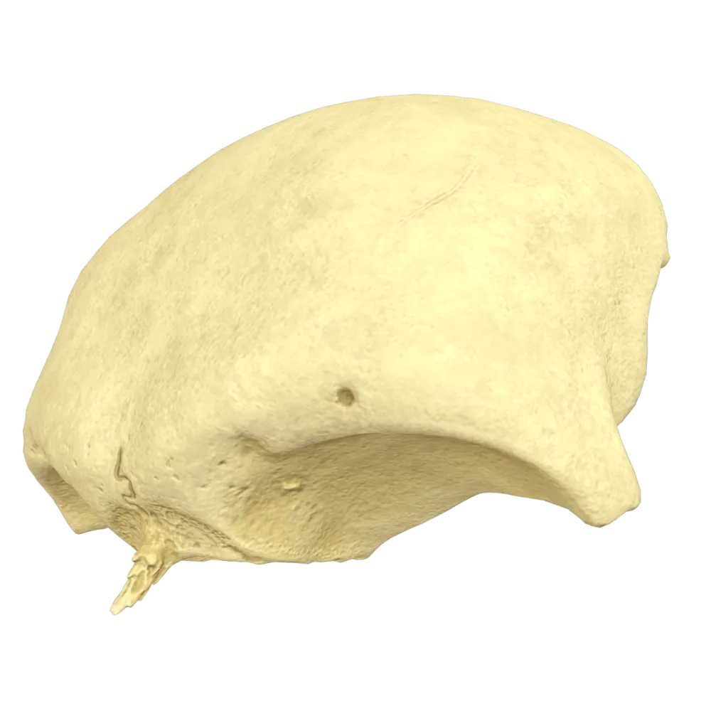
The frontal eminence (tuber frontale) is a paired rounded elevation on the external surface of the squamous part of the frontal bone corresponding to the center of primary ossification. It is more prominent in children than in adults. The superciliary arches are located inferiorly, above the superior margins of the orbits.
-
Frontal eminence
The frontal eminence (tuber frontale) is a paired rounded elevation on the external surface of the squamous part of the frontal bone corresponding to the center of primary ossification. It is more prominent in children than in adults. The superciliary arches are located inferiorly, above the superior margins of the orbits.
-
Superciliary arch
The frontal eminence (tuber frontale) is a paired rounded elevation on the external surface of the squamous part of the frontal bone corresponding to the center of primary ossification. It is more prominent in children than in adults. The superciliary arches are located inferiorly, above the superior margins of the orbits.
-
Zygomatic process
The frontal eminence (tuber frontale) is a paired rounded elevation on the external surface of the squamous part of the frontal bone corresponding to the center of primary ossification. It is more prominent in children than in adults. The superciliary arches are located inferiorly, above the superior margins of the orbits.
-
I find it difficult to answer
The frontal eminence (tuber frontale) is a paired rounded elevation on the external surface of the squamous part of the frontal bone corresponding to the center of primary ossification. It is more prominent in children than in adults. The superciliary arches are located inferiorly, above the superior margins of the orbits.
14. Which process of the frontal bone articulates with the zygomatic bone?
-
Nasal process
The zygomatic process (processus zygomaticus) of the frontal bone articulates with the zygomatic bone.
-
Zygomatic process
The zygomatic process (processus zygomaticus) of the frontal bone articulates with the zygomatic bone.
-
Orbital process
The zygomatic process (processus zygomaticus) of the frontal bone articulates with the zygomatic bone.
-
Mammillary process
The zygomatic process (processus zygomaticus) of the frontal bone articulates with the zygomatic bone.
-
I find it difficult to answer
The zygomatic process (processus zygomaticus) of the frontal bone articulates with the zygomatic bone.
15. What is the area between the superciliary arches on the external surface of the frontal bone called?
-
Frontal eminence
The glabella (glabella) is a smooth area on the external surface of the squamous part of the frontal bone, located between the superciliary arches along the median line. It is an important anthropometric landmark.
-
Glabella (glabella)
The glabella (glabella) is a smooth area on the external surface of the squamous part of the frontal bone, located between the superciliary arches along the median line. It is an important anthropometric landmark.
-
Supraorbital margin
The glabella (glabella) is a smooth area on the external surface of the squamous part of the frontal bone, located between the superciliary arches along the median line. It is an important anthropometric landmark.
-
Nasal part
The glabella (glabella) is a smooth area on the external surface of the squamous part of the frontal bone, located between the superciliary arches along the median line. It is an important anthropometric landmark.
-
I find it difficult to answer
The glabella (glabella) is a smooth area on the external surface of the squamous part of the frontal bone, located between the superciliary arches along the median line. It is an important anthropometric landmark.
16. Is there an air-filled cavity within the frontal bone, and what is it called?
-
Yes, it is the frontal sinus
The frontal bone is a pneumatic bone (ossa pneumatica). Within its squamous and nasal parts is the paired frontal sinus (sinus frontalis), which is lined with mucosa and filled with air.
-
No, the frontal bone is entirely cancellous and contains no cavities
The frontal bone is a pneumatic bone (ossa pneumatica). Within its squamous and nasal parts is the paired frontal sinus (sinus frontalis), which is lined with mucosa and filled with air.
-
Yes, it is the maxillary sinus
The frontal bone is a pneumatic bone (ossa pneumatica). Within its squamous and nasal parts is the paired frontal sinus (sinus frontalis), which is lined with mucosa and filled with air.
-
Yes, it is the sphenoidal sinus
The frontal bone is a pneumatic bone (ossa pneumatica). Within its squamous and nasal parts is the paired frontal sinus (sinus frontalis), which is lined with mucosa and filled with air.
-
I find it difficult to answer
The frontal bone is a pneumatic bone (ossa pneumatica). Within its squamous and nasal parts is the paired frontal sinus (sinus frontalis), which is lined with mucosa and filled with air.
17. Where does the frontal sinus open?
-
Into the superior nasal meatus.
The frontal sinus opens into the middle nasal meatus.
-
Into the middle nasal meatus.
The frontal sinus opens into the middle nasal meatus.
-
Into the inferior nasal meatus.
The frontal sinus opens into the middle nasal meatus.
-
Directly into the nasopharyngeal cavity
The frontal sinus opens into the middle nasal meatus.
-
I find it difficult to answer
The frontal sinus opens into the middle nasal meatus.
18. Which of the following functions is NOT performed by the paranasal sinuses (pneumatic cavities) of the bones of the cranium?
-
Humidification, warming, and purification of inhaled air
The paranasal sinuses perform 4 main functions: 1. Air conditioning: they warm, humidify, and purify inhaled air. 2. Reduction of cranial mass: they reduce the weight of the bones without compromising their strength. 3. Voice resonance: they impart timbre and volume to speech. 4. Protection (shock absorption): they cushion impacts in the facial region, protecting the brain. The sinuses do not produce hormones.
-
Production of hormones that regulate the growth of the bones of the viscerocranium
The paranasal sinuses perform 4 main functions: 1. Air conditioning: they warm, humidify, and purify inhaled air. 2. Reduction of cranial mass: they reduce the weight of the bones without compromising their strength. 3. Voice resonance: they impart timbre and volume to speech. 4. Protection (shock absorption): they cushion impacts in the facial region, protecting the brain. The sinuses do not produce hormones.
-
Reduction of the mass of the neurocranium
The paranasal sinuses perform 4 main functions: 1. Air conditioning: they warm, humidify, and purify inhaled air. 2. Reduction of cranial mass: they reduce the weight of the bones without compromising their strength. 3. Voice resonance: they impart timbre and volume to speech. 4. Protection (shock absorption): they cushion impacts in the facial region, protecting the brain. The sinuses do not produce hormones.
-
Contribution to the individual timbre and sound of the voice (resonator function)
The paranasal sinuses perform 4 main functions: 1. Air conditioning: they warm, humidify, and purify inhaled air. 2. Reduction of cranial mass: they reduce the weight of the bones without compromising their strength. 3. Voice resonance: they impart timbre and volume to speech. 4. Protection (shock absorption): they cushion impacts in the facial region, protecting the brain. The sinuses do not produce hormones.
-
I find it difficult to answer
The paranasal sinuses perform 4 main functions: 1. Air conditioning: they warm, humidify, and purify inhaled air. 2. Reduction of cranial mass: they reduce the weight of the bones without compromising their strength. 3. Voice resonance: they impart timbre and volume to speech. 4. Protection (shock absorption): they cushion impacts in the facial region, protecting the brain. The sinuses do not produce hormones.
19. With which bone does the frontal bone articulate via the sphenofrontal suture (sutura sphenofrontalis)?
-
Ethmoid bone
The sphenofrontal suture (sutura sphenofrontalis) connects the frontal bone with the greater wings of the sphenoid bone on the lateral wall of the cranium (in the temporal region).
-
Sphenoid bone
The sphenofrontal suture (sutura sphenofrontalis) connects the frontal bone with the greater wings of the sphenoid bone on the lateral wall of the cranium (in the temporal region).
-
Parietal bone
The sphenofrontal suture (sutura sphenofrontalis) connects the frontal bone with the greater wings of the sphenoid bone on the lateral wall of the cranium (in the temporal region).
-
Lacrimal bone
The sphenofrontal suture (sutura sphenofrontalis) connects the frontal bone with the greater wings of the sphenoid bone on the lateral wall of the cranium (in the temporal region).
-
I find it difficult to answer
The sphenofrontal suture (sutura sphenofrontalis) connects the frontal bone with the greater wings of the sphenoid bone on the lateral wall of the cranium (in the temporal region).
20. What are the depressions on the internal surface of the frontal bone produced by the adjacent cerebral gyri called?
-
Digital impressions
Digital impressions (impressiones digitatae, juga cerebralia) are depressions on the internal surface of the bones of the cranium formed by the cerebral gyri.
-
Arterial grooves
Digital impressions (impressiones digitatae, juga cerebralia) are depressions on the internal surface of the bones of the cranium formed by the cerebral gyri.
-
Groove for the superior sagittal sinus
Digital impressions (impressiones digitatae, juga cerebralia) are depressions on the internal surface of the bones of the cranium formed by the cerebral gyri.
-
Grooves for the venous sinuses
Digital impressions (impressiones digitatae, juga cerebralia) are depressions on the internal surface of the bones of the cranium formed by the cerebral gyri.
-
I find it difficult to answer
Digital impressions (impressiones digitatae, juga cerebralia) are depressions on the internal surface of the bones of the cranium formed by the cerebral gyri.
Retake this quiz?
Your current progress will be reset.
