
Ethmoid bone anatomy quiz
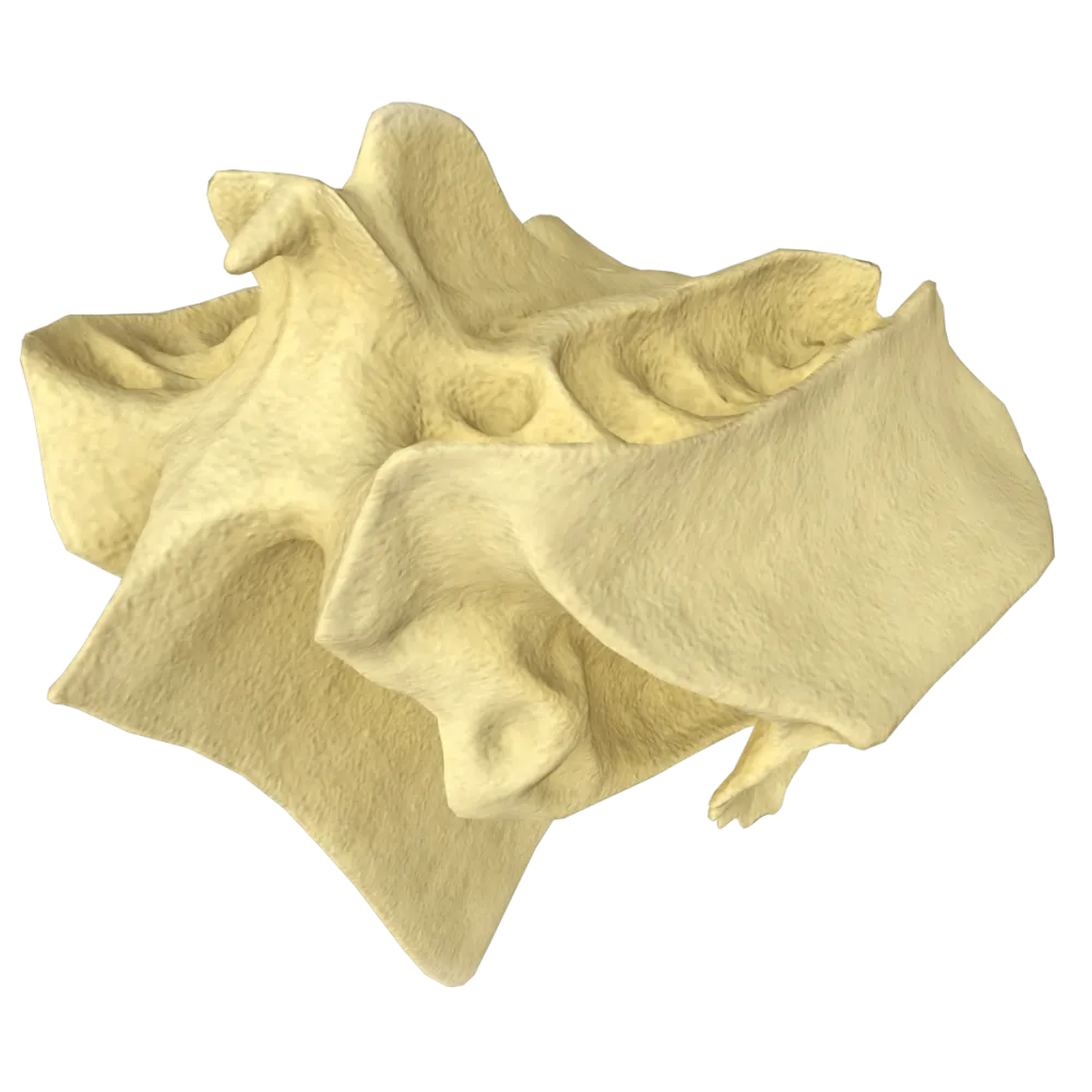
Normal anatomy quiz on the ethmoid bone: structure, parts, processes, foramina, and topographic relationships.
1/20
bold
text
1. To which group of bones of the cranium does the ethmoid bone belong?
-
Bones of the viscerocranium
The ethmoid bone (os ethmoidale) is part of the neurocranium, forming part of its base and contributing to the formation of the anterior portion of the cranial base.
-
Neurocranium Bones
The ethmoid bone (os ethmoidale) is part of the neurocranium, forming part of its base and contributing to the formation of the anterior portion of the cranial base.
-
Bones of the cranial base exclusively
The ethmoid bone (os ethmoidale) is part of the neurocranium, forming part of its base and contributing to the formation of the anterior portion of the cranial base.
-
Mixed bones that do not belong to any group
The ethmoid bone (os ethmoidale) is part of the neurocranium, forming part of its base and contributing to the formation of the anterior portion of the cranial base.
-
I find it difficult to answer
The ethmoid bone (os ethmoidale) is part of the neurocranium, forming part of its base and contributing to the formation of the anterior portion of the cranial base.
2. Which of the listed structures is part of the ethmoid bone?
-
Crista galli
The cock's comb (crista galli) is an unpaired vertical process of the ethmoid bone that projects into the cranial cavity and provides attachment for the falx cerebri.
-
Pterygoid process
The cock's comb (crista galli) is an unpaired vertical process of the ethmoid bone that projects into the cranial cavity and provides attachment for the falx cerebri.
-
Styloid process
The cock's comb (crista galli) is an unpaired vertical process of the ethmoid bone that projects into the cranial cavity and provides attachment for the falx cerebri.
-
Mammillary process
The cock's comb (crista galli) is an unpaired vertical process of the ethmoid bone that projects into the cranial cavity and provides attachment for the falx cerebri.
-
I find it difficult to answer
The cock's comb (crista galli) is an unpaired vertical process of the ethmoid bone that projects into the cranial cavity and provides attachment for the falx cerebri.
3. The cribriform plate (lamina cribrosa) of the ethmoid bone is part of the:
-
Lateral labyrinths
The cribriform plate (lamina cribrosa) is a horizontal plate of the ethmoid bone perforated by numerous foramina for the passage of the olfactory filaments (fila olfactoria).
-
Perpendicular plate
The cribriform plate (lamina cribrosa) is a horizontal plate of the ethmoid bone perforated by numerous foramina for the passage of the olfactory filaments (fila olfactoria).
-
Horizontal plate
The cribriform plate (lamina cribrosa) is a horizontal plate of the ethmoid bone perforated by numerous foramina for the passage of the olfactory filaments (fila olfactoria).
-
Orbital plate
The cribriform plate (lamina cribrosa) is a horizontal plate of the ethmoid bone perforated by numerous foramina for the passage of the olfactory filaments (fila olfactoria).
-
I find it difficult to answer
The cribriform plate (lamina cribrosa) is a horizontal plate of the ethmoid bone perforated by numerous foramina for the passage of the olfactory filaments (fila olfactoria).
4. The following pass through the foramina of the cribriform plate:
-
Branches of the trigeminal nerve
The numerous foramina of the lamina cribrosa transmit the olfactory filaments (fila olfactoria), the axons of olfactory cells in the nasal mucosa that constitute part of cranial nerve I.
-
Olfactory filaments (fila olfactoria)
The numerous foramina of the lamina cribrosa transmit the olfactory filaments (fila olfactoria), the axons of olfactory cells in the nasal mucosa that constitute part of cranial nerve I.
-
Branches of the facial nerve
The numerous foramina of the lamina cribrosa transmit the olfactory filaments (fila olfactoria), the axons of olfactory cells in the nasal mucosa that constitute part of cranial nerve I.
-
Optic nerve fibers
The numerous foramina of the lamina cribrosa transmit the olfactory filaments (fila olfactoria), the axons of olfactory cells in the nasal mucosa that constitute part of cranial nerve I.
-
I find it difficult to answer
The numerous foramina of the lamina cribrosa transmit the olfactory filaments (fila olfactoria), the axons of olfactory cells in the nasal mucosa that constitute part of cranial nerve I.
5. The perpendicular plate (lamina perpendicularis) of the ethmoid bone contributes to the formation of the:
-
Lateral wall of the nasal cavity
The perpendicular plate (lamina perpendicularis) of the ethmoid bone forms the superior part of the bony nasal septum, whereas the inferior part is formed by the vomer.
-
Superior wall of the nasal cavity
The perpendicular plate (lamina perpendicularis) of the ethmoid bone forms the superior part of the bony nasal septum, whereas the inferior part is formed by the vomer.
-
Bony nasal septum
The perpendicular plate (lamina perpendicularis) of the ethmoid bone forms the superior part of the bony nasal septum, whereas the inferior part is formed by the vomer.
-
Inferior wall of the orbit
The perpendicular plate (lamina perpendicularis) of the ethmoid bone forms the superior part of the bony nasal septum, whereas the inferior part is formed by the vomer.
-
I find it difficult to answer
The perpendicular plate (lamina perpendicularis) of the ethmoid bone forms the superior part of the bony nasal septum, whereas the inferior part is formed by the vomer.
6. The orbital plate (lamina orbitalis) of the ethmoid bone forms the:
-
Inferior wall of the orbit
The lamina orbitalis (orbital plate) of the ethmoid bone is the principal component of the medial wall of the orbit and borders the cells of the ethmoidal labyrinth.
-
Medial wall of the orbit
The lamina orbitalis (orbital plate) of the ethmoid bone is the principal component of the medial wall of the orbit and borders the cells of the ethmoidal labyrinth.
-
Lateral wall of the orbit
The lamina orbitalis (orbital plate) of the ethmoid bone is the principal component of the medial wall of the orbit and borders the cells of the ethmoidal labyrinth.
-
Superior wall of the orbit
The lamina orbitalis (orbital plate) of the ethmoid bone is the principal component of the medial wall of the orbit and borders the cells of the ethmoidal labyrinth.
-
I find it difficult to answer
The lamina orbitalis (orbital plate) of the ethmoid bone is the principal component of the medial wall of the orbit and borders the cells of the ethmoidal labyrinth.
7. The ethmoidal labyrinth (labyrinthus ethmoidalis) consists of:
-
Two large air-filled sinuses
The ethmoidal labyrinth is formed by numerous thin-walled air cells (cellulae ethmoidales) that communicate with one another and with the nasal cavity.
-
Numerous thin-walled air cells
The ethmoidal labyrinth is formed by numerous thin-walled air cells (cellulae ethmoidales) that communicate with one another and with the nasal cavity.
-
Dense cancellous bone without cavities
The ethmoidal labyrinth is formed by numerous thin-walled air cells (cellulae ethmoidales) that communicate with one another and with the nasal cavity.
-
A single large cavity divided by one septum
The ethmoidal labyrinth is formed by numerous thin-walled air cells (cellulae ethmoidales) that communicate with one another and with the nasal cavity.
-
I find it difficult to answer
The ethmoidal labyrinth is formed by numerous thin-walled air cells (cellulae ethmoidales) that communicate with one another and with the nasal cavity.
8. The superior nasal concha (concha nasalis superior) is an integral part of the:
-
Inferior turbinate
The superior nasal concha is a process of the ethmoid bone.
-
Ethmoid bone
The superior nasal concha is a process of the ethmoid bone.
-
Palatine bone
The superior nasal concha is a process of the ethmoid bone.
-
Sphenoid bone
The superior nasal concha is a process of the ethmoid bone.
-
I find it difficult to answer
The superior nasal concha is a process of the ethmoid bone.
9. The middle nasal concha (concha nasalis media) is classified as a:
-
Separate facial bone
The middle nasal concha (concha nasalis media) is a process of the ethmoid bone that bounds the middle nasal meatus inferiorly.
-
Process of the ethmoid bone
The middle nasal concha (concha nasalis media) is a process of the ethmoid bone that bounds the middle nasal meatus inferiorly.
-
Part of the palatine bone
The middle nasal concha (concha nasalis media) is a process of the ethmoid bone that bounds the middle nasal meatus inferiorly.
-
Part of the sphenoid bone
The middle nasal concha (concha nasalis media) is a process of the ethmoid bone that bounds the middle nasal meatus inferiorly.
-
I find it difficult to answer
The middle nasal concha (concha nasalis media) is a process of the ethmoid bone that bounds the middle nasal meatus inferiorly.
10. Which nasal meatus is located between the middle and superior nasal conchae?
-
Inferior nasal meatus
The superior nasal meatus (meatus nasi superior) is located between the middle and superior nasal conchae. The posterior cells of the ethmoidal labyrinth and the sphenoidal sinus open into it.
-
Common nasal meatus
The superior nasal meatus (meatus nasi superior) is located between the middle and superior nasal conchae. The posterior cells of the ethmoidal labyrinth and the sphenoidal sinus open into it.
-
Superior nasal meatus
The superior nasal meatus (meatus nasi superior) is located between the middle and superior nasal conchae. The posterior cells of the ethmoidal labyrinth and the sphenoidal sinus open into it.
-
Middle nasal meatus
The superior nasal meatus (meatus nasi superior) is located between the middle and superior nasal conchae. The posterior cells of the ethmoidal labyrinth and the sphenoidal sinus open into it.
-
I find it difficult to answer
The superior nasal meatus (meatus nasi superior) is located between the middle and superior nasal conchae. The posterior cells of the ethmoidal labyrinth and the sphenoidal sinus open into it.
11. All of the following open into the middle nasal meatus except:
-
Frontal sinus
The sphenoidal sinus opens into the superior nasal meatus, but not into the middle nasal meatus. The frontal sinus, maxillary sinus, and anterior cells of the ethmoidal labyrinth open into the middle nasal meatus.
-
Anterior cells of the ethmoidal labyrinth
The sphenoidal sinus opens into the superior nasal meatus, but not into the middle nasal meatus. The frontal sinus, maxillary sinus, and anterior cells of the ethmoidal labyrinth open into the middle nasal meatus.
-
Maxillary sinus
The sphenoidal sinus opens into the superior nasal meatus, but not into the middle nasal meatus. The frontal sinus, maxillary sinus, and anterior cells of the ethmoidal labyrinth open into the middle nasal meatus.
-
Sphenoidal sinus
The sphenoidal sinus opens into the superior nasal meatus, but not into the middle nasal meatus. The frontal sinus, maxillary sinus, and anterior cells of the ethmoidal labyrinth open into the middle nasal meatus.
-
I find it difficult to answer
The sphenoidal sinus opens into the superior nasal meatus, but not into the middle nasal meatus. The frontal sinus, maxillary sinus, and anterior cells of the ethmoidal labyrinth open into the middle nasal meatus.
12. The uncinate process (processus uncinatus) is part of the:
-
Sphenoid bone
The uncinate process (processus uncinatus) is a small, crescent-shaped bony projection of the ethmoid bone that contributes to the formation of the semilunar hiatus (hiatus semilunaris) of the middle nasal meatus.
-
Inferior turbinate
The uncinate process (processus uncinatus) is a small, crescent-shaped bony projection of the ethmoid bone that contributes to the formation of the semilunar hiatus (hiatus semilunaris) of the middle nasal meatus.
-
Ethmoid bone
The uncinate process (processus uncinatus) is a small, crescent-shaped bony projection of the ethmoid bone that contributes to the formation of the semilunar hiatus (hiatus semilunaris) of the middle nasal meatus.
-
Palatine bone
The uncinate process (processus uncinatus) is a small, crescent-shaped bony projection of the ethmoid bone that contributes to the formation of the semilunar hiatus (hiatus semilunaris) of the middle nasal meatus.
-
I find it difficult to answer
The uncinate process (processus uncinatus) is a small, crescent-shaped bony projection of the ethmoid bone that contributes to the formation of the semilunar hiatus (hiatus semilunaris) of the middle nasal meatus.
13. The ethmoidal bulla (bulla ethmoidalis) is:
-
The largest cell of the ethmoidal labyrinth, projecting into the middle nasal meatus
The bulla ethmoidalis is the largest and most constant air cell of the ethmoidal labyrinth, forming an elevation on the lateral wall of the middle nasal meatus above the uncinate process.
-
A foramen in the cribriform plate
The bulla ethmoidalis is the largest and most constant air cell of the ethmoidal labyrinth, forming an elevation on the lateral wall of the middle nasal meatus above the uncinate process.
-
Part of the crista galli
The bulla ethmoidalis is the largest and most constant air cell of the ethmoidal labyrinth, forming an elevation on the lateral wall of the middle nasal meatus above the uncinate process.
-
A cell that opens into the superior nasal meatus
The bulla ethmoidalis is the largest and most constant air cell of the ethmoidal labyrinth, forming an elevation on the lateral wall of the middle nasal meatus above the uncinate process.
-
I find it difficult to answer
The bulla ethmoidalis is the largest and most constant air cell of the ethmoidal labyrinth, forming an elevation on the lateral wall of the middle nasal meatus above the uncinate process.
14. With which bone does the ethmoid bone articulate anteriorly along the median line?
-
Sphenoid bone = Sphenoid = Sphenoidal bone
The nasal bones articulate with the superior border of the perpendicular plate of the ethmoid bone along the median line. The frontal bone articulates with the horizontal plate superiorly.
-
Frontal bone
The nasal bones articulate with the superior border of the perpendicular plate of the ethmoid bone along the median line. The frontal bone articulates with the horizontal plate superiorly.
-
Nasal bone
The nasal bones articulate with the superior border of the perpendicular plate of the ethmoid bone along the median line. The frontal bone articulates with the horizontal plate superiorly.
-
Vomer
The nasal bones articulate with the superior border of the perpendicular plate of the ethmoid bone along the median line. The frontal bone articulates with the horizontal plate superiorly.
-
I find it difficult to answer
The nasal bones articulate with the superior border of the perpendicular plate of the ethmoid bone along the median line. The frontal bone articulates with the horizontal plate superiorly.
15. The cock's comb (crista galli) serves as the attachment site for:
-
Dura mater in the region of the falx cerebri
The anterior border of the falx cerebri (falx cerebri)—a fold of dura mater separating the cerebral hemispheres—attaches to the cock's comb (crista galli).
-
Diaphragma sellae
The anterior border of the falx cerebri (falx cerebri)—a fold of dura mater separating the cerebral hemispheres—attaches to the cock's comb (crista galli).
-
Tentorium cerebelli
The anterior border of the falx cerebri (falx cerebri)—a fold of dura mater separating the cerebral hemispheres—attaches to the cock's comb (crista galli).
-
Arachnoid mater.
The anterior border of the falx cerebri (falx cerebri)—a fold of dura mater separating the cerebral hemispheres—attaches to the cock's comb (crista galli).
-
I find it difficult to answer
The anterior border of the falx cerebri (falx cerebri)—a fold of dura mater separating the cerebral hemispheres—attaches to the cock's comb (crista galli).
16. Which part of the ethmoid bone forms the superior part of the nasal septum?
-
Orbital plate (lamina orbitalis)
The perpendicular plate (lamina perpendicularis) of the ethmoid bone forms the superior part of the bony nasal septum.
-
Cribriform plate (lamina cribrosa)
The perpendicular plate (lamina perpendicularis) of the ethmoid bone forms the superior part of the bony nasal septum.
-
Cock's comb (crista galli)
The perpendicular plate (lamina perpendicularis) of the ethmoid bone forms the superior part of the bony nasal septum.
-
Perpendicular plate (lamina perpendicularis)
The perpendicular plate (lamina perpendicularis) of the ethmoid bone forms the superior part of the bony nasal septum.
-
I find it difficult to answer
The perpendicular plate (lamina perpendicularis) of the ethmoid bone forms the superior part of the bony nasal septum.
17. Why can trauma to or fracture of the ethmoid bone (os ethmoidale) result in anosmia (loss of smell)?
-
The olfactory nerve fibers passing through the cribriform plate are compressed or damaged
Fine filaments of the olfactory nerve (cranial nerve I) pass through the foramina of the horizontal (cribriform) plate of the ethmoid bone (lamina cribrosa), extending from receptors in the nasal cavity to the olfactory bulb. In the event of fracture or displacement of bone fragments, these nerve fibers are easily torn, compressed, or damaged, resulting in complete or partial loss of smell (anosmia/hyposmia).
-
The olfactory receptors in the mucosa of the inferior turbinate are destroyed
Fine filaments of the olfactory nerve (cranial nerve I) pass through the foramina of the horizontal (cribriform) plate of the ethmoid bone (lamina cribrosa), extending from receptors in the nasal cavity to the olfactory bulb. In the event of fracture or displacement of bone fragments, these nerve fibers are easily torn, compressed, or damaged, resulting in complete or partial loss of smell (anosmia/hyposmia).
-
The blood supply to the olfactory areas of the cerebral cortex is impaired
Fine filaments of the olfactory nerve (cranial nerve I) pass through the foramina of the horizontal (cribriform) plate of the ethmoid bone (lamina cribrosa), extending from receptors in the nasal cavity to the olfactory bulb. In the event of fracture or displacement of bone fragments, these nerve fibers are easily torn, compressed, or damaged, resulting in complete or partial loss of smell (anosmia/hyposmia).
-
The lumen of the nasolacrimal duct is obstructed
Fine filaments of the olfactory nerve (cranial nerve I) pass through the foramina of the horizontal (cribriform) plate of the ethmoid bone (lamina cribrosa), extending from receptors in the nasal cavity to the olfactory bulb. In the event of fracture or displacement of bone fragments, these nerve fibers are easily torn, compressed, or damaged, resulting in complete or partial loss of smell (anosmia/hyposmia).
-
I find it difficult to answer
Fine filaments of the olfactory nerve (cranial nerve I) pass through the foramina of the horizontal (cribriform) plate of the ethmoid bone (lamina cribrosa), extending from receptors in the nasal cavity to the olfactory bulb. In the event of fracture or displacement of bone fragments, these nerve fibers are easily torn, compressed, or damaged, resulting in complete or partial loss of smell (anosmia/hyposmia).
18. Does the ethmoid bone (os ethmoidale) contain a single ethmoidal sinus?
-
Yes, it contains one large, undivided ethmoidal sinus
The term “ethmoidal sinus” is often used in clinical practice and anatomy; anatomically, however, it is not a single cavity (such as the maxillary or frontal sinus), but a complex of small air-filled cavities—the ethmoidal cells (cellulae ethmoidales)—that form the ethmoidal labyrinth (labyrinthus ethmoidalis). These cells are divided into anterior, middle, and posterior groups and communicate with the nasal cavity.
-
No, the ethmoid bone is entirely solid and contains no air-filled cavities
The term “ethmoidal sinus” is often used in clinical practice and anatomy; anatomically, however, it is not a single cavity (such as the maxillary or frontal sinus), but a complex of small air-filled cavities—the ethmoidal cells (cellulae ethmoidales)—that form the ethmoidal labyrinth (labyrinthus ethmoidalis). These cells are divided into anterior, middle, and posterior groups and communicate with the nasal cavity.
-
No, instead of a single sinus, it contains a labyrinth of numerous small air cells that collectively constitute the ethmoidal sinus
The term “ethmoidal sinus” is often used in clinical practice and anatomy; anatomically, however, it is not a single cavity (such as the maxillary or frontal sinus), but a complex of small air-filled cavities—the ethmoidal cells (cellulae ethmoidales)—that form the ethmoidal labyrinth (labyrinthus ethmoidalis). These cells are divided into anterior, middle, and posterior groups and communicate with the nasal cavity.
-
Yes, but it is filled with bone marrow rather than air
The term “ethmoidal sinus” is often used in clinical practice and anatomy; anatomically, however, it is not a single cavity (such as the maxillary or frontal sinus), but a complex of small air-filled cavities—the ethmoidal cells (cellulae ethmoidales)—that form the ethmoidal labyrinth (labyrinthus ethmoidalis). These cells are divided into anterior, middle, and posterior groups and communicate with the nasal cavity.
-
I find it difficult to answer
The term “ethmoidal sinus” is often used in clinical practice and anatomy; anatomically, however, it is not a single cavity (such as the maxillary or frontal sinus), but a complex of small air-filled cavities—the ethmoidal cells (cellulae ethmoidales)—that form the ethmoidal labyrinth (labyrinthus ethmoidalis). These cells are divided into anterior, middle, and posterior groups and communicate with the nasal cavity.
19. Which structure of the ethmoid bone contributes to the boundaries of the semilunar hiatus (hiatus semilunaris)?
-
Perpendicular plate
The semilunar hiatus (hiatus semilunaris) is bounded inferiorly and anteriorly by the uncinate process (processus uncinatus), and superiorly and posteriorly by the ethmoidal bulla. The infundibulum and the middle nasal meatus communicate through it.
-
Crista galli
The semilunar hiatus (hiatus semilunaris) is bounded inferiorly and anteriorly by the uncinate process (processus uncinatus), and superiorly and posteriorly by the ethmoidal bulla. The infundibulum and the middle nasal meatus communicate through it.
-
Uncinate process
The semilunar hiatus (hiatus semilunaris) is bounded inferiorly and anteriorly by the uncinate process (processus uncinatus), and superiorly and posteriorly by the ethmoidal bulla. The infundibulum and the middle nasal meatus communicate through it.
-
Bony superior nasal concha
The semilunar hiatus (hiatus semilunaris) is bounded inferiorly and anteriorly by the uncinate process (processus uncinatus), and superiorly and posteriorly by the ethmoidal bulla. The infundibulum and the middle nasal meatus communicate through it.
-
I find it difficult to answer
The semilunar hiatus (hiatus semilunaris) is bounded inferiorly and anteriorly by the uncinate process (processus uncinatus), and superiorly and posteriorly by the ethmoidal bulla. The infundibulum and the middle nasal meatus communicate through it.
20. What is the ethmoidal infundibulum (infundibulum ethmoidale) in the structure of the ethmoid bone?
-
A bony projection that separates the left and right halves of the nasal cavity
The ethmoidal infundibulum (infundibulum ethmoidale) is a funnel-shaped, curved cleft or canal located in the anterosuperior part of the ethmoidal labyrinth. Through the ethmoidal infundibulum, the frontal sinus and anterior cells of the ethmoid bone communicate with the middle nasal meatus, providing ventilation and mucus drainage.
-
A foramen in the cribriform plate
The ethmoidal infundibulum (infundibulum ethmoidale) is a funnel-shaped, curved cleft or canal located in the anterosuperior part of the ethmoidal labyrinth. Through the ethmoidal infundibulum, the frontal sinus and anterior cells of the ethmoid bone communicate with the middle nasal meatus, providing ventilation and mucus drainage.
-
A narrow, funnel-shaped canal in the anterior part of the ethmoidal labyrinth that connects the frontal sinus and anterior ethmoidal cells with the middle nasal meatus
The ethmoidal infundibulum (infundibulum ethmoidale) is a funnel-shaped, curved cleft or canal located in the anterosuperior part of the ethmoidal labyrinth. Through the ethmoidal infundibulum, the frontal sinus and anterior cells of the ethmoid bone communicate with the middle nasal meatus, providing ventilation and mucus drainage.
-
Cartilaginous part of the nasal septum
The ethmoidal infundibulum (infundibulum ethmoidale) is a funnel-shaped, curved cleft or canal located in the anterosuperior part of the ethmoidal labyrinth. Through the ethmoidal infundibulum, the frontal sinus and anterior cells of the ethmoid bone communicate with the middle nasal meatus, providing ventilation and mucus drainage.
-
I find it difficult to answer
The ethmoidal infundibulum (infundibulum ethmoidale) is a funnel-shaped, curved cleft or canal located in the anterosuperior part of the ethmoidal labyrinth. Through the ethmoidal infundibulum, the frontal sinus and anterior cells of the ethmoid bone communicate with the middle nasal meatus, providing ventilation and mucus drainage.
Retake this quiz?
Your current progress will be reset.
