
Parietal bone anatomy quiz
The quiz covers the anatomy of the parietal bone: surfaces, borders, angles, grooves, foramina, and topographic relationships.
1/20
bold
text
1. Which of the following surfaces of the parietal bone faces the brain?
-
External (convex) surface
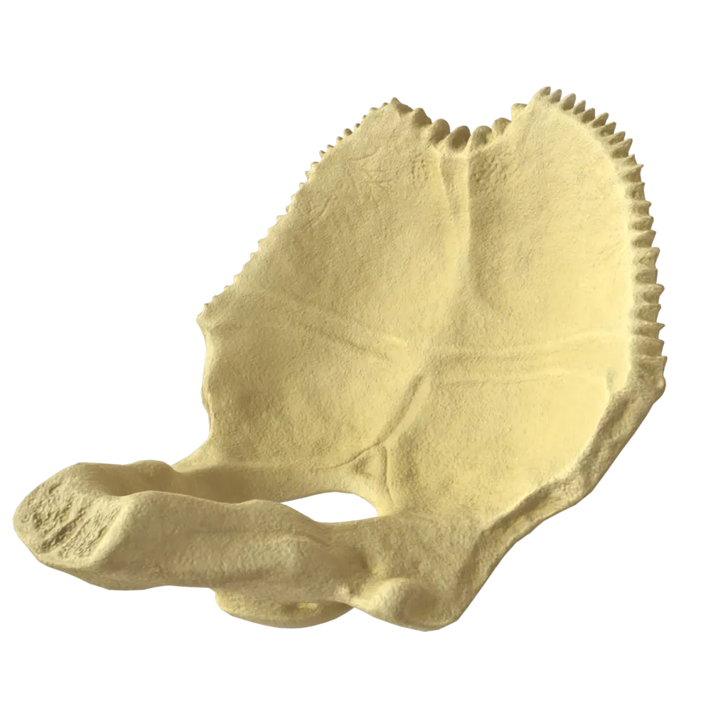
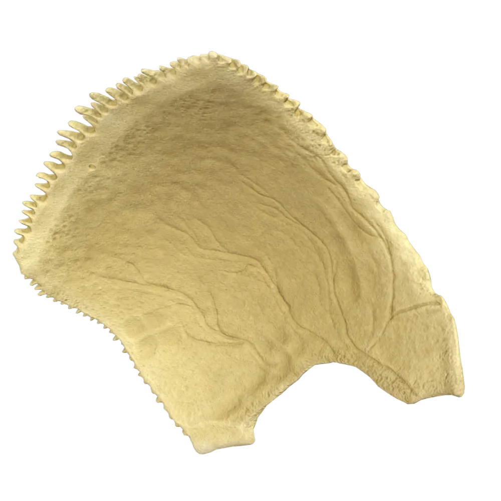
The internal (concave) surface—facies interna—faces the brain within the cranial cavity; the cerebral impressions and arterial grooves are located on this surface.
-
Internal (concave) surface
The internal (concave) surface—facies interna—faces the brain within the cranial cavity; the cerebral impressions and arterial grooves are located on this surface.
-
Sagittal border
The internal (concave) surface—facies interna—faces the brain within the cranial cavity; the cerebral impressions and arterial grooves are located on this surface.
-
Occipital border
The internal (concave) surface—facies interna—faces the brain within the cranial cavity; the cerebral impressions and arterial grooves are located on this surface.
-
I find it difficult to answer
The internal (concave) surface—facies interna—faces the brain within the cranial cavity; the cerebral impressions and arterial grooves are located on this surface.
2. What is the most prominent point on the external surface of the parietal bone called?
-
Parietal notch
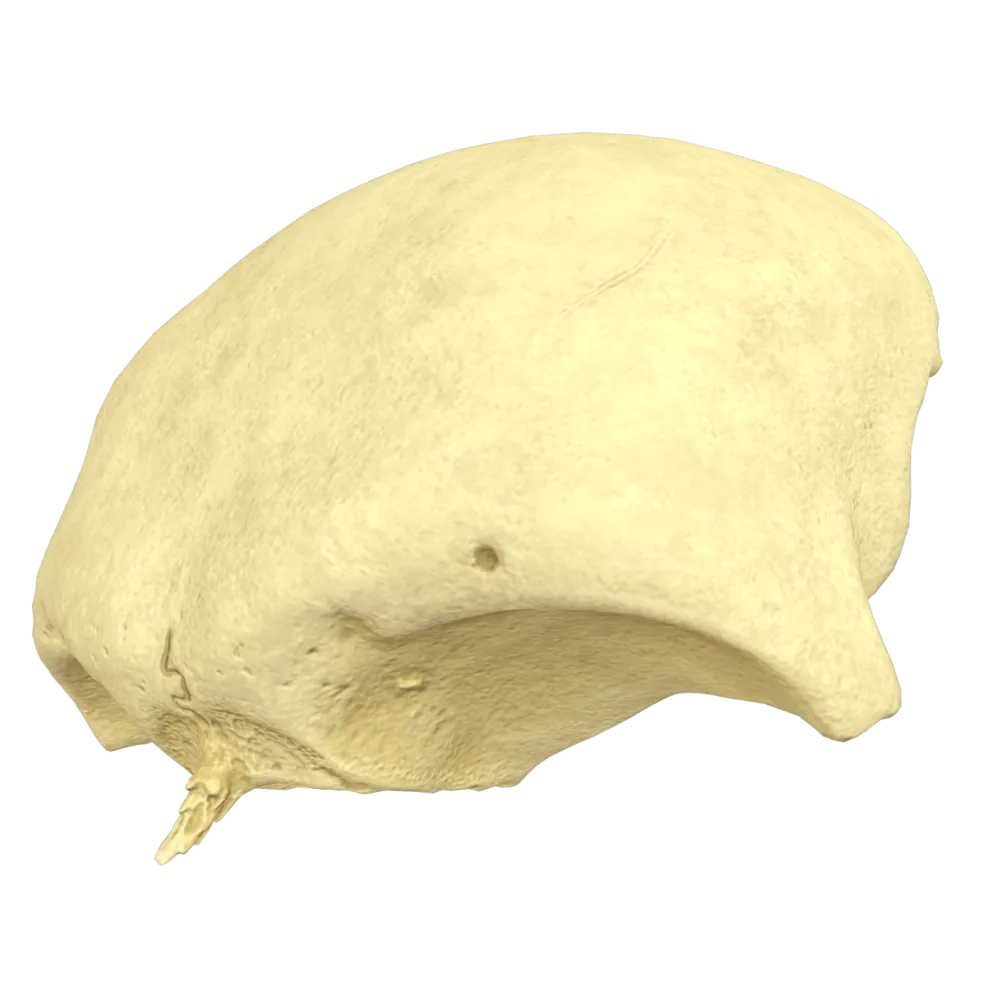
The parietal eminence (tuber parietale) is the most convex point on the external surface of the parietal bone and corresponds to the center of ossification.
-
Parietal eminence
The parietal eminence (tuber parietale) is the most convex point on the external surface of the parietal bone and corresponds to the center of ossification.
-
Parietal foramen
The parietal eminence (tuber parietale) is the most convex point on the external surface of the parietal bone and corresponds to the center of ossification.
-
Superior temporal line
The parietal eminence (tuber parietale) is the most convex point on the external surface of the parietal bone and corresponds to the center of ossification.
-
I find it difficult to answer
The parietal eminence (tuber parietale) is the most convex point on the external surface of the parietal bone and corresponds to the center of ossification.
3. Along which border does the parietal bone articulate with the squamous part of the frontal bone?
-
Sagittal border
The frontal (coronal) border—margo frontalis—of the parietal bone forms the coronal suture (sutura coronalis) by articulating with the squamous part of the frontal bone.
-
Occipital border
The frontal (coronal) border—margo frontalis—of the parietal bone forms the coronal suture (sutura coronalis) by articulating with the squamous part of the frontal bone.
-
Frontal border
The frontal (coronal) border—margo frontalis—of the parietal bone forms the coronal suture (sutura coronalis) by articulating with the squamous part of the frontal bone.
-
Squamous border
The frontal (coronal) border—margo frontalis—of the parietal bone forms the coronal suture (sutura coronalis) by articulating with the squamous part of the frontal bone.
-
I find it difficult to answer
The frontal (coronal) border—margo frontalis—of the parietal bone forms the coronal suture (sutura coronalis) by articulating with the squamous part of the frontal bone.
4. With which bone does the parietal bone articulate along its sagittal border?
-
With the occipital bone.
The sagittal border (margo sagittalis), the medial and longest border of the parietal bone, articulates with the corresponding border of the contralateral bone.
-
With the corresponding parietal bone on the opposite side
The sagittal border (margo sagittalis), the medial and longest border of the parietal bone, articulates with the corresponding border of the contralateral bone.
-
With the frontal bone
The sagittal border (margo sagittalis), the medial and longest border of the parietal bone, articulates with the corresponding border of the contralateral bone.
-
With the temporal bone
The sagittal border (margo sagittalis), the medial and longest border of the parietal bone, articulates with the corresponding border of the contralateral bone.
-
I find it difficult to answer
The sagittal border (margo sagittalis), the medial and longest border of the parietal bone, articulates with the corresponding border of the contralateral bone.
5. Which suture is formed by the articulation of the parietal bone with the occipital bone?
-
Coronal suture
The occipital border (margo occipitalis) of the parietal bone articulates with the occipital bone, forming the lambdoid suture (sutura lambdoidea), named after its resemblance to the Greek letter λ.
-
Sagittal suture
The occipital border (margo occipitalis) of the parietal bone articulates with the occipital bone, forming the lambdoid suture (sutura lambdoidea), named after its resemblance to the Greek letter λ.
-
Lambdoid suture
The occipital border (margo occipitalis) of the parietal bone articulates with the occipital bone, forming the lambdoid suture (sutura lambdoidea), named after its resemblance to the Greek letter λ.
-
Squamous suture
The occipital border (margo occipitalis) of the parietal bone articulates with the occipital bone, forming the lambdoid suture (sutura lambdoidea), named after its resemblance to the Greek letter λ.
-
I find it difficult to answer
The occipital border (margo occipitalis) of the parietal bone articulates with the occipital bone, forming the lambdoid suture (sutura lambdoidea), named after its resemblance to the Greek letter λ.
6. What type of suture is formed between the parietal and temporal bones?
-
Serrate suture
The squamous suture (sutura squamosa) is formed by the beveled, overlapping borders of the parietal bone and the squamous part of the temporal bone, providing a substantial contact area with minimal thickness.
-
Plane suture
The squamous suture (sutura squamosa) is formed by the beveled, overlapping borders of the parietal bone and the squamous part of the temporal bone, providing a substantial contact area with minimal thickness.
-
Squamous suture
The squamous suture (sutura squamosa) is formed by the beveled, overlapping borders of the parietal bone and the squamous part of the temporal bone, providing a substantial contact area with minimal thickness.
-
Sphenosquamosal suture
The squamous suture (sutura squamosa) is formed by the beveled, overlapping borders of the parietal bone and the squamous part of the temporal bone, providing a substantial contact area with minimal thickness.
-
I find it difficult to answer
The squamous suture (sutura squamosa) is formed by the beveled, overlapping borders of the parietal bone and the squamous part of the temporal bone, providing a substantial contact area with minimal thickness.
7. Where is the parietal foramen located on the parietal bone?
-
In the region of the parietal eminence
The parietal foramen (foramen parietale) is located near the sagittal border in the posterior part of the bone; the parietal emissary vein (v. emissaria parietalis), which connects the superior sagittal sinus with the superficial veins of the head, passes through it.
-
Near the sagittal border, posteriorly
The parietal foramen (foramen parietale) is located near the sagittal border in the posterior part of the bone; the parietal emissary vein (v. emissaria parietalis), which connects the superior sagittal sinus with the superficial veins of the head, passes through it.
-
In the center of the internal surface
The parietal foramen (foramen parietale) is located near the sagittal border in the posterior part of the bone; the parietal emissary vein (v. emissaria parietalis), which connects the superior sagittal sinus with the superficial veins of the head, passes through it.
-
Near the inferior (squamous) border
The parietal foramen (foramen parietale) is located near the sagittal border in the posterior part of the bone; the parietal emissary vein (v. emissaria parietalis), which connects the superior sagittal sinus with the superficial veins of the head, passes through it.
-
I find it difficult to answer
The parietal foramen (foramen parietale) is located near the sagittal border in the posterior part of the bone; the parietal emissary vein (v. emissaria parietalis), which connects the superior sagittal sinus with the superficial veins of the head, passes through it.
8. Which vessel forms a groove on the internal surface of the parietal bone?
-
Middle meningeal artery
The middle meningeal artery (a. meningea media) and its branches leave well-defined arterial grooves (sulci arteriosi) on the internal surface of the parietal bone.
-
Anterior meningeal artery
The middle meningeal artery (a. meningea media) and its branches leave well-defined arterial grooves (sulci arteriosi) on the internal surface of the parietal bone.
-
Posterior meningeal artery
The middle meningeal artery (a. meningea media) and its branches leave well-defined arterial grooves (sulci arteriosi) on the internal surface of the parietal bone.
-
Superficial temporal artery
The middle meningeal artery (a. meningea media) and its branches leave well-defined arterial grooves (sulci arteriosi) on the internal surface of the parietal bone.
-
I find it difficult to answer
The middle meningeal artery (a. meningea media) and its branches leave well-defined arterial grooves (sulci arteriosi) on the internal surface of the parietal bone.
9. What is the groove on the internal surface of the parietal bone corresponding to the superior sagittal sinus called?
-
Groove for transverse sinus
The groove for the superior sagittal sinus (sulcus sinus sagittalis superioris) is located along the sagittal border on the internal surfaces of both parietal bones.
-
Groove for sigmoid sinus
The groove for the superior sagittal sinus (sulcus sinus sagittalis superioris) is located along the sagittal border on the internal surfaces of both parietal bones.
-
Groove for the superior sagittal sinus
The groove for the superior sagittal sinus (sulcus sinus sagittalis superioris) is located along the sagittal border on the internal surfaces of both parietal bones.
-
Groove for the cavernous sinus
The groove for the superior sagittal sinus (sulcus sinus sagittalis superioris) is located along the sagittal border on the internal surfaces of both parietal bones.
-
I find it difficult to answer
The groove for the superior sagittal sinus (sulcus sinus sagittalis superioris) is located along the sagittal border on the internal surfaces of both parietal bones.
10. How many angles does the human parietal bone have?
-
2
The parietal bone (os parietale) is a paired quadrilateral bone of the calvaria; it therefore comprises 4 angles: the frontal angle (angulus frontalis), occipital angle (angulus occipitalis), sphenoidal angle (angulus sphenoidalis), and mastoid angle (angulus mastoideus).
-
3
The parietal bone (os parietale) is a paired quadrilateral bone of the calvaria; it therefore comprises 4 angles: the frontal angle (angulus frontalis), occipital angle (angulus occipitalis), sphenoidal angle (angulus sphenoidalis), and mastoid angle (angulus mastoideus).
-
4
The parietal bone (os parietale) is a paired quadrilateral bone of the calvaria; it therefore comprises 4 angles: the frontal angle (angulus frontalis), occipital angle (angulus occipitalis), sphenoidal angle (angulus sphenoidalis), and mastoid angle (angulus mastoideus).
-
5
The parietal bone (os parietale) is a paired quadrilateral bone of the calvaria; it therefore comprises 4 angles: the frontal angle (angulus frontalis), occipital angle (angulus occipitalis), sphenoidal angle (angulus sphenoidalis), and mastoid angle (angulus mastoideus).
-
I find it difficult to answer
The parietal bone (os parietale) is a paired quadrilateral bone of the calvaria; it therefore comprises 4 angles: the frontal angle (angulus frontalis), occipital angle (angulus occipitalis), sphenoidal angle (angulus sphenoidalis), and mastoid angle (angulus mastoideus).
11. Does the human parietal bone have a groove for the sigmoid sinus (sulcus sinus sigmoidei), and where exactly is it located?
-
No, this groove is found only on the occipital and temporal bones
The groove for the sigmoid sinus (sulcus sinus sigmoidei) is present on the parietal bone and is located in the region of its posteroinferior, or mastoid, angle (angulus mastoideus).
-
Yes, it runs precisely along the superior (sagittal) border of the bone
The groove for the sigmoid sinus (sulcus sinus sigmoidei) is present on the parietal bone and is located in the region of its posteroinferior, or mastoid, angle (angulus mastoideus).
-
No, the only sinus groove on the parietal bone is the groove for the transverse sinus
The groove for the sigmoid sinus (sulcus sinus sigmoidei) is present on the parietal bone and is located in the region of its posteroinferior, or mastoid, angle (angulus mastoideus).
-
Yes, it is located on the internal surface in the region of the mastoid angle
The groove for the sigmoid sinus (sulcus sinus sigmoidei) is present on the parietal bone and is located in the region of its posteroinferior, or mastoid, angle (angulus mastoideus).
-
I find it difficult to answer
The groove for the sigmoid sinus (sulcus sinus sigmoidei) is present on the parietal bone and is located in the region of its posteroinferior, or mastoid, angle (angulus mastoideus).
12. Which of the following borders is absent from the human parietal bone?
-
Mastoid border
The parietal bone (os parietale) has a convex quadrilateral shape and is bounded by 4 borders: the sagittal (superior) border (margo sagittalis), frontal (anterior) border (margo frontalis), occipital (posterior) border (margo occipitalis), and squamous (inferior) border (margo squamosus)
-
Occipital border
The parietal bone (os parietale) has a convex quadrilateral shape and is bounded by 4 borders: the sagittal (superior) border (margo sagittalis), frontal (anterior) border (margo frontalis), occipital (posterior) border (margo occipitalis), and squamous (inferior) border (margo squamosus)
-
Frontal border
The parietal bone (os parietale) has a convex quadrilateral shape and is bounded by 4 borders: the sagittal (superior) border (margo sagittalis), frontal (anterior) border (margo frontalis), occipital (posterior) border (margo occipitalis), and squamous (inferior) border (margo squamosus)
-
Squamous border
The parietal bone (os parietale) has a convex quadrilateral shape and is bounded by 4 borders: the sagittal (superior) border (margo sagittalis), frontal (anterior) border (margo frontalis), occipital (posterior) border (margo occipitalis), and squamous (inferior) border (margo squamosus)
-
I find it difficult to answer
The parietal bone (os parietale) has a convex quadrilateral shape and is bounded by 4 borders: the sagittal (superior) border (margo sagittalis), frontal (anterior) border (margo frontalis), occipital (posterior) border (margo occipitalis), and squamous (inferior) border (margo squamosus)
13. Which angle of the parietal bone should be used as the primary landmark to orient it correctly in anatomical position relative to oneself?
-
The frontal angle (angulus frontalis)—it is the most nearly right-angled
The sphenoidal angle (angulus sphenoidalis) is anteroinferior. It is the sharpest, longest, and most elongated of the four angles and is directed anteriorly and inferiorly toward the sphenoid bone. The deepest arterial groove begins from it on the internal surface.
-
The occipital angle (angulus occipitalis)—it is the most obtuse
The sphenoidal angle (angulus sphenoidalis) is anteroinferior. It is the sharpest, longest, and most elongated of the four angles and is directed anteriorly and inferiorly toward the sphenoid bone. The deepest arterial groove begins from it on the internal surface.
-
The mastoid angle (angulus mastoideus)—it is completely smooth
The sphenoidal angle (angulus sphenoidalis) is anteroinferior. It is the sharpest, longest, and most elongated of the four angles and is directed anteriorly and inferiorly toward the sphenoid bone. The deepest arterial groove begins from it on the internal surface.
-
The sphenoidal angle (angulus sphenoidalis)—it is the most pointed and elongated
The sphenoidal angle (angulus sphenoidalis) is anteroinferior. It is the sharpest, longest, and most elongated of the four angles and is directed anteriorly and inferiorly toward the sphenoid bone. The deepest arterial groove begins from it on the internal surface.
-
I find it difficult to answer
The sphenoidal angle (angulus sphenoidalis) is anteroinferior. It is the sharpest, longest, and most elongated of the four angles and is directed anteriorly and inferiorly toward the sphenoid bone. The deepest arterial groove begins from it on the internal surface.
14. Which lines are located on the external surface of the parietal bone?
-
Inferior and superior nuchal lines
The inferior and superior temporal lines (lineae temporales inferior et superior) run across the external surface of the parietal bone and bound the temporal surface of the bone.
-
Inferior and superior temporal lines
The inferior and superior temporal lines (lineae temporales inferior et superior) run across the external surface of the parietal bone and bound the temporal surface of the bone.
-
Inferior and superior semicircular lines
The inferior and superior temporal lines (lineae temporales inferior et superior) run across the external surface of the parietal bone and bound the temporal surface of the bone.
-
Inferior and superior rough lines
The inferior and superior temporal lines (lineae temporales inferior et superior) run across the external surface of the parietal bone and bound the temporal surface of the bone.
-
I find it difficult to answer
The inferior and superior temporal lines (lineae temporales inferior et superior) run across the external surface of the parietal bone and bound the temporal surface of the bone.
15. Which muscle attaches to the inferior temporal line of the parietal bone?
-
Masseter
The temporalis muscle (m. temporalis) originates from the inferior temporal line and the area below it, within the temporal fossa.
-
Medial pterygoid muscle
The temporalis muscle (m. temporalis) originates from the inferior temporal line and the area below it, within the temporal fossa.
-
Temporalis muscle
The temporalis muscle (m. temporalis) originates from the inferior temporal line and the area below it, within the temporal fossa.
-
Lateral pterygoid muscle
The temporalis muscle (m. temporalis) originates from the inferior temporal line and the area below it, within the temporal fossa.
-
I find it difficult to answer
The temporalis muscle (m. temporalis) originates from the inferior temporal line and the area below it, within the temporal fossa.
16. What attaches to the superior temporal line of the parietal bone?
-
Temporalis muscle
The epicranial aponeurosis (galea aponeurotica) and the superficial layer of the temporal fascia (fascia temporalis), which covers the temporalis muscle, attach to the superior temporal line.
-
Epicranial aponeurosis and temporal fascia
The epicranial aponeurosis (galea aponeurotica) and the superficial layer of the temporal fascia (fascia temporalis), which covers the temporalis muscle, attach to the superior temporal line.
-
Masseteric fascia.
The epicranial aponeurosis (galea aponeurotica) and the superficial layer of the temporal fascia (fascia temporalis), which covers the temporalis muscle, attach to the superior temporal line.
-
Auricular muscle
The epicranial aponeurosis (galea aponeurotica) and the superficial layer of the temporal fascia (fascia temporalis), which covers the temporalis muscle, attach to the superior temporal line.
-
I find it difficult to answer
The epicranial aponeurosis (galea aponeurotica) and the superficial layer of the temporal fascia (fascia temporalis), which covers the temporalis muscle, attach to the superior temporal line.
17. What appearance does the internal surface of the parietal bone have in the region where the cerebral gyri are apposed to it?
-
Smooth and even
The internal surface of the parietal bone comprises cerebral impressions—depressions corresponding to the cerebral gyri—and cerebral elevations between them.
-
It bears cerebral impressions and cerebral elevations
The internal surface of the parietal bone comprises cerebral impressions—depressions corresponding to the cerebral gyri—and cerebral elevations between them.
-
It bears numerous nutrient foramina
The internal surface of the parietal bone comprises cerebral impressions—depressions corresponding to the cerebral gyri—and cerebral elevations between them.
-
It has a cellular structure
The internal surface of the parietal bone comprises cerebral impressions—depressions corresponding to the cerebral gyri—and cerebral elevations between them.
-
I find it difficult to answer
The internal surface of the parietal bone comprises cerebral impressions—depressions corresponding to the cerebral gyri—and cerebral elevations between them.
18. Does the parietal bone belong to the neurocranial part of the skull (neurocranium)?
-
No, the parietal bone belongs to the facial part of the skull (viscerocranium)
The neurocranial part (neurocranium) contains and protects the brain. It comprises 8 bones: 2 parietal bones, 2 temporal bones, the frontal bone, occipital bone, sphenoid bone, and ethmoid bone.
-
Yes, the parietal bone is a paired bone of the neurocranial part of the skull
The neurocranial part (neurocranium) contains and protects the brain. It comprises 8 bones: 2 parietal bones, 2 temporal bones, the frontal bone, occipital bone, sphenoid bone, and ethmoid bone.
-
Yes, but only in children until the fontanelles close, after which it becomes part of the facial skeleton
The neurocranial part (neurocranium) contains and protects the brain. It comprises 8 bones: 2 parietal bones, 2 temporal bones, the frontal bone, occipital bone, sphenoid bone, and ethmoid bone.
-
No, it forms a separate part of the cranial base and does not belong to any of the principal parts
The neurocranial part (neurocranium) contains and protects the brain. It comprises 8 bones: 2 parietal bones, 2 temporal bones, the frontal bone, occipital bone, sphenoid bone, and ethmoid bone.
-
I find it difficult to answer
The neurocranial part (neurocranium) contains and protects the brain. It comprises 8 bones: 2 parietal bones, 2 temporal bones, the frontal bone, occipital bone, sphenoid bone, and ethmoid bone.
19. What forms the diploë of the parietal bone?
-
Compact bone only
The diploë (diploë) of the parietal bone is a layer of cancellous (trabecular) bone located between the external and internal plates of compact bone; it contains channels for the diploic veins.
-
Cancellous bone enclosed between two plates of compact bone
The diploë (diploë) of the parietal bone is a layer of cancellous (trabecular) bone located between the external and internal plates of compact bone; it contains channels for the diploic veins.
-
Cancellous bone only, not enclosed between compact plates
The diploë (diploë) of the parietal bone is a layer of cancellous (trabecular) bone located between the external and internal plates of compact bone; it contains channels for the diploic veins.
-
Cartilaginous tissue between the bony plates
The diploë (diploë) of the parietal bone is a layer of cancellous (trabecular) bone located between the external and internal plates of compact bone; it contains channels for the diploic veins.
-
I find it difficult to answer
The diploë (diploë) of the parietal bone is a layer of cancellous (trabecular) bone located between the external and internal plates of compact bone; it contains channels for the diploic veins.
20. Which of the following elements is an inconstant (variable) structure of the parietal bone?
-
Parietal eminence
The parietal foramen (foramen parietale) is an inconstant structure: it may be absent on one or both sides. The other listed structures are constant anatomical elements of the parietal bone.
-
Superior temporal line
The parietal foramen (foramen parietale) is an inconstant structure: it may be absent on one or both sides. The other listed structures are constant anatomical elements of the parietal bone.
-
Parietal foramen
The parietal foramen (foramen parietale) is an inconstant structure: it may be absent on one or both sides. The other listed structures are constant anatomical elements of the parietal bone.
-
Sagittal border
The parietal foramen (foramen parietale) is an inconstant structure: it may be absent on one or both sides. The other listed structures are constant anatomical elements of the parietal bone.
-
I find it difficult to answer
The parietal foramen (foramen parietale) is an inconstant structure: it may be absent on one or both sides. The other listed structures are constant anatomical elements of the parietal bone.
Retake this quiz?
Your current progress will be reset.
