Acute Sinusitis (Acute Rhinosinusitis): Classification, Clinical Manifestations, Diagnosis, and Treatment
A detailed review of rhinosinusitis, including classification, symptoms, diagnostic approaches, and current treatment strategies.
Anesthesia
Pain management and sedation techniques
Angiology
Arterial and venous pathologies
Cardiology
Acquired and congenital heart diseases
Dentistry
Diseases of teeth, gums, and the oral cavity
Dermatology
Disorders of the skin and subcutaneous tissue
Endocrinology
Disorders of the glands and hormonal imbalance
Gastroenterology
Stomach, intestinal, and digestive diseases
Gynecology
Diseases of female reproductive organs
Hepatology
Liver, gallbladder, and biliary tract diseases
Neurology
Brain, spinal cord, and peripheral nerve disorders
Obstetrics
Pregnancy complications and abnormal fetal positions
Oncology
Cancer types, benign and malignant tumors
Ophthalmology
Conditions affecting the eyes and vision
Otorhinolaryngology
Ear, nose, and throat diseases
Pediatrics
Child health, development, and clinical conditions
Physiology
Biological processes within organs and systems
Pulmonology
Lung and respiratory tract diseases
Traumatology
Acute injuries and musculoskeletal trauma
Urology
Urinary tract and male reproductive disorders

This article is for informational purposes only
The content on this website, including text, graphics, and other materials, is provided for informational purposes only. It is not intended as advice or guidance. Regarding your specific medical condition or treatment, please consult your healthcare provider.
Nasal bone fracture is a violation of the structural integrity of the nasal osteocartilaginous framework due to mechanical impact.
This condition is the most common among all types of injuries, especially among injuries to the facial area. The causes of fractures are home and traffic accidents, falls in the street, and sports injuries.
Nasal bone fractures do not have a universally approved classification. We have attempted to compile the most common ones used in clinical practice.
This type of injury affects the nasal bones, the frontal processes of maxilla, and the nasal septum.
The extent and nature of the damage depend on the strength and direction of the damaging factor, as described below.

The division of fractures into open and closed still remains ambiguous.
Some authors believe that open fractures can be considered those in the event of a violation of the skin integrity, but without communication between the bone part and the skin wound via a wound channel.
Other authors, on the contrary, agree that fractures should be considered open if the bone is in contact with the external environment.
The nasal mucosa may remain intact, or it may be also injured. In this case, ruptures occur, associated with moderate bleeding; as well, subcutaneous emphysema may develop.
When using the Johnson’s classification of nasal bone fractures, which is quite common in European countries, the following features should be noted:
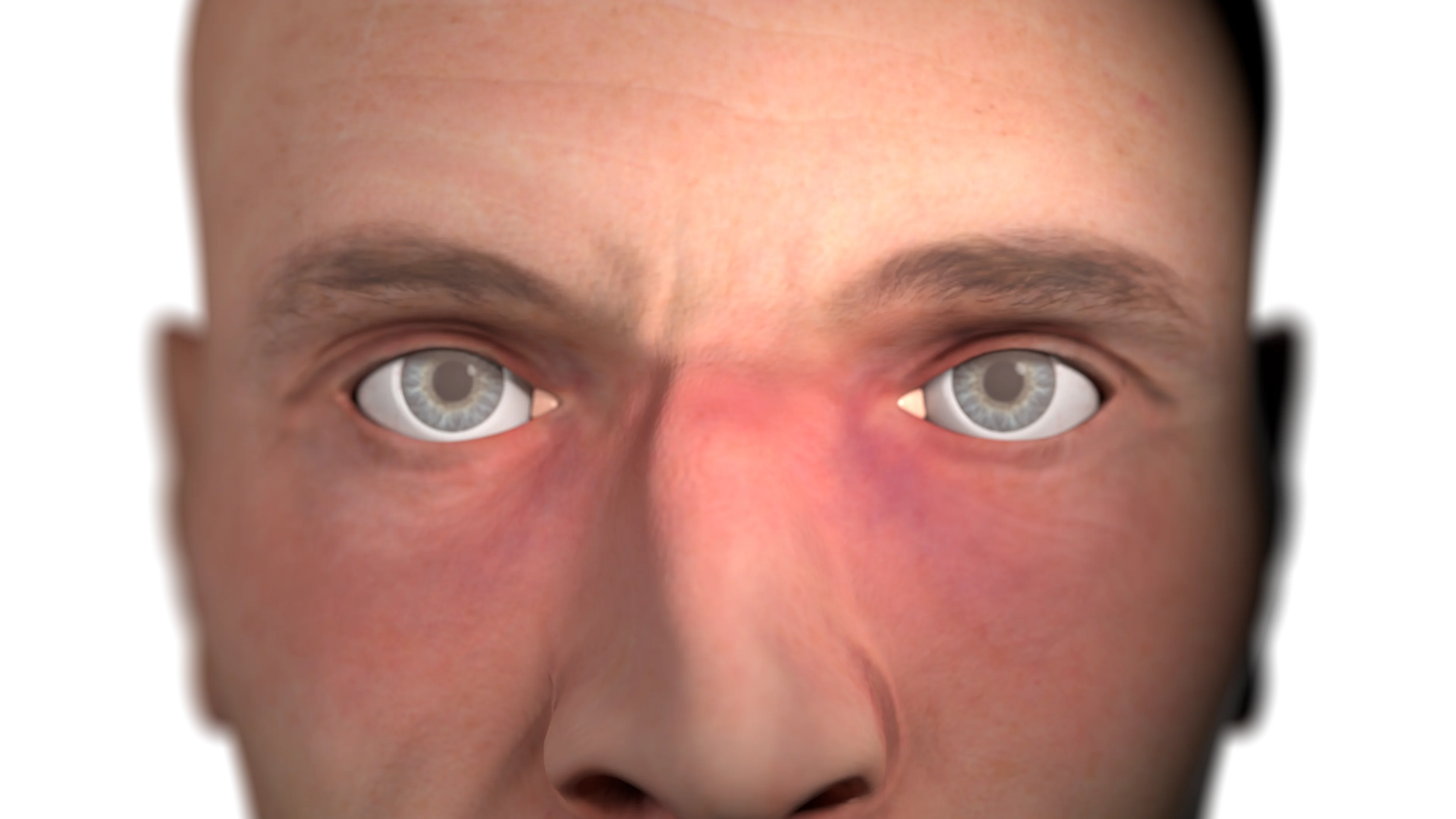
The following symptoms are typical for the external nose fractures:
A thorough medical history is collected, and the circumstances of the injury are clarified.
Typical findings on rhinoscopy in the nasal cavity:
Fracture-specific signs:
Palpation and external examination:
Palpation reveals severe tenderness, bony crepitus and pathological mobility.
If possible, the shape of the external nose, the presence of hematoma, and subcutaneous emphysema should be assessed. However, due to the severe swelling of the soft tissues, it is often not possible to objectively assess whether there is a nasal deformity.
All patients with suspected nasal bone fractures should be referred to lateral nasal bone radiography.
If X-ray shows controversial result, or to determine the location and nature of the injury more precisely, a CT scan of the facial skull is performed.
Find more scientifically accurate content on our social media


Conservative therapy is used, aiming to maintain breathing function.
Local vasoconstrictors, regular nasal irrigation with saline solutions, and systemic analgesics are prescribed.
It is recommended to limit physical exertion and avoid mechanical impact on the external nose.
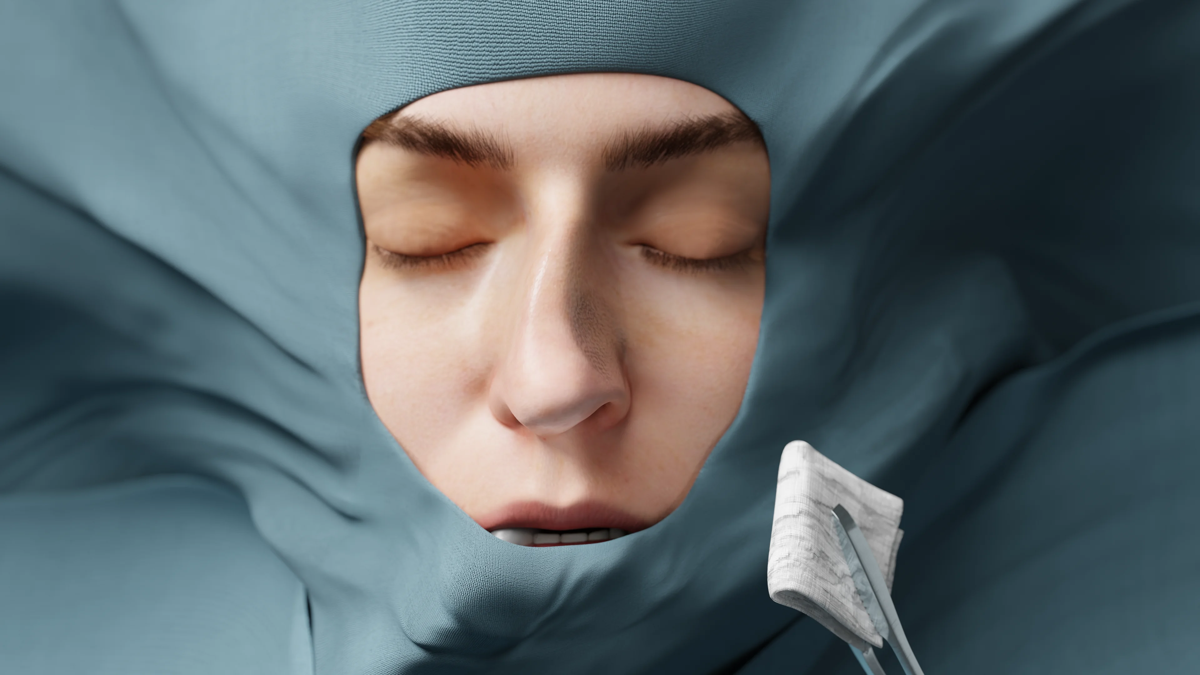
Initial surgical debridement for the wound is provided. Tetanus preventive vaccination is mandatory.
Surgical treatment is performed, aiming to reposition and fix bone fragments, restore breathing function, and eliminate acquired deformities of the external nose.
There are three options of surgical treatment: early (up to 7–10 days), delayed (10–14 days), and elective.
Repositioning of the nasal bones can be performed using external (manually by fingers) or endonasal (using elevators) methods.
Then, a tight nasal packing is performed with gauze turundas, and, if necessary, external fixation with a plaster cast.
No earlier than 6 months after the injury, septo- or rhinoseptoplasty is performed to restore the shape of the external nose and the breathing function.
1. How is a nasal bone fracture diagnosed?
2. What is the difference between an open and closed nasal fracture?
3. How is the severity of a displaced nasal fracture assessed?
4. How long does it take for a nasal fracture to heal?
5. What are the consequences of a nasal fracture?
6. What to do if the fusion of the nasal bones has occurred incorrectly?
References
1.
VOKA 3D Anatomy & Pathology – Complete Anatomy and Pathology 3D Atlas [Internet]. VOKA 3D Anatomy & Pathology.
Available from: https://catalog.voka.io/
2.
Total Otolaryngology—Head and Neck Surgery, Anthony P. Sclafani, Robin A. Dyleski, Michael J. Pitman, Stimson P. Schantz. Thieme Medical Publishers, Inc., 2015. ISBN 978-1-60406-646-3.
3.
Бербом Х. Болезни уха, горла и носа / Ханс Бербом, Оливер Кашке, Тадеус Навка, Эндрю Свифт; пер. с англ. – 2-е изд. – М. : МЕДпреcс-информ, 2016. – 776 с. : ил. ISBN 978-5-00030- 322-1.
4.
Landeen KC, Kimura K, Stephan SJ. Nasal Fractures. Facial Plast Surg Clin North Am. 2022 Feb;30(1):23-30. doi: 10.1016/j.fsc.2021.08.002. PMID: 34809884.
5.
Marston AP, O’Brien EK, Hamilton GS 3rd. Nasal Injuries in Sports. Clin Sports Med. 2017 Apr;36(2):337-353. doi: 10.1016/j.csm.2016.11.004. PMID: 28314421.
6.
Alvi S, Patel BC. Nasal Fracture Reduction. [Updated 2023 Apr 3]. In: StatPearls [Internet]. Treasure Island (FL): StatPearls Publishing; 2025 Jan-.
Available from: https://www.ncbi.nlm.nih.gov/books/NBK538299/
7.
Ramachandra T, Ries WR. Management of Nasal and Perinasal Soft Tissue Injuries. Facial Plast Surg. 2015 Jun;31(3):194-200. doi: 10.1055/s-0035-1555619. Epub 2015 Jun 30. PMID: 26126216.
8.
Kelley BP, Downey CR, Stal S. Evaluation and reduction of nasal trauma. Semin Plast Surg. 2010 Nov;24(4):339-47. doi: 10.1055/s-0030-1269763. PMID: 22550458; PMCID: PMC3324218.
Loading test 6 questions
Table of Contents


Summarize article with AI
Choose your preferable AI assistant:
Link successfully copied to clipboard
Thank you!
Your message is sent!
Our experts will contact you shortly. If you have any additional questions, please contact us at info@voka.io