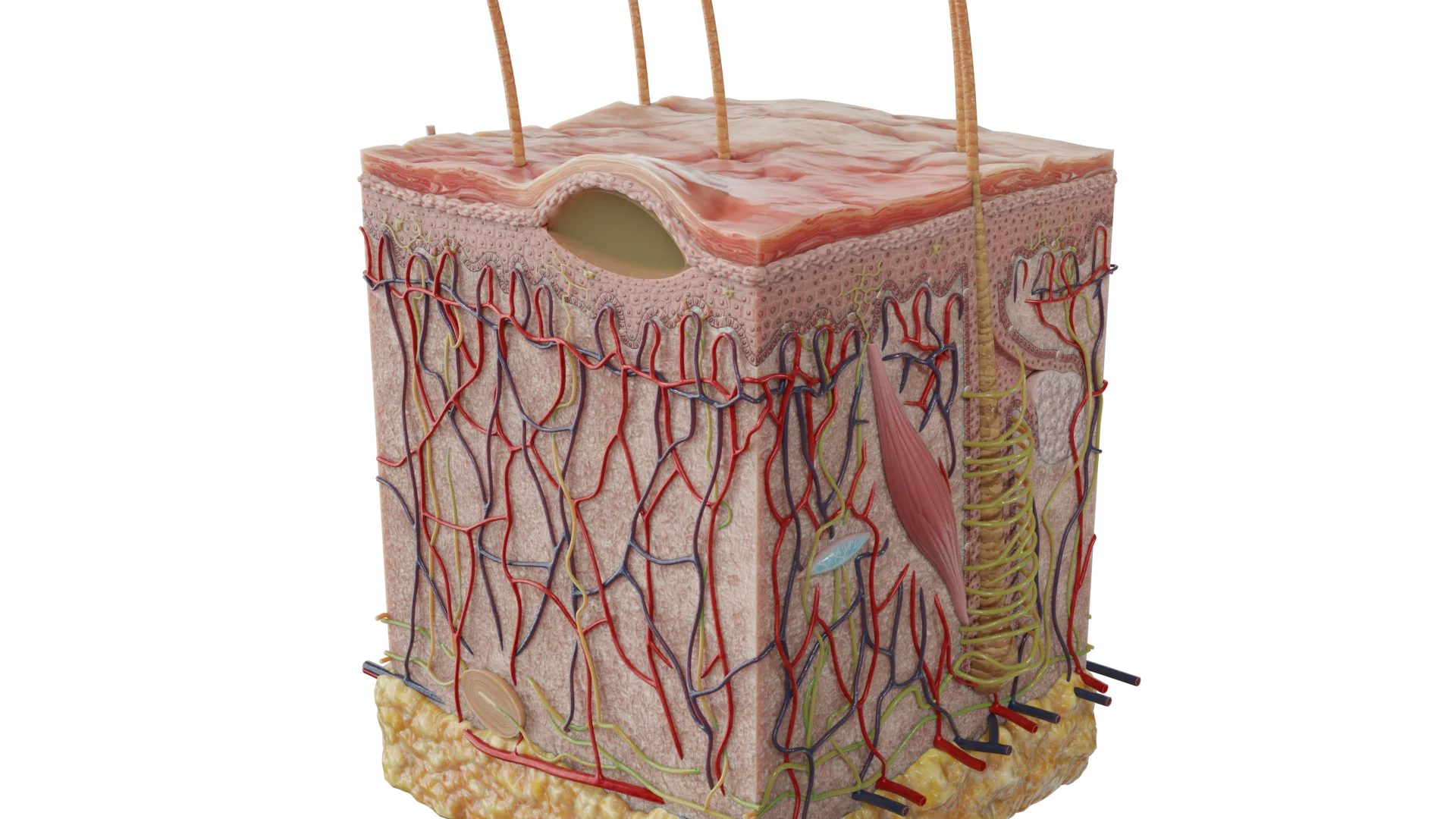
Skin Biopsy in Dermatology: Classification, Technique, and Diagnostic Value
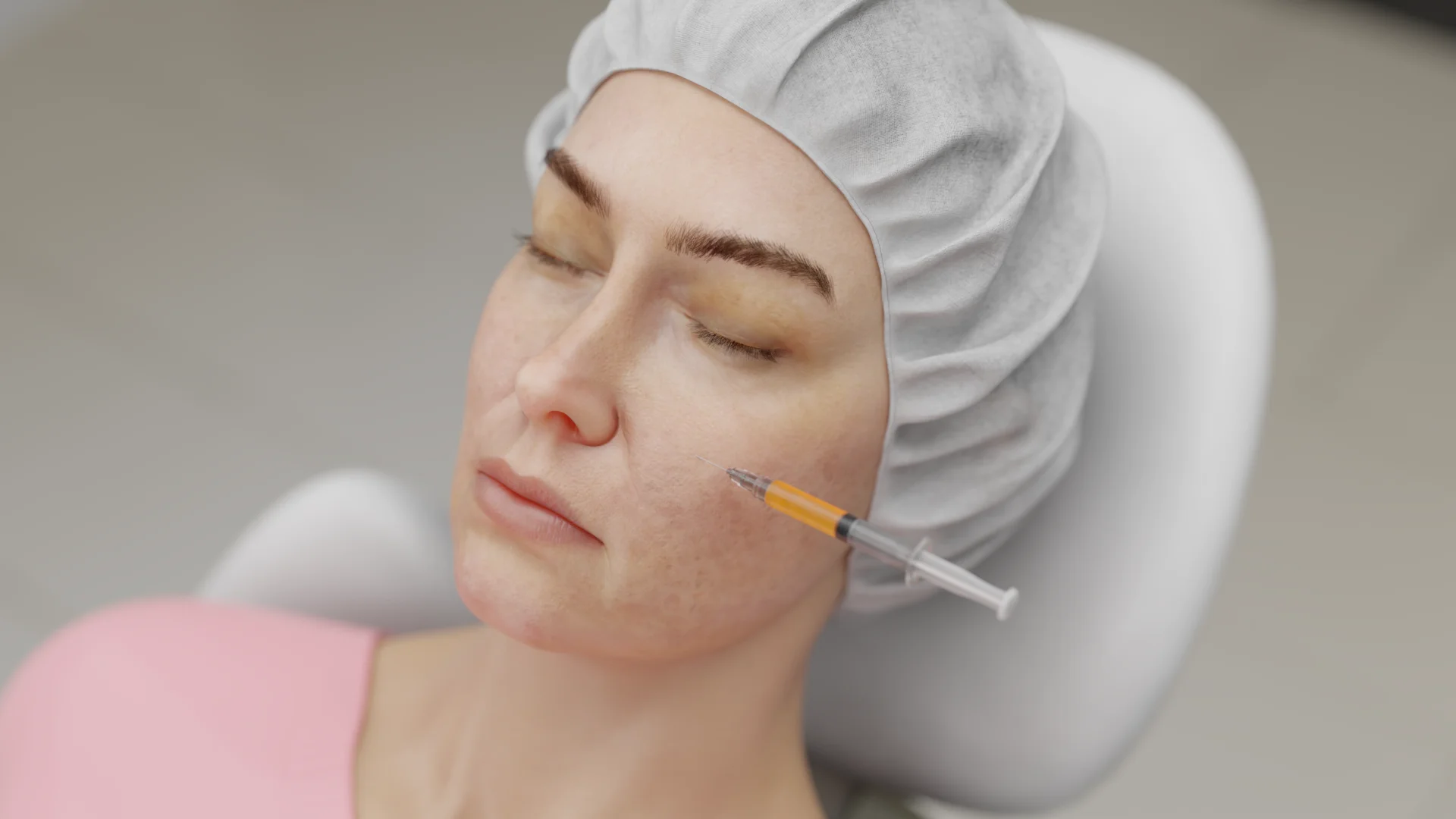
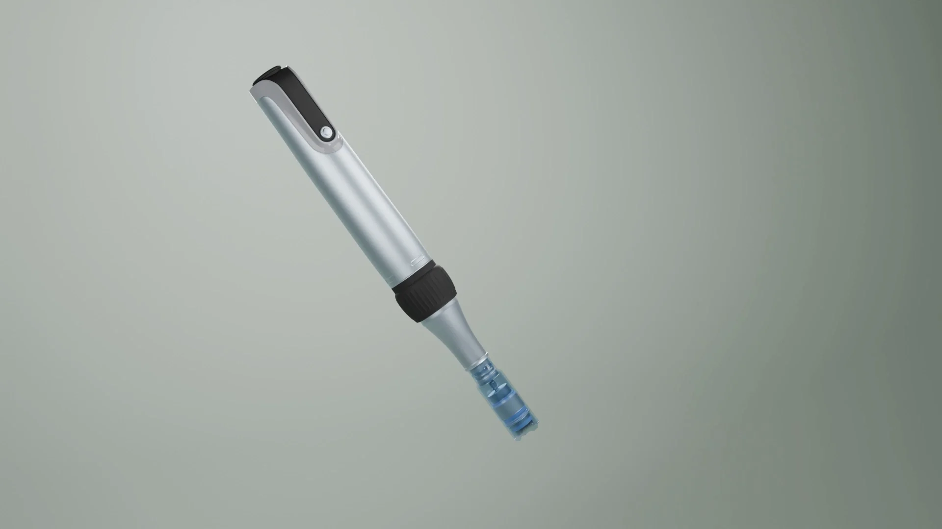
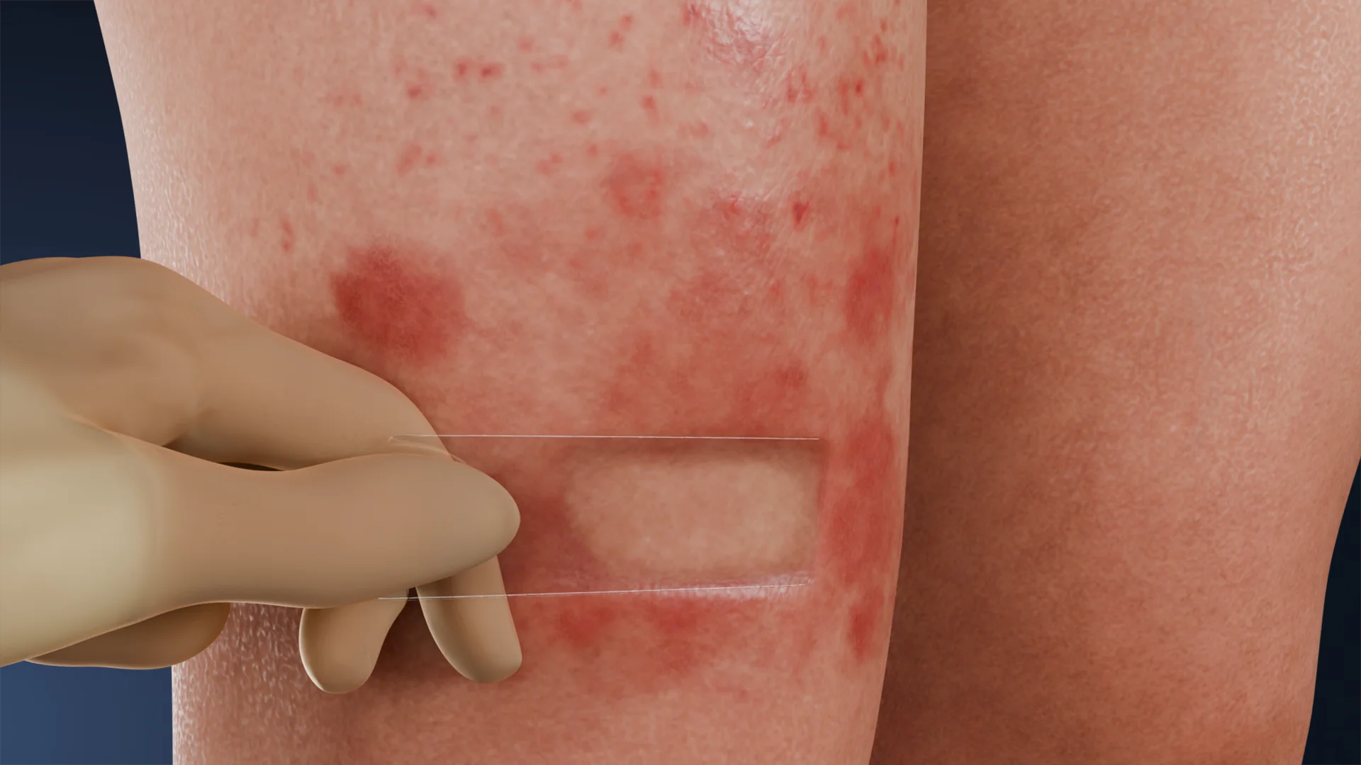
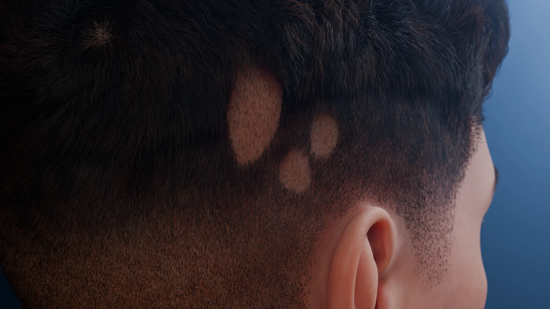
Skin biopsy. This article covers punch, shave, and excisional techniques, specimen handling and fixation, and the role of biopsy in dermatology and oncology.


Anesthesia
Pain management and sedation techniques

Angiology
Arterial and venous pathologies

Cardiology
Acquired and congenital heart diseases

Dentistry
Diseases of teeth, gums, and the oral cavity

Dermatology
Disorders of the skin and subcutaneous tissue

Embryology
Stages of embryogenesis and congenital anomalies

Endocrinology
Disorders of the glands and hormonal imbalance

Gastroenterology
Stomach, intestinal, and digestive diseases

Gynecology
Diseases of female reproductive organs

Hematology
Hematopoiesis and blood-related disorders

Hepatology
Liver, gallbladder, and biliary tract diseases

Histology
Microscopic tissue and cell structures

Infectious diseases
Bacterial, viral, and parasitic infections

Neurology
Brain, spinal cord, and peripheral nerve disorders

Obstetrics
Pregnancy complications and abnormal fetal positions

Oncology
Cancer types, benign and malignant tumors

Ophthalmology
Conditions affecting the eyes and vision

Orthopedics
Bone, joint, and soft tissue disorders

Otorhinolaryngology
Ear, nose, and throat diseases

Pediatrics
Child health, development, and clinical conditions

Physiology
Biological processes within organs and systems

Plastic surgery
Reconstructive and aesthetic procedures

Pulmonology
Lung and respiratory tract diseases

Traumatology
Acute injuries and musculoskeletal trauma

Urology
Urinary tract and male reproductive disorders
Anesthesia
Pain management and sedation techniques
Angiology
Arterial and venous pathologies
Cardiology
Acquired and congenital heart diseases
Dentistry
Diseases of teeth, gums, and the oral cavity
Dermatology
Disorders of the skin and subcutaneous tissue
Embryology
Stages of embryogenesis and congenital anomalies
Endocrinology
Disorders of the glands and hormonal imbalance
Gastroenterology
Stomach, intestinal, and digestive diseases
Gynecology
Diseases of female reproductive organs
Hematology
Hematopoiesis and blood-related disorders
Hepatology
Liver, gallbladder, and biliary tract diseases
Histology
Microscopic tissue and cell structures
Infectious diseases
Bacterial, viral, and parasitic infections
Neurology
Brain, spinal cord, and peripheral nerve disorders
Obstetrics
Pregnancy complications and abnormal fetal positions
Oncology
Cancer types, benign and malignant tumors
Ophthalmology
Conditions affecting the eyes and vision
Orthopedics
Bone, joint, and soft tissue disorders
Otorhinolaryngology
Ear, nose, and throat diseases
Pediatrics
Child health, development, and clinical conditions
Physiology
Biological processes within organs and systems
Plastic surgery
Reconstructive and aesthetic procedures
Pulmonology
Lung and respiratory tract diseases
Traumatology
Acute injuries and musculoskeletal trauma
Urology
Urinary tract and male reproductive disorders
Specialties
AnesthesiaAngiologyCardiologyDentistryDermatologyEmbryologyEndocrinologyGastroenterologyGynecologyHematologyHepatologyHistologyInfectious diseasesNeurologyObstetricsOncologyOphthalmologyOrthopedicsOtorhinolaryngologyPediatricsPhysiologyPlastic surgeryPulmonologyTraumatologyUrology
This article is for informational purposes only
The content on this website, including text, graphics, and other materials, is provided for informational purposes only. It is not intended as advice or guidance. Regarding your specific medical condition or treatment, please consult your healthcare provider.
Primary morphological elements are changes in the skin and mucous membranes, induced by various pathological processes.
Five primary sexless morphological elements are distinguished:
3D Models of Primary Morphological Elements without Cavity:
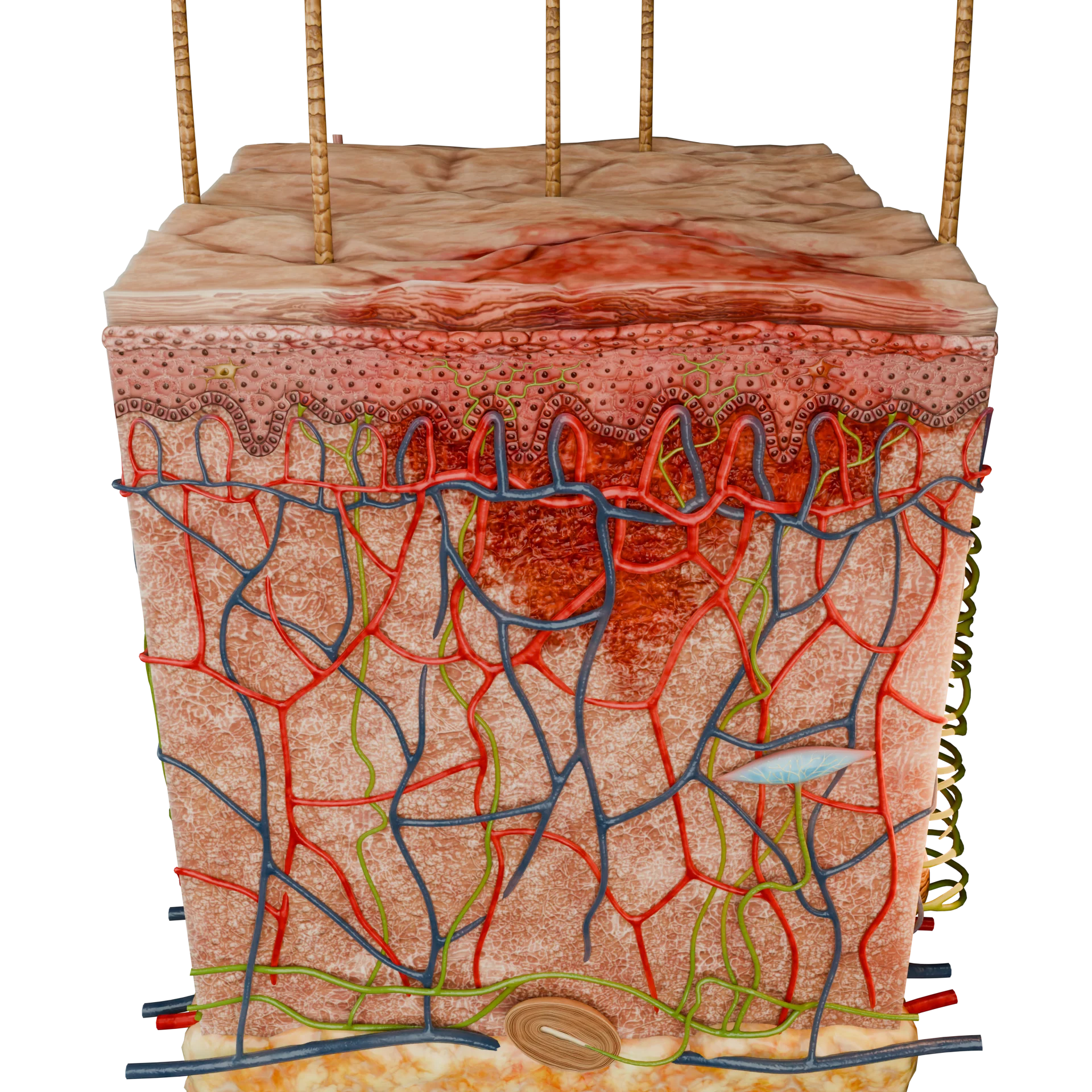 Vascular Inflammatory Macule
 Vascular Non-Inflammatory Macule
 Vascular Hemorrhagic Macule
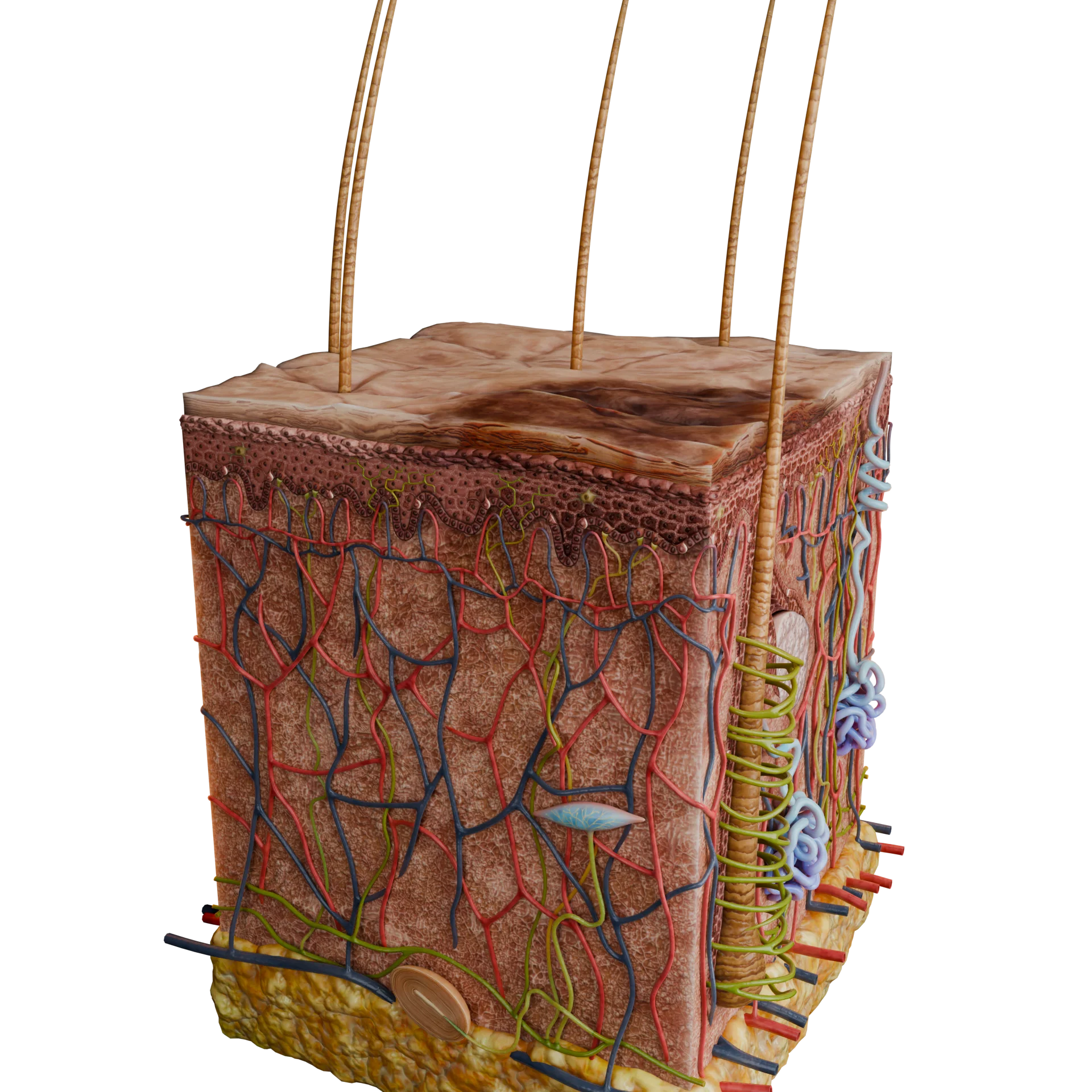 Hyperpigmented Macule
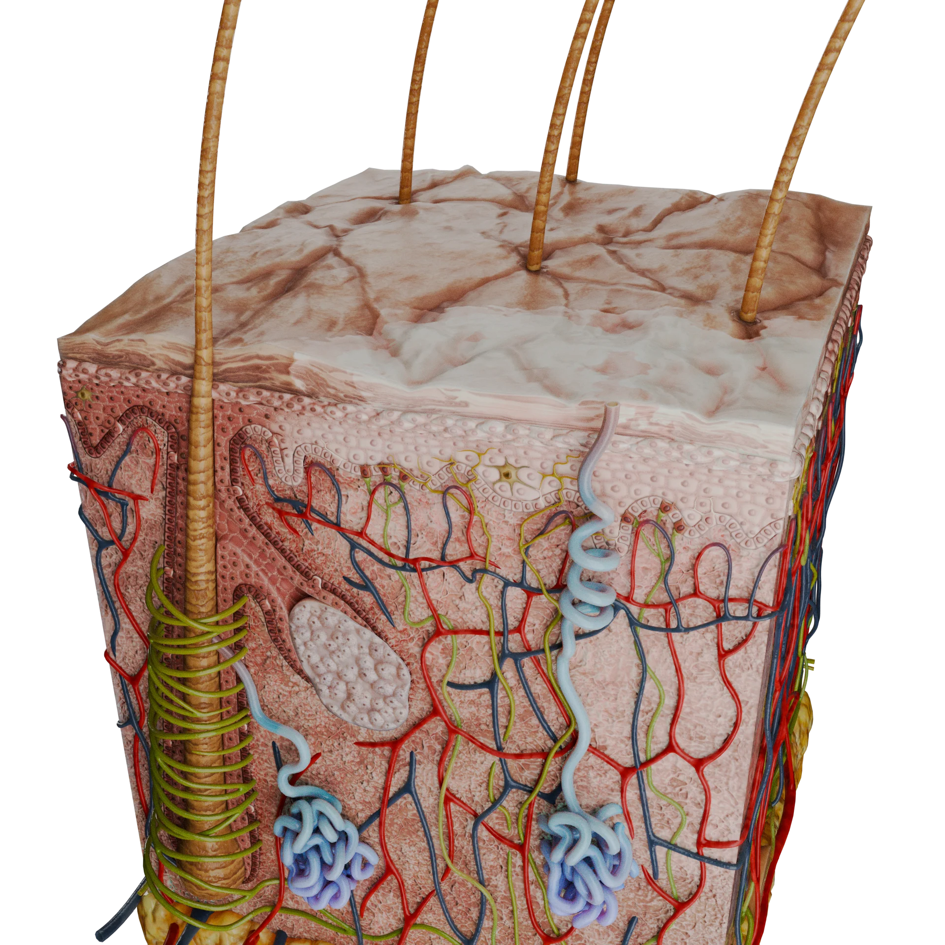 Hypopigmented Macule
A macule is a primary morphological element without cavity that appears on the skin or mucous membranes characterized by color change within a limited area. Macules are always flat and do not leave scars on the skin after resolution.
International sources classify macules by size as follows:
Depending on the nature, macules may be:
By origin, macules are categorized into:
Vascular inflammatory macules appear as a result of dermal vascular dilation against the backdrop of an inflammatory reaction, which disappear when pressure is applied (diascopy).
Macules may be pale pink to bright red, typically irregular in shape and margins undefined.
Macules up to 1 cm in diameter are called rozeolas, whereas spots over 1 cm are called erythemas.
These macules can occur in diseases such as syphilitic and infectious roseola, eczema, dermatitis, rosacea, and lupus erythematosus.
Vascular non-inflammatory macules result from temporary or permanent enlargement of skin vessels without an inflammatory response that do not disappear when pressure is applied (diascopy).
Examples include emotional erythema or flushing, hemangiomas, or telangiectasias.
Vascular hemorrhagic macules are non-inflammatory macules that occur when blood elements are released from the vascular bed into the surrounding tissues due to mechanical rupture of the vessel wall (trauma, bites) or an increase in its permeability (vasculitis, blood diseases).
By size, these macules are classified into:
Massive outpourings of blood into tissues are called hematomas.
When pressed upon, such macules do not disappear. During recovery, they may change color from bright red to yellow and green.
Non-inflammatory macules associated with excessive deposition of melanin are called hyperpigmented macules.
By origin, they are classified into:
Non-inflammatory macules associated with partial or complete absence of melanin deposition are called hypopigmented macules.
Skin hypopigmentation may be:
A papule is a primary morphological element of a skin rash without a cavity that has a dense-elastic consistency and protrudes above the skin surface. After resolution, they do not cause scarring, but may leave temporary or permanent pigmented spots.
3D Models of Primary Morphological Elements Without Cavity:
 Epidermal Papule
 Dermal-Epidermal Papule
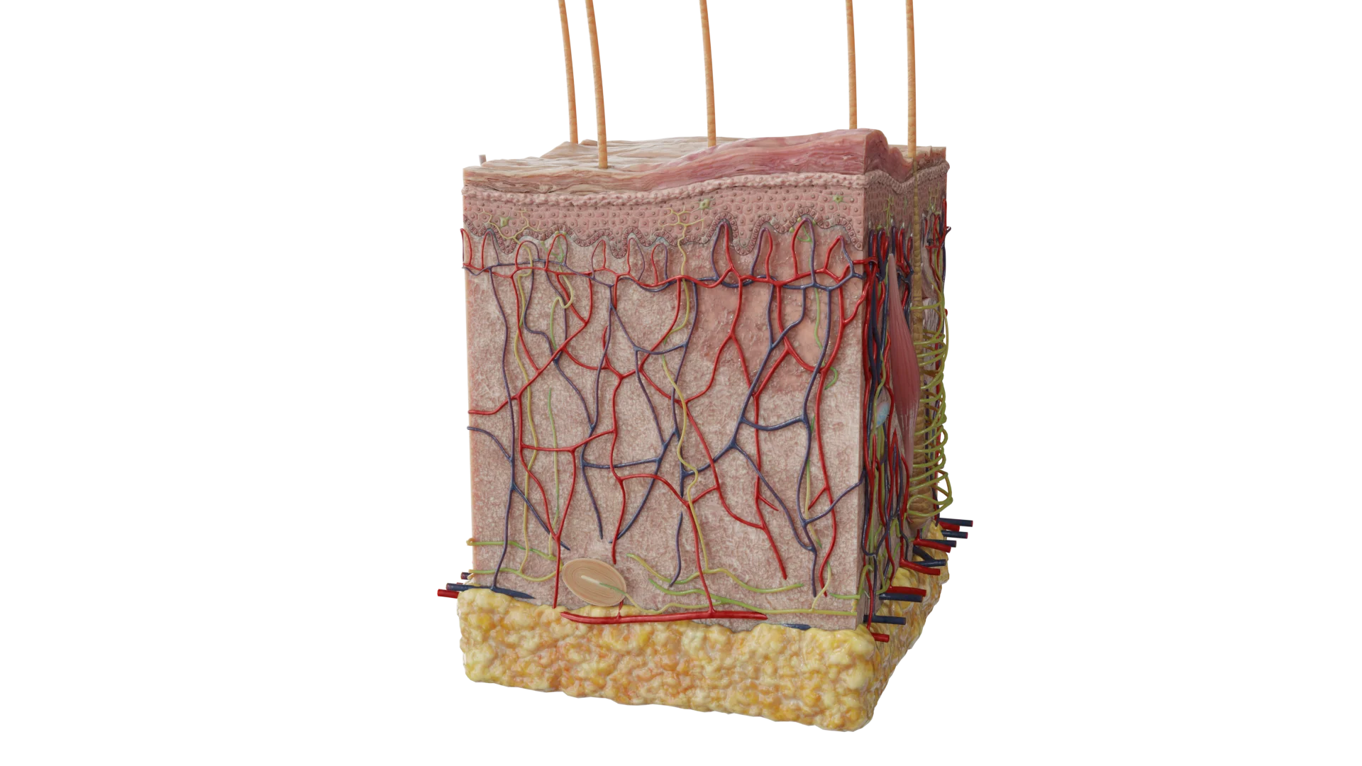 Dermal Papule
By depth:
By size:
By origin:
By shape:
By color:
Find more scientifically accurate content on our social media



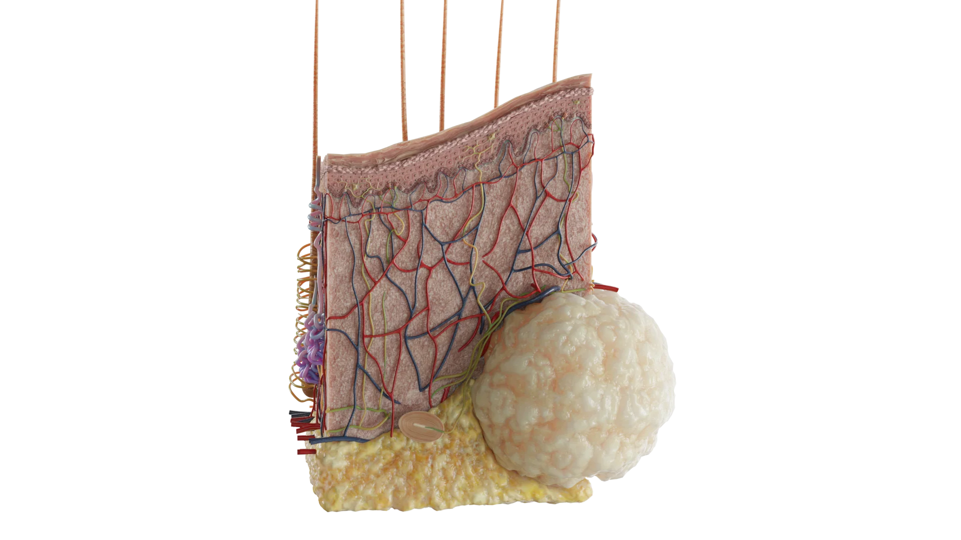
A nodule is a primary morphological element of a skin rash without a cavity, usually round, with a diameter starting from 1 cm. It is characterized by varying depth (most often in the deeper layers). On palpation, a nodule feels firmer than a papule. It may ulcerate, leaving a scar.
A nodule most commonly occurs in lipoma, and may also be seen in tertiary syphilis, tuberculosis, leprosy, cutaneous leishmaniasis, erythema nodosum, intradermal nevi, and cysts.
Classification of Nodules by Depth:
1. What are morphological elements without cavity?
2. Why do hemorrhagic macules not disappear when pressed upon?
3. How do a macule and a patch differ?
4. What is the difference between a papule and a nodule?
5. Can a nodule be a benign lesion?
6. Why is it important to distinguish types of primary lesions?
References
1.
VOKA 3D Anatomy & Pathology – Complete Anatomy and Pathology 3D Atlas. VOKA 3D Anatomy & Pathology.
Available from: https://catalog.voka.io/
2.
Carter KF, Dufour LT, Ballard CN. Identifying primary skin lesions. Nursing. 2003 Dec;33(12):68-69.
3.
Burgin S. An approach to primary lesions. In: Burgin S, ed. Guidebook to dermatologic diagnosis. McGraw-Hill Education; 2021. Accessed 2025 Apr 1.
4.
Mowlabaccus WB. Common benign skin lesions [Internet]. DermNet; 2020.
Available from: https://dermnetnz.org
5.
Soutor C, Hordinsky MK, eds. Clinical dermatology: diagnosis and management of common disorders. 2nd ed. McGraw-Hill Education; 2022.
6.
Hunter JAA, Savin J, Dahl MV. Clinical dermatology. 3rd ed.
Make VOKA your preferred source
See more VOKA articles in Google Search
Table of Contents









Summarize article with AI
Choose your preferable AI assistant:
Link successfully copied to clipboard
Thank you!
Your message is sent!
Our experts will contact you shortly. If you have any additional questions, please contact us at info@voka.io