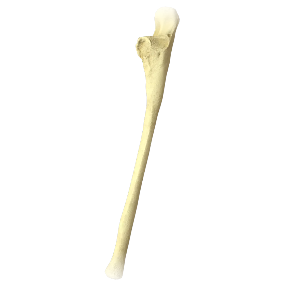
Ulna anatomy quiz
Ulna anatomy quiz: structure, processes, surfaces, articular fossae, and anatomical landmarks.
1/20
bold
text
1. Which bone forms the skeleton of the forearm together with the ulna?
-
Humerus
The skeleton of the forearm is formed by two long bones: the radius (radius) and the ulna (ulna). The radius is located laterally, and the ulna medially.
-
Radius
The skeleton of the forearm is formed by two long bones: the radius (radius) and the ulna (ulna). The radius is located laterally, and the ulna medially.
-
Carpal bone
The skeleton of the forearm is formed by two long bones: the radius (radius) and the ulna (ulna). The radius is located laterally, and the ulna medially.
-
Scaphoid
The skeleton of the forearm is formed by two long bones: the radius (radius) and the ulna (ulna). The radius is located laterally, and the ulna medially.
-
I find it difficult to answer
The skeleton of the forearm is formed by two long bones: the radius (radius) and the ulna (ulna). The radius is located laterally, and the ulna medially.
2. What is the large proximal process of the ulna that forms the posterior contour of the elbow joint called?
-
Styloid process
The olecranon (olecranon) is a large proximal process of the ulna. It projects posteriorly, forms the posterior contour of the elbow joint, and serves as the attachment site for the tendon of the triceps brachii muscle.
-
Coronoid process
The olecranon (olecranon) is a large proximal process of the ulna. It projects posteriorly, forms the posterior contour of the elbow joint, and serves as the attachment site for the tendon of the triceps brachii muscle.
-
Olecranon
The olecranon (olecranon) is a large proximal process of the ulna. It projects posteriorly, forms the posterior contour of the elbow joint, and serves as the attachment site for the tendon of the triceps brachii muscle.
-
Uncinate process
The olecranon (olecranon) is a large proximal process of the ulna. It projects posteriorly, forms the posterior contour of the elbow joint, and serves as the attachment site for the tendon of the triceps brachii muscle.
-
I find it difficult to answer
The olecranon (olecranon) is a large proximal process of the ulna. It projects posteriorly, forms the posterior contour of the elbow joint, and serves as the attachment site for the tendon of the triceps brachii muscle.
3. Which articular surface of the ulna articulates with the trochlea of the humerus?
-
Radial notch
The trochlear notch (incisura trochlearis) is a large articular fossa at the proximal end of the ulna that articulates with the trochlea (trochlea) of the humerus, forming the humeroulnar joint.
-
Trochlear notch
The trochlear notch (incisura trochlearis) is a large articular fossa at the proximal end of the ulna that articulates with the trochlea (trochlea) of the humerus, forming the humeroulnar joint.
-
Head of the ulna.
The trochlear notch (incisura trochlearis) is a large articular fossa at the proximal end of the ulna that articulates with the trochlea (trochlea) of the humerus, forming the humeroulnar joint.
-
Carpal articular surface
The trochlear notch (incisura trochlearis) is a large articular fossa at the proximal end of the ulna that articulates with the trochlea (trochlea) of the humerus, forming the humeroulnar joint.
-
I find it difficult to answer
The trochlear notch (incisura trochlearis) is a large articular fossa at the proximal end of the ulna that articulates with the trochlea (trochlea) of the humerus, forming the humeroulnar joint.
4. Where is the radial notch of the ulna (incisura radialis) located?
-
On the medial surface of the shaft of the ulna
The radial notch (incisura radialis) is located on the lateral side of the coronoid process of the ulna. It is the articular surface for articulation with the head of the radius in the proximal radioulnar joint.
-
On the lateral side of the coronoid process
The radial notch (incisura radialis) is located on the lateral side of the coronoid process of the ulna. It is the articular surface for articulation with the head of the radius in the proximal radioulnar joint.
-
At the distal end of the ulna
The radial notch (incisura radialis) is located on the lateral side of the coronoid process of the ulna. It is the articular surface for articulation with the head of the radius in the proximal radioulnar joint.
-
On the posterior surface of the olecranon
The radial notch (incisura radialis) is located on the lateral side of the coronoid process of the ulna. It is the articular surface for articulation with the head of the radius in the proximal radioulnar joint.
-
I find it difficult to answer
The radial notch (incisura radialis) is located on the lateral side of the coronoid process of the ulna. It is the articular surface for articulation with the head of the radius in the proximal radioulnar joint.
5. What is the anterior process of the proximal end of the ulna that bounds the trochlear notch inferiorly called?
-
Olecranon
The coronoid process (processus coronoideus) is the anterior process of the proximal end of the ulna. It bounds the trochlear notch inferiorly (anteriorly) and contributes to the formation of the humeroulnar joint.
-
Styloid process
The coronoid process (processus coronoideus) is the anterior process of the proximal end of the ulna. It bounds the trochlear notch inferiorly (anteriorly) and contributes to the formation of the humeroulnar joint.
-
Coronoid process
The coronoid process (processus coronoideus) is the anterior process of the proximal end of the ulna. It bounds the trochlear notch inferiorly (anteriorly) and contributes to the formation of the humeroulnar joint.
-
Ulnar tubercle
The coronoid process (processus coronoideus) is the anterior process of the proximal end of the ulna. It bounds the trochlear notch inferiorly (anteriorly) and contributes to the formation of the humeroulnar joint.
-
I find it difficult to answer
The coronoid process (processus coronoideus) is the anterior process of the proximal end of the ulna. It bounds the trochlear notch inferiorly (anteriorly) and contributes to the formation of the humeroulnar joint.
6. On which surface of the shaft of the ulna is the interosseous border (margo interosseus) located?
-
Posterior surface
The interosseous border (margo interosseus) of the ulna faces laterally, toward the radius. The interosseous membrane of the forearm (membrana interossea antebrachii) attaches to it.
-
Medial surface
The interosseous border (margo interosseus) of the ulna faces laterally, toward the radius. The interosseous membrane of the forearm (membrana interossea antebrachii) attaches to it.
-
Lateral surface
The interosseous border (margo interosseus) of the ulna faces laterally, toward the radius. The interosseous membrane of the forearm (membrana interossea antebrachii) attaches to it.
-
Anterior surface
The interosseous border (margo interosseus) of the ulna faces laterally, toward the radius. The interosseous membrane of the forearm (membrana interossea antebrachii) attaches to it.
-
I find it difficult to answer
The interosseous border (margo interosseus) of the ulna faces laterally, toward the radius. The interosseous membrane of the forearm (membrana interossea antebrachii) attaches to it.
7. What is the distal end of the ulna that contributes to the formation of the distal radioulnar joint called?
-
Styloid process
The head of the ulna (caput ulnae) is the distal end of the ulna. The articular circumference of the head articulates with the ulnar notch of the radius, forming the distal radioulnar joint.
-
Head of the ulna
The head of the ulna (caput ulnae) is the distal end of the ulna. The articular circumference of the head articulates with the ulnar notch of the radius, forming the distal radioulnar joint.
-
Coronoid process
The head of the ulna (caput ulnae) is the distal end of the ulna. The articular circumference of the head articulates with the ulnar notch of the radius, forming the distal radioulnar joint.
-
Ulnar tubercle
The head of the ulna (caput ulnae) is the distal end of the ulna. The articular circumference of the head articulates with the ulnar notch of the radius, forming the distal radioulnar joint.
-
I find it difficult to answer
The head of the ulna (caput ulnae) is the distal end of the ulna. The articular circumference of the head articulates with the ulnar notch of the radius, forming the distal radioulnar joint.
8. What is the small bony projection on the distomedial border of the head of the ulna called?
-
Coronoid process
The styloid process (processus styloideus) arises from the mediodistal border of the head of the ulna. The ulnar collateral ligament of the wrist attaches to it.
-
Olecranon
The styloid process (processus styloideus) arises from the mediodistal border of the head of the ulna. The ulnar collateral ligament of the wrist attaches to it.
-
Styloid process
The styloid process (processus styloideus) arises from the mediodistal border of the head of the ulna. The ulnar collateral ligament of the wrist attaches to it.
-
Epicondyle
The styloid process (processus styloideus) arises from the mediodistal border of the head of the ulna. The ulnar collateral ligament of the wrist attaches to it.
-
I find it difficult to answer
The styloid process (processus styloideus) arises from the mediodistal border of the head of the ulna. The ulnar collateral ligament of the wrist attaches to it.
9. Which process of the ulna has a tuberosity to which the brachialis muscle (m. brachialis) attaches?
-
Olecranon
The ulnar tuberosity (tuberositas ulnae) is located on the anteroinferior surface of the coronoid process and serves as the attachment site for the brachialis muscle (m. brachialis), the principal flexor of the forearm.
-
Styloid process
The ulnar tuberosity (tuberositas ulnae) is located on the anteroinferior surface of the coronoid process and serves as the attachment site for the brachialis muscle (m. brachialis), the principal flexor of the forearm.
-
Coronoid process
The ulnar tuberosity (tuberositas ulnae) is located on the anteroinferior surface of the coronoid process and serves as the attachment site for the brachialis muscle (m. brachialis), the principal flexor of the forearm.
-
Interosseous crest
The ulnar tuberosity (tuberositas ulnae) is located on the anteroinferior surface of the coronoid process and serves as the attachment site for the brachialis muscle (m. brachialis), the principal flexor of the forearm.
-
I find it difficult to answer
The ulnar tuberosity (tuberositas ulnae) is located on the anteroinferior surface of the coronoid process and serves as the attachment site for the brachialis muscle (m. brachialis), the principal flexor of the forearm.
10. What type of bone is the ulna according to its classification?
-
Short cancellous bone
The ulna (ulna) is classified as a long bone (ossa longa). It comprises a shaft (diaphysis) and two ends (epiphyses): the proximal end (with the olecranon and coronoid process) and the distal end (the head with the styloid process).
-
Flat bone
The ulna (ulna) is classified as a long bone (ossa longa). It comprises a shaft (diaphysis) and two ends (epiphyses): the proximal end (with the olecranon and coronoid process) and the distal end (the head with the styloid process).
-
Long bone
The ulna (ulna) is classified as a long bone (ossa longa). It comprises a shaft (diaphysis) and two ends (epiphyses): the proximal end (with the olecranon and coronoid process) and the distal end (the head with the styloid process).
-
Mixed bone
The ulna (ulna) is classified as a long bone (ossa longa). It comprises a shaft (diaphysis) and two ends (epiphyses): the proximal end (with the olecranon and coronoid process) and the distal end (the head with the styloid process).
-
I find it difficult to answer
The ulna (ulna) is classified as a long bone (ossa longa). It comprises a shaft (diaphysis) and two ends (epiphyses): the proximal end (with the olecranon and coronoid process) and the distal end (the head with the styloid process).
11. Which surface of the shaft of the ulna is the widest and faces anteriorly?
-
Posterior surface
The anterior surface (facies anterior) of the shaft of the ulna faces anteriorly and is the widest of the three surfaces of the diaphysis. The nutrient foramen (foramen nutricium) and the origin of the flexor digitorum profundus are located on it.
-
Medial surface
The anterior surface (facies anterior) of the shaft of the ulna faces anteriorly and is the widest of the three surfaces of the diaphysis. The nutrient foramen (foramen nutricium) and the origin of the flexor digitorum profundus are located on it.
-
Anterior surface
The anterior surface (facies anterior) of the shaft of the ulna faces anteriorly and is the widest of the three surfaces of the diaphysis. The nutrient foramen (foramen nutricium) and the origin of the flexor digitorum profundus are located on it.
-
Lateral surface
The anterior surface (facies anterior) of the shaft of the ulna faces anteriorly and is the widest of the three surfaces of the diaphysis. The nutrient foramen (foramen nutricium) and the origin of the flexor digitorum profundus are located on it.
-
I find it difficult to answer
The anterior surface (facies anterior) of the shaft of the ulna faces anteriorly and is the widest of the three surfaces of the diaphysis. The nutrient foramen (foramen nutricium) and the origin of the flexor digitorum profundus are located on it.
12. What is the fossa on the posterior surface of the humerus into which the olecranon enters during extension of the forearm called?
-
Coronoid fossa
The olecranon fossa (fossa olecrani) is located on the posterior surface of the distal end of the humerus. During full extension of the forearm, the olecranon enters this fossa, limiting further extension.
-
Radial fossa
The olecranon fossa (fossa olecrani) is located on the posterior surface of the distal end of the humerus. During full extension of the forearm, the olecranon enters this fossa, limiting further extension.
-
Olecranon fossa
The olecranon fossa (fossa olecrani) is located on the posterior surface of the distal end of the humerus. During full extension of the forearm, the olecranon enters this fossa, limiting further extension.
-
Trochlear notch
The olecranon fossa (fossa olecrani) is located on the posterior surface of the distal end of the humerus. During full extension of the forearm, the olecranon enters this fossa, limiting further extension.
-
I find it difficult to answer
The olecranon fossa (fossa olecrani) is located on the posterior surface of the distal end of the humerus. During full extension of the forearm, the olecranon enters this fossa, limiting further extension.
13. Which ligament attaches to the supinator crest (crista musculi supinatoris) of the ulna?
-
Annular ligament of the radius
The annular ligament of the radius (lig. anulare radii) attaches to the anterior and posterior margins of the radial notch of the ulna, as well as to the supinator crest (crista musculi supinatoris), retaining the head of the radius in the proximal radioulnar joint.
-
Quadrate ligament
The annular ligament of the radius (lig. anulare radii) attaches to the anterior and posterior margins of the radial notch of the ulna, as well as to the supinator crest (crista musculi supinatoris), retaining the head of the radius in the proximal radioulnar joint.
-
Oblique cord
The annular ligament of the radius (lig. anulare radii) attaches to the anterior and posterior margins of the radial notch of the ulna, as well as to the supinator crest (crista musculi supinatoris), retaining the head of the radius in the proximal radioulnar joint.
-
Dorsal ulnocarpal ligament
The annular ligament of the radius (lig. anulare radii) attaches to the anterior and posterior margins of the radial notch of the ulna, as well as to the supinator crest (crista musculi supinatoris), retaining the head of the radius in the proximal radioulnar joint.
-
I find it difficult to answer
The annular ligament of the radius (lig. anulare radii) attaches to the anterior and posterior margins of the radial notch of the ulna, as well as to the supinator crest (crista musculi supinatoris), retaining the head of the radius in the proximal radioulnar joint.
14. Which muscle originates from the supinator crest and the proximal part of the shaft of the ulna?
-
Flexor carpi ulnaris
The supinator muscle (m. supinator) originates from the lateral epicondyle of the humerus, the radial collateral ligament, the annular ligament, and the supinator crest and supinator fossa (fossa supinatoris) of the ulna.
-
Extensor hallucis longus
The supinator muscle (m. supinator) originates from the lateral epicondyle of the humerus, the radial collateral ligament, the annular ligament, and the supinator crest and supinator fossa (fossa supinatoris) of the ulna.
-
Supinator muscle
The supinator muscle (m. supinator) originates from the lateral epicondyle of the humerus, the radial collateral ligament, the annular ligament, and the supinator crest and supinator fossa (fossa supinatoris) of the ulna.
-
Extensor carpi ulnaris
The supinator muscle (m. supinator) originates from the lateral epicondyle of the humerus, the radial collateral ligament, the annular ligament, and the supinator crest and supinator fossa (fossa supinatoris) of the ulna.
-
I find it difficult to answer
The supinator muscle (m. supinator) originates from the lateral epicondyle of the humerus, the radial collateral ligament, the annular ligament, and the supinator crest and supinator fossa (fossa supinatoris) of the ulna.
15. What is the cartilage-covered articular surface at the distal end of the ulna that faces the wrist called?
-
Trochlear notch
The inferior surface of the head of the ulna has an articular surface (facies articularis) covered with cartilage. It faces distally toward the wrist and participates (through the articular disc) in the formation of the radiocarpal joint.
-
Carpal articular surface of the head
The inferior surface of the head of the ulna has an articular surface (facies articularis) covered with cartilage. It faces distally toward the wrist and participates (through the articular disc) in the formation of the radiocarpal joint.
-
Radial notch
The inferior surface of the head of the ulna has an articular surface (facies articularis) covered with cartilage. It faces distally toward the wrist and participates (through the articular disc) in the formation of the radiocarpal joint.
-
Lunate surface
The inferior surface of the head of the ulna has an articular surface (facies articularis) covered with cartilage. It faces distally toward the wrist and participates (through the articular disc) in the formation of the radiocarpal joint.
-
I find it difficult to answer
The inferior surface of the head of the ulna has an articular surface (facies articularis) covered with cartilage. It faces distally toward the wrist and participates (through the articular disc) in the formation of the radiocarpal joint.
16. Which joint is formed by the trochlear notch of the ulna and the trochlea of the humerus?
-
Proximal radioulnar joint
The humeroulnar joint (articulatio humeroulnaris) is formed by the trochlea (trochlea) of the humerus and the trochlear notch (incisura trochlearis) of the ulna. It is a hinge joint in shape, permitting flexion and extension of the forearm.
-
Radiocapitellar joint
The humeroulnar joint (articulatio humeroulnaris) is formed by the trochlea (trochlea) of the humerus and the trochlear notch (incisura trochlearis) of the ulna. It is a hinge joint in shape, permitting flexion and extension of the forearm.
-
Humeroulnar joint
The humeroulnar joint (articulatio humeroulnaris) is formed by the trochlea (trochlea) of the humerus and the trochlear notch (incisura trochlearis) of the ulna. It is a hinge joint in shape, permitting flexion and extension of the forearm.
-
Distal radioulnar joint
The humeroulnar joint (articulatio humeroulnaris) is formed by the trochlea (trochlea) of the humerus and the trochlear notch (incisura trochlearis) of the ulna. It is a hinge joint in shape, permitting flexion and extension of the forearm.
-
I find it difficult to answer
The humeroulnar joint (articulatio humeroulnaris) is formed by the trochlea (trochlea) of the humerus and the trochlear notch (incisura trochlearis) of the ulna. It is a hinge joint in shape, permitting flexion and extension of the forearm.
17. Which muscle attaches to the posterior surface of the olecranon and is the principal extensor of the forearm?
-
Biceps brachii
The triceps brachii muscle (m. triceps brachii) attaches by its common tendon to the posterior surface and apex of the olecranon. It is the principal extensor of the forearm at the elbow joint.
-
Brachialis muscle
The triceps brachii muscle (m. triceps brachii) attaches by its common tendon to the posterior surface and apex of the olecranon. It is the principal extensor of the forearm at the elbow joint.
-
Triceps brachii muscle
The triceps brachii muscle (m. triceps brachii) attaches by its common tendon to the posterior surface and apex of the olecranon. It is the principal extensor of the forearm at the elbow joint.
-
Anconeus muscle
The triceps brachii muscle (m. triceps brachii) attaches by its common tendon to the posterior surface and apex of the olecranon. It is the principal extensor of the forearm at the elbow joint.
-
I find it difficult to answer
The triceps brachii muscle (m. triceps brachii) attaches by its common tendon to the posterior surface and apex of the olecranon. It is the principal extensor of the forearm at the elbow joint.
18. Where is the nutrient foramen (foramen nutricium) located on the ulna?
-
On the posterior surface of the olecranon
The nutrient foramen (foramen nutricium) of the ulna is located on the anterior surface of the shaft in the upper third of the diaphysis. The nutrient artery (a. nutricia ulnae) enters the medullary canal through it, supplying the diaphysis with blood.
-
On the anterior surface of the shaft in the upper third of the diaphysis
The nutrient foramen (foramen nutricium) of the ulna is located on the anterior surface of the shaft in the upper third of the diaphysis. The nutrient artery (a. nutricia ulnae) enters the medullary canal through it, supplying the diaphysis with blood.
-
On the medial surface of the distal epiphysis
The nutrient foramen (foramen nutricium) of the ulna is located on the anterior surface of the shaft in the upper third of the diaphysis. The nutrient artery (a. nutricia ulnae) enters the medullary canal through it, supplying the diaphysis with blood.
-
On the radial notch of the coronoid process
The nutrient foramen (foramen nutricium) of the ulna is located on the anterior surface of the shaft in the upper third of the diaphysis. The nutrient artery (a. nutricia ulnae) enters the medullary canal through it, supplying the diaphysis with blood.
-
I find it difficult to answer
The nutrient foramen (foramen nutricium) of the ulna is located on the anterior surface of the shaft in the upper third of the diaphysis. The nutrient artery (a. nutricia ulnae) enters the medullary canal through it, supplying the diaphysis with blood.
19. The radius is positioned relative to the ulna in the forearm as follows:
-
Lateral and anterior to the ulna
The radius is located laterally relative to the ulna and runs parallel to it along the entire forearm. In the anatomical position of the body (supinatio), both bones lie in the same plane; the ulna is on the side of the little finger (medially).
-
Lateral and parallel to the ulna
The radius is located laterally relative to the ulna and runs parallel to it along the entire forearm. In the anatomical position of the body (supinatio), both bones lie in the same plane; the ulna is on the side of the little finger (medially).
-
Lateral and posterior to the ulna
The radius is located laterally relative to the ulna and runs parallel to it along the entire forearm. In the anatomical position of the body (supinatio), both bones lie in the same plane; the ulna is on the side of the little finger (medially).
-
Anteriorly and medially in the lower third of the forearm
The radius is located laterally relative to the ulna and runs parallel to it along the entire forearm. In the anatomical position of the body (supinatio), both bones lie in the same plane; the ulna is on the side of the little finger (medially).
-
I find it difficult to answer
The radius is located laterally relative to the ulna and runs parallel to it along the entire forearm. In the anatomical position of the body (supinatio), both bones lie in the same plane; the ulna is on the side of the little finger (medially).
20. The ulna is positioned relative to the radius in the forearm as follows:
-
Lateral and anterior to the radius
The ulna (ulna) is located medially relative to the radius (radius) and runs parallel to it along the entire forearm. In the anatomical position of the body (supinatio), both bones lie in the same plane; the ulna is on the side of the little finger (medially).
-
Medial and parallel to the radius
The ulna (ulna) is located medially relative to the radius (radius) and runs parallel to it along the entire forearm. In the anatomical position of the body (supinatio), both bones lie in the same plane; the ulna is on the side of the little finger (medially).
-
Lateral and posterior to the radius
The ulna (ulna) is located medially relative to the radius (radius) and runs parallel to it along the entire forearm. In the anatomical position of the body (supinatio), both bones lie in the same plane; the ulna is on the side of the little finger (medially).
-
Anteriorly and medially in the lower third of the forearm
The ulna (ulna) is located medially relative to the radius (radius) and runs parallel to it along the entire forearm. In the anatomical position of the body (supinatio), both bones lie in the same plane; the ulna is on the side of the little finger (medially).
-
I find it difficult to answer
The ulna (ulna) is located medially relative to the radius (radius) and runs parallel to it along the entire forearm. In the anatomical position of the body (supinatio), both bones lie in the same plane; the ulna is on the side of the little finger (medially).
Retake this quiz?
Your current progress will be reset.
