1/20
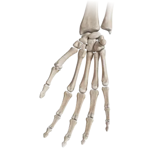
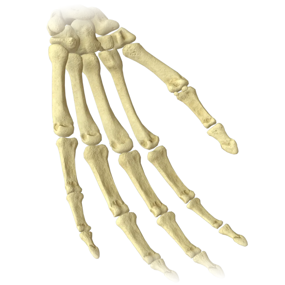
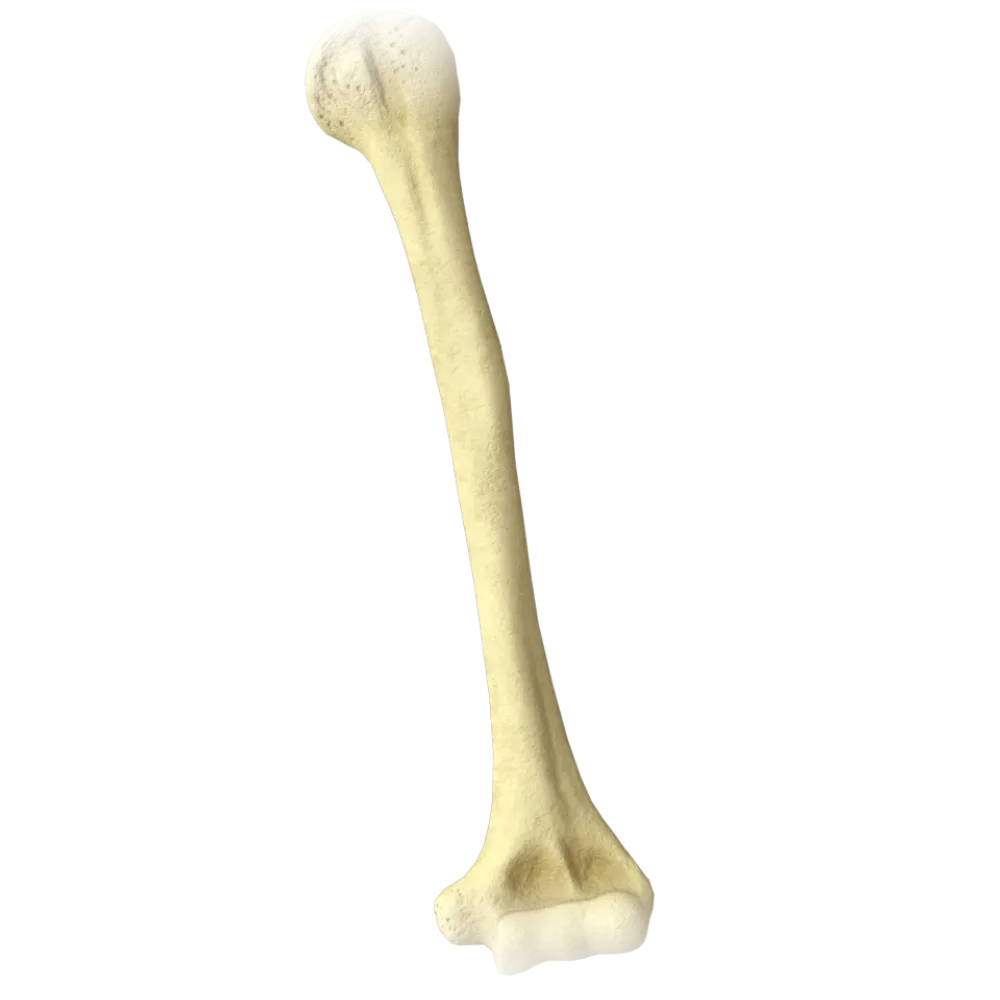
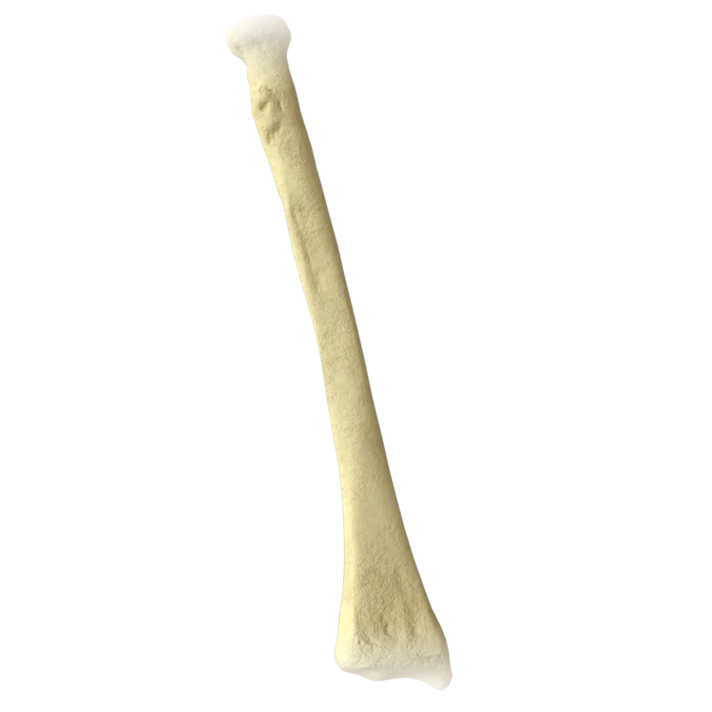
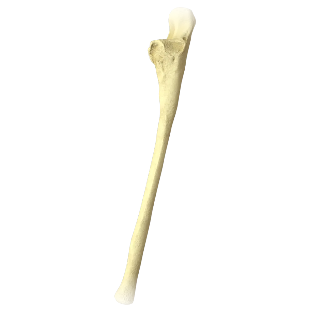
Quiz on the Anatomy of the Bones of the Free Part of the Upper Limb
Test on the normal anatomy of the free upper limb bones: arm, forearm, hand.
1. Which bone forms the skeleton of the arm?
-
Radius
The skeleton of the arm is represented by one bone – the humerus. The radius and ulna form the skeleton of the forearm, while the clavicle belongs to the pectoral girdle.
-
Humerus
The skeleton of the arm is represented by one bone – the humerus. The radius and ulna form the skeleton of the forearm, while the clavicle belongs to the pectoral girdle.
-
Ulna
The skeleton of the arm is represented by one bone – the humerus. The radius and ulna form the skeleton of the forearm, while the clavicle belongs to the pectoral girdle.
-
Clavicle
The skeleton of the arm is represented by one bone – the humerus. The radius and ulna form the skeleton of the forearm, while the clavicle belongs to the pectoral girdle.
-
I find it difficult to answer
The skeleton of the arm is represented by one bone – the humerus. The radius and ulna form the skeleton of the forearm, while the clavicle belongs to the pectoral girdle.
2. Which anatomical feature of the humerus serves as the attachment point for the deltoid muscle?
-
Lesser tubercle
The deltoid muscle attaches to the deltoid tuberosity on the lateral surface of the body of humerus, approximately at its midpoint.
-
Lateral epicondyle.
The deltoid muscle attaches to the deltoid tuberosity on the lateral surface of the body of humerus, approximately at its midpoint.
-
Deltoid tuberosity
The deltoid muscle attaches to the deltoid tuberosity on the lateral surface of the body of humerus, approximately at its midpoint.
-
Crest of greater tubercle
The deltoid muscle attaches to the deltoid tuberosity on the lateral surface of the body of humerus, approximately at its midpoint.
-
I find it difficult to answer
The deltoid muscle attaches to the deltoid tuberosity on the lateral surface of the body of humerus, approximately at its midpoint.
3. Which structure of the humerus serves as a fulcrum for the lateral rotation of the arm and attaches to the supraspinatus muscle?
-
Lesser tubercle
The greater tubercle of the humerus serves as the attachment site for the supraspinatus, infraspinatus, and teres minor muscles, facilitating external rotation of the arm.
-
Greater tubercle.
The greater tubercle of the humerus serves as the attachment site for the supraspinatus, infraspinatus, and teres minor muscles, facilitating external rotation of the arm.
-
Head of the humerus
The greater tubercle of the humerus serves as the attachment site for the supraspinatus, infraspinatus, and teres minor muscles, facilitating external rotation of the arm.
-
Lateral epicondyle.
The greater tubercle of the humerus serves as the attachment site for the supraspinatus, infraspinatus, and teres minor muscles, facilitating external rotation of the arm.
-
I find it difficult to answer
The greater tubercle of the humerus serves as the attachment site for the supraspinatus, infraspinatus, and teres minor muscles, facilitating external rotation of the arm.
4. In which groove of the humerus does the tendon of the long head of the biceps brachii lie?
-
Radial nerve groove
The tendon of the long head of the biceps brachii passes through the intertubercular groove, located between the greater and lesser tubercles of the humerus.
-
Ulnar nerve groove
The tendon of the long head of the biceps brachii passes through the intertubercular groove, located between the greater and lesser tubercles of the humerus.
-
Intertubercular groove
The tendon of the long head of the biceps brachii passes through the intertubercular groove, located between the greater and lesser tubercles of the humerus.
-
Bicipital groove
The tendon of the long head of the biceps brachii passes through the intertubercular groove, located between the greater and lesser tubercles of the humerus.
-
I find it difficult to answer
The tendon of the long head of the biceps brachii passes through the intertubercular groove, located between the greater and lesser tubercles of the humerus.
5. Which anatomical structure on the humerus accomodates the olecranon?
-
Coronoid fossa
The olecranon fossa (fossa olecrani) is located on the posterior surface of the distal epiphysis of the humerus and accommodates the olecranon during extension of the forearm.
-
Radial fossa.
The olecranon fossa (fossa olecrani) is located on the posterior surface of the distal epiphysis of the humerus and accommodates the olecranon during extension of the forearm.
-
Olecranon fossa.
The olecranon fossa (fossa olecrani) is located on the posterior surface of the distal epiphysis of the humerus and accommodates the olecranon during extension of the forearm.
-
Condylar fossa
The olecranon fossa (fossa olecrani) is located on the posterior surface of the distal epiphysis of the humerus and accommodates the olecranon during extension of the forearm.
-
I find it difficult to answer
The olecranon fossa (fossa olecrani) is located on the posterior surface of the distal epiphysis of the humerus and accommodates the olecranon during extension of the forearm.
6. Which bone of the forearm has a trochlear notch for articulation with the trochlea of the humerus?
-
Radius
The trochlear notch is part of the ulna and encompasses the trochlea of the humerus, forming the primary axis of the humeroulnar joint.
-
Ulna
The trochlear notch is part of the ulna and encompasses the trochlea of the humerus, forming the primary axis of the humeroulnar joint.
-
Both bones of the forearm
The trochlear notch is part of the ulna and encompasses the trochlea of the humerus, forming the primary axis of the humeroulnar joint.
-
Neither bone of the forearm
The trochlear notch is part of the ulna and encompasses the trochlea of the humerus, forming the primary axis of the humeroulnar joint.
-
I find it difficult to answer
The trochlear notch is part of the ulna and encompasses the trochlea of the humerus, forming the primary axis of the humeroulnar joint.
7. Which process limits the trochlear notch posteriorly?
-
Coronoid process
The olecranon is a large posterior process of the proximal end of the ulna, limiting the trochlear notch posteriorly and superiorly.
-
Styloid process
The olecranon is a large posterior process of the proximal end of the ulna, limiting the trochlear notch posteriorly and superiorly.
-
Olecranon
The olecranon is a large posterior process of the proximal end of the ulna, limiting the trochlear notch posteriorly and superiorly.
-
Uncinate process
The olecranon is a large posterior process of the proximal end of the ulna, limiting the trochlear notch posteriorly and superiorly.
-
I find it difficult to answer
The olecranon is a large posterior process of the proximal end of the ulna, limiting the trochlear notch posteriorly and superiorly.
8. Where on the radius is the articular circumference, which articulates with the radial notch of the ulna, located?
-
On the head of the radius.
The articular circumference is located on the lateral surface of the head of the radius and articulates with the radial notch of the ulna, forming the proximal radioulnar joint.
-
On the neck of the radius
The articular circumference is located on the lateral surface of the head of the radius and articulates with the radial notch of the ulna, forming the proximal radioulnar joint.
-
On the body of the radius
The articular circumference is located on the lateral surface of the head of the radius and articulates with the radial notch of the ulna, forming the proximal radioulnar joint.
-
On the distal epiphysis of the radius
The articular circumference is located on the lateral surface of the head of the radius and articulates with the radial notch of the ulna, forming the proximal radioulnar joint.
-
I find it difficult to answer
The articular circumference is located on the lateral surface of the head of the radius and articulates with the radial notch of the ulna, forming the proximal radioulnar joint.
9. Which of the listed structures is part of the distal end of the radius?
-
Coronoid process
The styloid process of the radius extends from the lateral edge of the distal end of the radius and serves as an easily palpable landmark.
-
Styloid process of the radius
The styloid process of the radius extends from the lateral edge of the distal end of the radius and serves as an easily palpable landmark.
-
Olecranon
The styloid process of the radius extends from the lateral edge of the distal end of the radius and serves as an easily palpable landmark.
-
Uncinate process
The styloid process of the radius extends from the lateral edge of the distal end of the radius and serves as an easily palpable landmark.
-
I find it difficult to answer
The styloid process of the radius extends from the lateral edge of the distal end of the radius and serves as an easily palpable landmark.
10. How many carpal bones are located in the proximal row (from the radial to the ulnar side)?
-
3 bones
The proximal row of the carpus includes 4 bones: the scaphoid, lunate, triquetrum, and pisiform.
-
4 bones
The proximal row of the carpus includes 4 bones: the scaphoid, lunate, triquetrum, and pisiform.
-
5 bones
The proximal row of the carpus includes 4 bones: the scaphoid, lunate, triquetrum, and pisiform.
-
2 bones
The proximal row of the carpus includes 4 bones: the scaphoid, lunate, triquetrum, and pisiform.
-
I find it difficult to answer
The proximal row of the carpus includes 4 bones: the scaphoid, lunate, triquetrum, and pisiform.
11. Which carpal bone located in the proximal row on the radial side is most frequently injured when weight is applied to an extended hand?
-
Lunate bone
The scaphoid bone occupies the radial side of the proximal row of the carpus and is most vulnerable to axial loads due to its anatomical position.
-
Navicular bone
The scaphoid bone occupies the radial side of the proximal row of the carpus and is most vulnerable to axial loads due to its anatomical position.
-
Trapezoid
The scaphoid bone occupies the radial side of the proximal row of the carpus and is most vulnerable to axial loads due to its anatomical position.
-
Capitate bone
The scaphoid bone occupies the radial side of the proximal row of the carpus and is most vulnerable to axial loads due to its anatomical position.
-
I find it difficult to answer
The scaphoid bone occupies the radial side of the proximal row of the carpus and is most vulnerable to axial loads due to its anatomical position.
12. Which bone of the distal carpal row is the largest?
-
Trapezoid
The capitate bone is the largest carpal bone, located at the center of the distal row; it articulates primarily with the third metacarpal bone.
-
Trapezoid and hamate equally
The capitate bone is the largest carpal bone, located at the center of the distal row; it articulates primarily with the third metacarpal bone.
-
Capitate bone
The capitate bone is the largest carpal bone, located at the center of the distal row; it articulates primarily with the third metacarpal bone.
-
Uncinate bone
The capitate bone is the largest carpal bone, located at the center of the distal row; it articulates primarily with the third metacarpal bone.
-
I find it difficult to answer
The capitate bone is the largest carpal bone, located at the center of the distal row; it articulates primarily with the third metacarpal bone.
13. Which process of the hamate bone is well-defined upon palpation from the palmar side of the wrist?
-
Tubercle of the scaphoid bone
The hook of the hamate bone projects from the palmar surface of the distal row of the carpus and serves as a lateral landmark of the carpal tunnel.
-
Hook of hamate
The hook of the hamate bone projects from the palmar surface of the distal row of the carpus and serves as a lateral landmark of the carpal tunnel.
-
Styloid process of the ulna
The hook of the hamate bone projects from the palmar surface of the distal row of the carpus and serves as a lateral landmark of the carpal tunnel.
-
Pisiform bone
The hook of the hamate bone projects from the palmar surface of the distal row of the carpus and serves as a lateral landmark of the carpal tunnel.
-
I find it difficult to answer
The hook of the hamate bone projects from the palmar surface of the distal row of the carpus and serves as a lateral landmark of the carpal tunnel.
14. How many metacarpal bones form the metacarpal skeleton?
-
4
The metacarpal skeleton is formed by five metacarpal bones (ossa metacarpi I–V), each having a base, a body, and a head.
-
6
The metacarpal skeleton is formed by five metacarpal bones (ossa metacarpi I–V), each having a base, a body, and a head.
-
5
The metacarpal skeleton is formed by five metacarpal bones (ossa metacarpi I–V), each having a base, a body, and a head.
-
3
The metacarpal skeleton is formed by five metacarpal bones (ossa metacarpi I–V), each having a base, a body, and a head.
-
I find it difficult to answer
The metacarpal skeleton is formed by five metacarpal bones (ossa metacarpi I–V), each having a base, a body, and a head.
15. Which surface of the head of the metacarpal bones II–V contributes to the formation of the metacarpophalangeal joints?
-
Proximal articular surface
The heads of the metacarpal bones II–V possess a spherical articular surface, which articulates with the concave proximal articular surface of the proximal phalanges, forming the metacarpophalangeal joints.
-
Distal articular surface in the shape of a ball
The heads of the metacarpal bones II–V possess a spherical articular surface, which articulates with the concave proximal articular surface of the proximal phalanges, forming the metacarpophalangeal joints.
-
Saddle-shaped articular surface
The heads of the metacarpal bones II–V possess a spherical articular surface, which articulates with the concave proximal articular surface of the proximal phalanges, forming the metacarpophalangeal joints.
-
Trochlear articular surface
The heads of the metacarpal bones II–V possess a spherical articular surface, which articulates with the concave proximal articular surface of the proximal phalanges, forming the metacarpophalangeal joints.
-
I find it difficult to answer
The heads of the metacarpal bones II–V possess a spherical articular surface, which articulates with the concave proximal articular surface of the proximal phalanges, forming the metacarpophalangeal joints.
16. How does the first metacarpal bone differ from the metacarpal bones II–V in terms of the shape of the proximal articular surface?
-
It has a spherical articular surface
The first metacarpal bone has a saddle-shaped proximal articular surface, allowing a wide range of motion in the carpometacarpal joint of the thumb (saddle-shaped joint).
-
It has a saddle-shaped articular surface
The first metacarpal bone has a saddle-shaped proximal articular surface, allowing a wide range of motion in the carpometacarpal joint of the thumb (saddle-shaped joint).
-
It has a flat articular surface
The first metacarpal bone has a saddle-shaped proximal articular surface, allowing a wide range of motion in the carpometacarpal joint of the thumb (saddle-shaped joint).
-
It has a trochlear articular surface
The first metacarpal bone has a saddle-shaped proximal articular surface, allowing a wide range of motion in the carpometacarpal joint of the thumb (saddle-shaped joint).
-
I find it difficult to answer
The first metacarpal bone has a saddle-shaped proximal articular surface, allowing a wide range of motion in the carpometacarpal joint of the thumb (saddle-shaped joint).
17. How many phalanges does the hand's thumb have?
-
3 phalanges
The thumb of the hand has only 2 phalanges — proximal and distal, while digits II–V have three phalanges (proximal, middle, and distal).
-
1 phalanx
The thumb of the hand has only 2 phalanges — proximal and distal, while digits II–V have three phalanges (proximal, middle, and distal).
-
2 phalanges
The thumb of the hand has only 2 phalanges — proximal and distal, while digits II–V have three phalanges (proximal, middle, and distal).
-
4 phalanges
The thumb of the hand has only 2 phalanges — proximal and distal, while digits II–V have three phalanges (proximal, middle, and distal).
-
I find it difficult to answer
The thumb of the hand has only 2 phalanges — proximal and distal, while digits II–V have three phalanges (proximal, middle, and distal).
18. How many phalanges does the hand's index finger have?
-
3 phalanges
The index finger of the hand has 3 phalanges — proximal, middle, and distal.
-
1 phalanx
The index finger of the hand has 3 phalanges — proximal, middle, and distal.
-
2 phalanges
The index finger of the hand has 3 phalanges — proximal, middle, and distal.
-
4 phalanges
The index finger of the hand has 3 phalanges — proximal, middle, and distal.
-
I find it difficult to answer
The index finger of the hand has 3 phalanges — proximal, middle, and distal.
19. The interosseous membrane of the forearm connects the forearm bones between which anatomical structures?
-
With the bodies of the radius and ulna along the interosseous borders
The interosseous membrane of the forearm is stretched between the interosseous borders of the bodies of the radius and ulna, dividing the forearm into anterior and posterior muscular compartments.
-
With the heads of the radius and ulna
The interosseous membrane of the forearm is stretched between the interosseous borders of the bodies of the radius and ulna, dividing the forearm into anterior and posterior muscular compartments.
-
With the distal epiphyses of the radius and ulna
The interosseous membrane of the forearm is stretched between the interosseous borders of the bodies of the radius and ulna, dividing the forearm into anterior and posterior muscular compartments.
-
With the necks of the radius and ulna
The interosseous membrane of the forearm is stretched between the interosseous borders of the bodies of the radius and ulna, dividing the forearm into anterior and posterior muscular compartments.
-
I find it difficult to answer
The interosseous membrane of the forearm is stretched between the interosseous borders of the bodies of the radius and ulna, dividing the forearm into anterior and posterior muscular compartments.
20. Which carpal bone of the distal row articulates with the first metacarpal bone, forming the carpometacarpal joint of the thumb?
-
Navicular bone
The trapezium bone – the most radial bone of the distal carpal row – articulates with the base of the first metacarpal, forming a saddle-shaped carpometacarpal joint of the thumb.
-
Trapezoid
The trapezium bone – the most radial bone of the distal carpal row – articulates with the base of the first metacarpal, forming a saddle-shaped carpometacarpal joint of the thumb.
-
Trapezoid bone
The trapezium bone – the most radial bone of the distal carpal row – articulates with the base of the first metacarpal, forming a saddle-shaped carpometacarpal joint of the thumb.
-
Trapezium bone
The trapezium bone – the most radial bone of the distal carpal row – articulates with the base of the first metacarpal, forming a saddle-shaped carpometacarpal joint of the thumb.
-
I find it difficult to answer
The trapezium bone – the most radial bone of the distal carpal row – articulates with the base of the first metacarpal, forming a saddle-shaped carpometacarpal joint of the thumb.
Retake this quiz?
Your current progress will be reset.
Bones of free part of upper limb
Ulna
0/20

Cookie Consent
We use cookies to enhance your browsing experience, analyze site traffic, and deliver content. Please choose whether you accept all cookies or wish to reject non-essential tracking.
Cookie Preferences
Manage your cookie preferences below:
Essential cookies enable basic functions and are necessary for the proper function of the website.
Name
Description
Duration
Geolocation Config
This cookie is used to store the consent settings based on the visitor's location.
30 days
Cookie Preferences
This cookie is used to store the user's cookie consent preferences.
30 days
Google reCAPTCHA helps protect websites from spam and abuse by verifying user interactions through challenges.
Name
Description
Duration
_GRECAPTCHA
Google reCAPTCHA sets a necessary cookie (_GRECAPTCHA) when executed for the purpose of providing its risk analysis.
179 days
Statistics cookies collect information anonymously. This information helps us understand how visitors use our website.
Google Analytics is a powerful tool that tracks and analyzes website traffic for informed marketing decisions.
Service URL: policies.google.com (opens in a new window)
Name
Description
Duration
__utma
ID used to identify users and sessions
2 years after last activity
__utmt
Used to monitor number of Google Analytics server requests
10 minutes
__utmb
Used to distinguish new sessions and visits. This cookie is set when the GA.js javascript library is loaded and there is no existing __utmb cookie. The cookie is updated every time data is sent to the Google Analytics server.
30 minutes after last activity
__utmc
Used only with old Urchin versions of Google Analytics and not with GA.js. Was used to distinguish between new sessions and visits at the end of a session.
End of session (browser)
__utmz
Contains information about the traffic source or campaign that directed user to the website. The cookie is set when the GA.js javascript is loaded and updated when data is sent to the Google Anaytics server
6 months after last activity
__utmv
Contains custom information set by the web developer via the _setCustomVar method in Google Analytics. This cookie is updated every time new data is sent to the Google Analytics server.
2 years after last activity
__utmx
Used to determine whether a user is included in an A / B or Multivariate test.
18 months
_ga
ID used to identify users
2 years
_gali
Used by Google Analytics to determine which links on a page are being clicked
30 seconds
_ga_
ID used to identify users
2 years
_gid
ID used to identify users for 24 hours after last activity
24 hours
_gat
Used to monitor number of Google Analytics server requests when using Google Tag Manager
1 minute
_gac_
Contains information related to marketing campaigns of the user. These are shared with Google AdWords / Google Ads when the Google Ads and Google Analytics accounts are linked together.
90 days
Clarity is a web analytics service that tracks and reports website traffic.
Service URL: clarity.microsoft.com (opens in a new window)
Name
Description
Duration
_clsk
Connects multiple page views by a user into a single Clarity session recording.
1 day
_clck
Stores a unique user identifier to analyze visitor behavior through heatmaps and session recordings.
1 year
ANONCHK
Indicates whether MUID is transferred to ANID, a cookie used for advertising. Clarity doesn't use ANID and so this is always set to 0.
Session
CLID
Identifies the first-time Clarity saw this user on any site using Clarity.
12 months
_clsk
Connects multiple page views by a user into a single Clarity session recording.
12 months
_clck
Persists the Clarity User ID and preferences, unique to that site is attributed to the same user ID.
12 months