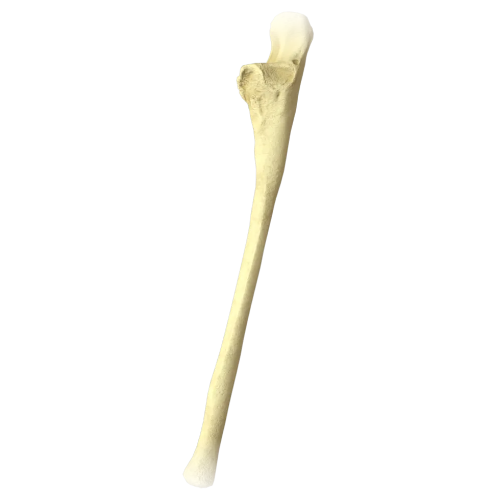
Humerus anatomy quiz
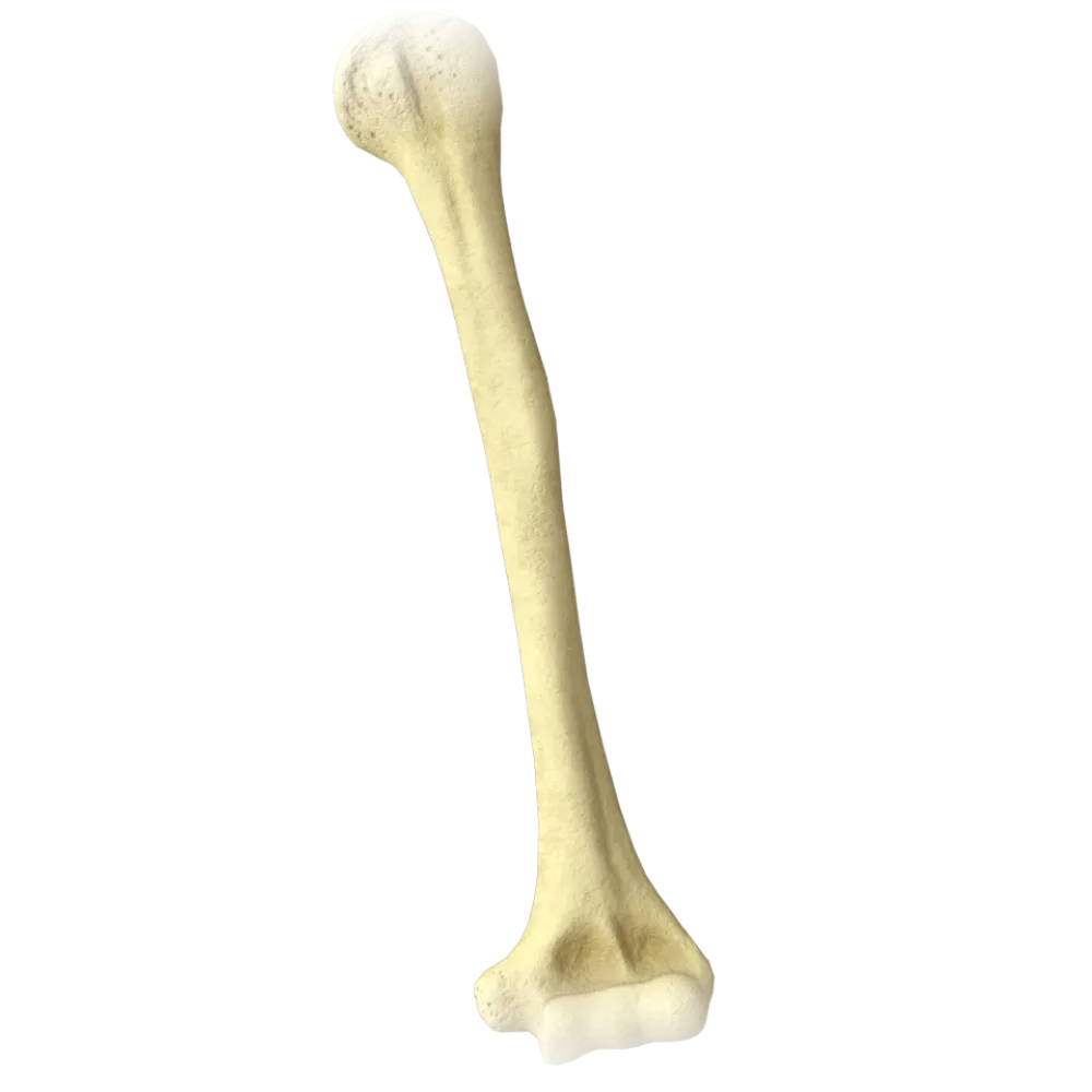
The quiz covers the structure of the humerus: divisions, anatomical structures, muscle attachment sites, articular surfaces, and bony landmarks.
1/20
bold
text
1. Which anatomical structure is located on the proximal epiphysis of the humerus and serves as the articular surface for the scapula?
-
Capitulum of the humerus
Caput humeri (head of the humerus) is the hemispherical articular surface of the proximal epiphysis that articulates with the cavitas glenoidalis of the scapula in the glenohumeral joint.
-
Head of the humerus
Caput humeri (head of the humerus) is the hemispherical articular surface of the proximal epiphysis that articulates with the cavitas glenoidalis of the scapula in the glenohumeral joint.
-
Trochlea of the humerus
Caput humeri (head of the humerus) is the hemispherical articular surface of the proximal epiphysis that articulates with the cavitas glenoidalis of the scapula in the glenohumeral joint.
-
Greater tubercle
Caput humeri (head of the humerus) is the hemispherical articular surface of the proximal epiphysis that articulates with the cavitas glenoidalis of the scapula in the glenohumeral joint.
-
I find it difficult to answer
Caput humeri (head of the humerus) is the hemispherical articular surface of the proximal epiphysis that articulates with the cavitas glenoidalis of the scapula in the glenohumeral joint.
2. What is the constriction of the humerus that separates the head from the greater and lesser tubercles called?
-
Sulcus intertubercularis
Collum anatomicum (anatomical neck) is the groove separating the head of the humerus from the tubercles. The collum chirurgicum is located more distally, at the junction of the epiphysis and shaft.
-
Anatomical neck
Collum anatomicum (anatomical neck) is the groove separating the head of the humerus from the tubercles. The collum chirurgicum is located more distally, at the junction of the epiphysis and shaft.
-
Surgical neck
Collum anatomicum (anatomical neck) is the groove separating the head of the humerus from the tubercles. The collum chirurgicum is located more distally, at the junction of the epiphysis and shaft.
-
Corpus humeri
Collum anatomicum (anatomical neck) is the groove separating the head of the humerus from the tubercles. The collum chirurgicum is located more distally, at the junction of the epiphysis and shaft.
-
I find it difficult to answer
Collum anatomicum (anatomical neck) is the groove separating the head of the humerus from the tubercles. The collum chirurgicum is located more distally, at the junction of the epiphysis and shaft.
3. To which tubercle of the humerus does the m. supraspinatus?
-
Lesser tubercle
M. supraspinatus attaches to the superior facet of the tuberculum majus (greater tubercle). The m. also attach to this tubercle. infraspinatus and m. teres minor, each to its respective facet.
-
Crest of the lesser tubercle
M. supraspinatus attaches to the superior facet of the tuberculum majus (greater tubercle). The m. also attach to this tubercle. infraspinatus and m. teres minor, each to its respective facet.
-
Greater tubercle
M. supraspinatus attaches to the superior facet of the tuberculum majus (greater tubercle). The m. also attach to this tubercle. infraspinatus and m. teres minor, each to its respective facet.
-
Crest of the greater tubercle
M. supraspinatus attaches to the superior facet of the tuberculum majus (greater tubercle). The m. also attach to this tubercle. infraspinatus and m. teres minor, each to its respective facet.
-
I find it difficult to answer
M. supraspinatus attaches to the superior facet of the tuberculum majus (greater tubercle). The m. also attach to this tubercle. infraspinatus and m. teres minor, each to its respective facet.
4. Which muscle attaches to the tuberculum minus of the humerus?
-
Teres major muscle
M. subscapularis attaches to the tuberculum minus (lesser tubercle) of the humerus. It is the only rotator cuff muscle attaching to the lesser tubercle.
-
M. subscapularis
M. subscapularis attaches to the tuberculum minus (lesser tubercle) of the humerus. It is the only rotator cuff muscle attaching to the lesser tubercle.
-
Infraspinatus muscle
M. subscapularis attaches to the tuberculum minus (lesser tubercle) of the humerus. It is the only rotator cuff muscle attaching to the lesser tubercle.
-
M. deltoideus
M. subscapularis attaches to the tuberculum minus (lesser tubercle) of the humerus. It is the only rotator cuff muscle attaching to the lesser tubercle.
-
I find it difficult to answer
M. subscapularis attaches to the tuberculum minus (lesser tubercle) of the humerus. It is the only rotator cuff muscle attaching to the lesser tubercle.
5. Which structure passes through the sulcus intertubercularis (intertubercular groove) of the humerus?
-
Axillary nerve
The tendon of the long head of the biceps brachii (tendo m. bicipitis brachii, caput longum) passes through the sulcus intertubercularis and is retained by the lig. transverse humeral.
-
Tendon of m. biceps brachii (long head)
The tendon of the long head of the biceps brachii (tendo m. bicipitis brachii, caput longum) passes through the sulcus intertubercularis and is retained by the lig. transverse humeral.
-
Anterior circumflex humeral artery
The tendon of the long head of the biceps brachii (tendo m. bicipitis brachii, caput longum) passes through the sulcus intertubercularis and is retained by the lig. transverse humeral.
-
Tendon of m. coracobrachialis
The tendon of the long head of the biceps brachii (tendo m. bicipitis brachii, caput longum) passes through the sulcus intertubercularis and is retained by the lig. transverse humeral.
-
I find it difficult to answer
The tendon of the long head of the biceps brachii (tendo m. bicipitis brachii, caput longum) passes through the sulcus intertubercularis and is retained by the lig. transverse humeral.
6. The following attaches to the crista tuberculi majoris (crest of the greater tubercle):
-
Pectoralis major muscle
M. pectoralis major attaches to the crista tuberculi majoris of the humerus. This site is distal to the tuberculum majus, on the shaft, and forms the characteristic crest for this muscle.
-
Teres minor muscle
M. pectoralis major attaches to the crista tuberculi majoris of the humerus. This site is distal to the tuberculum majus, on the shaft, and forms the characteristic crest for this muscle.
-
Coracobrachialis
M. pectoralis major attaches to the crista tuberculi majoris of the humerus. This site is distal to the tuberculum majus, on the shaft, and forms the characteristic crest for this muscle.
-
Supraspinatus muscle
M. pectoralis major attaches to the crista tuberculi majoris of the humerus. This site is distal to the tuberculum majus, on the shaft, and forms the characteristic crest for this muscle.
-
I find it difficult to answer
M. pectoralis major attaches to the crista tuberculi majoris of the humerus. This site is distal to the tuberculum majus, on the shaft, and forms the characteristic crest for this muscle.
7. Which structure of the humeral shaft is the attachment site for the m. deltoideus?
-
Deltoid tuberosity
The tuberositas deltoidea (deltoid tuberosity) is located on the lateral surface of the middle of the humeral shaft and is the attachment site of the m. deltoideus.
-
Sulcus nervi radialis
The tuberositas deltoidea (deltoid tuberosity) is located on the lateral surface of the middle of the humeral shaft and is the attachment site of the m. deltoideus.
-
Lateral supracondylar ridge
The tuberositas deltoidea (deltoid tuberosity) is located on the lateral surface of the middle of the humeral shaft and is the attachment site of the m. deltoideus.
-
Anterior border
The tuberositas deltoidea (deltoid tuberosity) is located on the lateral surface of the middle of the humeral shaft and is the attachment site of the m. deltoideus.
-
I find it difficult to answer
The tuberositas deltoidea (deltoid tuberosity) is located on the lateral surface of the middle of the humeral shaft and is the attachment site of the m. deltoideus.
8. The sulcus nervi radialis (radial nerve groove) on the humerus runs:
-
Along the anterior surface of the shaft from superior to inferior
The sulcus nervi radialis runs spirally along the posterior surface of the humeral shaft, from the medial border of the proximal part to the lateral border of the distal part. It contains the n. radialis and a. profunda brachii.
-
Spirally along the posterior surface of the shaft, from superomedial to inferolateral
The sulcus nervi radialis runs spirally along the posterior surface of the humeral shaft, from the medial border of the proximal part to the lateral border of the distal part. It contains the n. radialis and a. profunda brachii.
-
Along the medial epicondyle
The sulcus nervi radialis runs spirally along the posterior surface of the humeral shaft, from the medial border of the proximal part to the lateral border of the distal part. It contains the n. radialis and a. profunda brachii.
-
Vertically along the medial surface of the shaft
The sulcus nervi radialis runs spirally along the posterior surface of the humeral shaft, from the medial border of the proximal part to the lateral border of the distal part. It contains the n. radialis and a. profunda brachii.
-
I find it difficult to answer
The sulcus nervi radialis runs spirally along the posterior surface of the humeral shaft, from the medial border of the proximal part to the lateral border of the distal part. It contains the n. radialis and a. profunda brachii.
9. Which structure of the distal epiphysis of the humerus articulates with the caput radii?
-
Trochlea of the humerus
Capitulum humeri (capitulum of the humerus) is the rounded articular surface on the lateral part of the distal epiphysis that articulates with the fovea articularis of the head of the radius.
-
Medial epicondyle
Capitulum humeri (capitulum of the humerus) is the rounded articular surface on the lateral part of the distal epiphysis that articulates with the fovea articularis of the head of the radius.
-
Capitulum of the humerus
Capitulum humeri (capitulum of the humerus) is the rounded articular surface on the lateral part of the distal epiphysis that articulates with the fovea articularis of the head of the radius.
-
Olecranon fossa
Capitulum humeri (capitulum of the humerus) is the rounded articular surface on the lateral part of the distal epiphysis that articulates with the fovea articularis of the head of the radius.
-
I find it difficult to answer
Capitulum humeri (capitulum of the humerus) is the rounded articular surface on the lateral part of the distal epiphysis that articulates with the fovea articularis of the head of the radius.
10. With which structure of the ulna does the trochlea humeri articulate?
-
Styloid process of the ulna
The trochlea humeri (trochlea of the humerus) articulates with the incisura trochlearis (trochlear notch) of the ulna. This articulation permits flexion and extension at the elbow joint.
-
Trochlear notch of the ulna
The trochlea humeri (trochlea of the humerus) articulates with the incisura trochlearis (trochlear notch) of the ulna. This articulation permits flexion and extension at the elbow joint.
-
Head of the ulna
The trochlea humeri (trochlea of the humerus) articulates with the incisura trochlearis (trochlear notch) of the ulna. This articulation permits flexion and extension at the elbow joint.
-
Tuberosity of the ulna
The trochlea humeri (trochlea of the humerus) articulates with the incisura trochlearis (trochlear notch) of the ulna. This articulation permits flexion and extension at the elbow joint.
-
I find it difficult to answer
The trochlea humeri (trochlea of the humerus) articulates with the incisura trochlearis (trochlear notch) of the ulna. This articulation permits flexion and extension at the elbow joint.
11. The fossa olecrani is located on the:
-
Anterior surface of the distal epiphysis above the trochlea
The fossa olecrani (olecranon fossa) is located on the posterior surface of the distal epiphysis of the humerus, above the trochlea. The olecranon of the ulna enters it during extension at the elbow joint.
-
Posterior surface of the distal epiphysis above the trochlea
The fossa olecrani (olecranon fossa) is located on the posterior surface of the distal epiphysis of the humerus, above the trochlea. The olecranon of the ulna enters it during extension at the elbow joint.
-
Lateral surface of the shaft
The fossa olecrani (olecranon fossa) is located on the posterior surface of the distal epiphysis of the humerus, above the trochlea. The olecranon of the ulna enters it during extension at the elbow joint.
-
Medial surface of the proximal epiphysis
The fossa olecrani (olecranon fossa) is located on the posterior surface of the distal epiphysis of the humerus, above the trochlea. The olecranon of the ulna enters it during extension at the elbow joint.
-
I find it difficult to answer
The fossa olecrani (olecranon fossa) is located on the posterior surface of the distal epiphysis of the humerus, above the trochlea. The olecranon of the ulna enters it during extension at the elbow joint.
12. The fossa coronoidea of the humerus is intended for:
-
Accommodation of the processus coronoideus ulnae during flexion
The fossa coronoidea (coronoid fossa) is located on the anterior surface of the distal epiphysis above the trochlea and receives the processus coronoideus of the ulna during maximal flexion at the elbow joint.
-
Accommodation of the caput radii during pronation
The fossa coronoidea (coronoid fossa) is located on the anterior surface of the distal epiphysis above the trochlea and receives the processus coronoideus of the ulna during maximal flexion at the elbow joint.
-
Attachment of the lig. collaterale ulnare
The fossa coronoidea (coronoid fossa) is located on the anterior surface of the distal epiphysis above the trochlea and receives the processus coronoideus of the ulna during maximal flexion at the elbow joint.
-
Passage of the a. brachialis
The fossa coronoidea (coronoid fossa) is located on the anterior surface of the distal epiphysis above the trochlea and receives the processus coronoideus of the ulna during maximal flexion at the elbow joint.
-
I find it difficult to answer
The fossa coronoidea (coronoid fossa) is located on the anterior surface of the distal epiphysis above the trochlea and receives the processus coronoideus of the ulna during maximal flexion at the elbow joint.
13. The following attaches to the crista tuberculi minoris (crest of the lesser tubercle):
-
M. latissimus dorsi
The m. attach to the crista tuberculi minoris. teres major and m. latissimus dorsi muscle. They are located adjacent to each other, and both muscles share an attachment site on this crest of the humerus.
-
Teres major muscle
The m. attach to the crista tuberculi minoris. teres major and m. latissimus dorsi muscle. They are located adjacent to each other, and both muscles share an attachment site on this crest of the humerus.
-
M. subscapularis
The m. attach to the crista tuberculi minoris. teres major and m. latissimus dorsi muscle. They are located adjacent to each other, and both muscles share an attachment site on this crest of the humerus.
-
Both: m. latissimus dorsi and m. teres major
The m. attach to the crista tuberculi minoris. teres major and m. latissimus dorsi muscle. They are located adjacent to each other, and both muscles share an attachment site on this crest of the humerus.
-
I find it difficult to answer
The m. attach to the crista tuberculi minoris. teres major and m. latissimus dorsi muscle. They are located adjacent to each other, and both muscles share an attachment site on this crest of the humerus.
14. The epicondylus medialis humeri is the site of origin of the:
-
Mm. extensores carpi and digitorum
The muscles of the superficial flexor layer of the forearm originate from the epicondylus medialis: pronator teres muscle, flexor carpi radialis muscle, palmaris longus muscle, flexor carpi ulnaris muscle, superficial flexor of the fingers.
-
Mm. flexores antebrachii (superficial group)
The muscles of the superficial flexor layer of the forearm originate from the epicondylus medialis: pronator teres muscle, flexor carpi radialis muscle, palmaris longus muscle, flexor carpi ulnaris muscle, superficial flexor of the fingers.
-
M. brachioradialis
The muscles of the superficial flexor layer of the forearm originate from the epicondylus medialis: pronator teres muscle, flexor carpi radialis muscle, palmaris longus muscle, flexor carpi ulnaris muscle, superficial flexor of the fingers.
-
M. anconeus
The muscles of the superficial flexor layer of the forearm originate from the epicondylus medialis: pronator teres muscle, flexor carpi radialis muscle, palmaris longus muscle, flexor carpi ulnaris muscle, superficial flexor of the fingers.
-
I find it difficult to answer
The muscles of the superficial flexor layer of the forearm originate from the epicondylus medialis: pronator teres muscle, flexor carpi radialis muscle, palmaris longus muscle, flexor carpi ulnaris muscle, superficial flexor of the fingers.
15. Which epicondyle of the humerus is adjacent to the sulcus nervi ulnaris?
-
Lateral epicondyle
The sulcus nervi ulnaris (ulnar nerve groove) passes along the posterior surface of the medial epicondyle (epicondylus medialis) of the humerus, where the n. ulnaris is palpable.
-
Medial epicondyle
The sulcus nervi ulnaris (ulnar nerve groove) passes along the posterior surface of the medial epicondyle (epicondylus medialis) of the humerus, where the n. ulnaris is palpable.
-
Lateral supracondylar ridge
The sulcus nervi ulnaris (ulnar nerve groove) passes along the posterior surface of the medial epicondyle (epicondylus medialis) of the humerus, where the n. ulnaris is palpable.
-
Medial supracondylar ridge
The sulcus nervi ulnaris (ulnar nerve groove) passes along the posterior surface of the medial epicondyle (epicondylus medialis) of the humerus, where the n. ulnaris is palpable.
-
I find it difficult to answer
The sulcus nervi ulnaris (ulnar nerve groove) passes along the posterior surface of the medial epicondyle (epicondylus medialis) of the humerus, where the n. ulnaris is palpable.
16. The following originate from the epicondylus lateralis humeri:
-
Mm. forearm flexors
The muscles of the superficial extensor layer of the forearm originate from the epicondylus lateralis: m. extensor carpi radialis brevis muscle, extensor digitorum muscle, extensor digiti minimi muscle, extensor carpi ulnaris muscle, anconeus muscle.
-
M. pronator teres and m. flexor carpi ulnaris
The muscles of the superficial extensor layer of the forearm originate from the epicondylus lateralis: m. extensor carpi radialis brevis muscle, extensor digitorum muscle, extensor digiti minimi muscle, extensor carpi ulnaris muscle, anconeus muscle.
-
Mm. extensores antebrachii (superficial group)
The muscles of the superficial extensor layer of the forearm originate from the epicondylus lateralis: m. extensor carpi radialis brevis muscle, extensor digitorum muscle, extensor digiti minimi muscle, extensor carpi ulnaris muscle, anconeus muscle.
-
M. brachialis
The muscles of the superficial extensor layer of the forearm originate from the epicondylus lateralis: m. extensor carpi radialis brevis muscle, extensor digitorum muscle, extensor digiti minimi muscle, extensor carpi ulnaris muscle, anconeus muscle.
-
I find it difficult to answer
The muscles of the superficial extensor layer of the forearm originate from the epicondylus lateralis: m. extensor carpi radialis brevis muscle, extensor digitorum muscle, extensor digiti minimi muscle, extensor carpi ulnaris muscle, anconeus muscle.
17. The fossa radialis of the humerus is located:
-
On the posterior surface of the distal epiphysis, laterally above the capitulum
The fossa radialis (radial fossa) is located on the anterior surface of the distal epiphysis, laterally, above the capitulum of the humerus. The caput radii enters it during flexion at the elbow joint.
-
On the anterior surface of the distal epiphysis, laterally above the capitulum
The fossa radialis (radial fossa) is located on the anterior surface of the distal epiphysis, laterally, above the capitulum of the humerus. The caput radii enters it during flexion at the elbow joint.
-
On the medial surface of the proximal epiphysis
The fossa radialis (radial fossa) is located on the anterior surface of the distal epiphysis, laterally, above the capitulum of the humerus. The caput radii enters it during flexion at the elbow joint.
-
On the posterior surface of the shaft
The fossa radialis (radial fossa) is located on the anterior surface of the distal epiphysis, laterally, above the capitulum of the humerus. The caput radii enters it during flexion at the elbow joint.
-
I find it difficult to answer
The fossa radialis (radial fossa) is located on the anterior surface of the distal epiphysis, laterally, above the capitulum of the humerus. The caput radii enters it during flexion at the elbow joint.
18. To which morphological classification group of bones does the humerus belong?
-
Cancellous bone (os spongiosum)
The humerus (humerus) is a long bone (os longum). It comprises a body (corpus/diaphysis) and two ends (epiphyses: proximal and distal), which meets the criteria for long bones.
-
Flat bone (os planum)
The humerus (humerus) is a long bone (os longum). It comprises a body (corpus/diaphysis) and two ends (epiphyses: proximal and distal), which meets the criteria for long bones.
-
Long bone (os longum)
The humerus (humerus) is a long bone (os longum). It comprises a body (corpus/diaphysis) and two ends (epiphyses: proximal and distal), which meets the criteria for long bones.
-
Irregular bone (os mixtum)
The humerus (humerus) is a long bone (os longum). It comprises a body (corpus/diaphysis) and two ends (epiphyses: proximal and distal), which meets the criteria for long bones.
-
I find it difficult to answer
The humerus (humerus) is a long bone (os longum). It comprises a body (corpus/diaphysis) and two ends (epiphyses: proximal and distal), which meets the criteria for long bones.
19. The collum chirurgicum of the humerus is located:
-
Between the head and tubercles
The collum chirurgicum (surgical neck) is located distal to the tubercles, at the junction of the proximal epiphysis and shaft of the humerus. The n. passes through this region. axillary and a. circumflex humeral posterior.
-
Distal to the tubercles, at the junction of the proximal epiphysis and shaft
The collum chirurgicum (surgical neck) is located distal to the tubercles, at the junction of the proximal epiphysis and shaft of the humerus. The n. passes through this region. axillary and a. circumflex humeral posterior.
-
At the level of the tuberositas deltoidea
The collum chirurgicum (surgical neck) is located distal to the tubercles, at the junction of the proximal epiphysis and shaft of the humerus. The n. passes through this region. axillary and a. circumflex humeral posterior.
-
Immediately above the medial epicondyle
The collum chirurgicum (surgical neck) is located distal to the tubercles, at the junction of the proximal epiphysis and shaft of the humerus. The n. passes through this region. axillary and a. circumflex humeral posterior.
-
I find it difficult to answer
The collum chirurgicum (surgical neck) is located distal to the tubercles, at the junction of the proximal epiphysis and shaft of the humerus. The n. passes through this region. axillary and a. circumflex humeral posterior.
20. Which structure of the proximal epiphysis of the humerus separates the head from the tubercles?
-
Surgical neck
The collum anatomicum (anatomical neck) is a circumferential groove that directly separates the caput humeri from the tuberculum majus and tuberculum minus. It is the anatomical boundary of the articular cartilage of the head.
-
Sulcus intertubercularis
The collum anatomicum (anatomical neck) is a circumferential groove that directly separates the caput humeri from the tuberculum majus and tuberculum minus. It is the anatomical boundary of the articular cartilage of the head.
-
Anatomical neck
The collum anatomicum (anatomical neck) is a circumferential groove that directly separates the caput humeri from the tuberculum majus and tuberculum minus. It is the anatomical boundary of the articular cartilage of the head.
-
Crest of the greater tubercle
The collum anatomicum (anatomical neck) is a circumferential groove that directly separates the caput humeri from the tuberculum majus and tuberculum minus. It is the anatomical boundary of the articular cartilage of the head.
-
I find it difficult to answer
The collum anatomicum (anatomical neck) is a circumferential groove that directly separates the caput humeri from the tuberculum majus and tuberculum minus. It is the anatomical boundary of the articular cartilage of the head.
Retake this quiz?
Your current progress will be reset.
