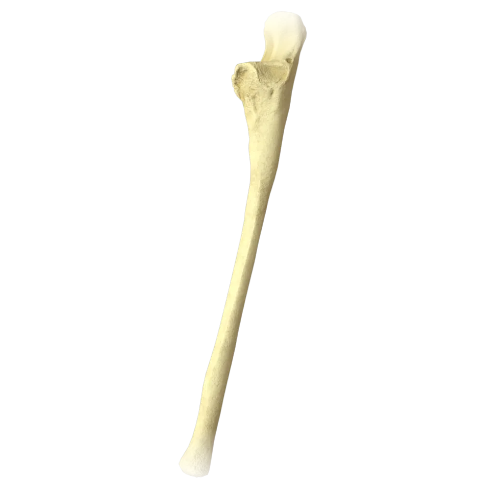
Radius anatomy test
The test covers the anatomy of the radius: structure, joints, muscles, vessels, and nerves within the framework of systemic anatomy.
1/20
bold
text
1. Which bone forms the proximal radioulnar joint together with the radius?
-
Humerus
The proximal radioulnar joint (articulatio radioulnaris proximalis) is formed by the articular circumference of the head of the radius and the radial notch of the ulna.
-
Ulna
The proximal radioulnar joint (articulatio radioulnaris proximalis) is formed by the articular circumference of the head of the radius and the radial notch of the ulna.
-
Scaphoid
The proximal radioulnar joint (articulatio radioulnaris proximalis) is formed by the articular circumference of the head of the radius and the radial notch of the ulna.
-
Lunate bone
The proximal radioulnar joint (articulatio radioulnaris proximalis) is formed by the articular circumference of the head of the radius and the radial notch of the ulna.
-
I find it difficult to answer
The proximal radioulnar joint (articulatio radioulnaris proximalis) is formed by the articular circumference of the head of the radius and the radial notch of the ulna.
2. What is the articular surface on the head of the radius that articulates with the capitulum of the humerus called?
-
Articular circumference
Fovea articularis (fovea of the head of the radius) is the concave articular surface at the apex of the head of the radius that articulates with the capitulum (capitulum humeri) of the humerus.
-
Fovea of the head of the radius
Fovea articularis (fovea of the head of the radius) is the concave articular surface at the apex of the head of the radius that articulates with the capitulum (capitulum humeri) of the humerus.
-
Styloid process
Fovea articularis (fovea of the head of the radius) is the concave articular surface at the apex of the head of the radius that articulates with the capitulum (capitulum humeri) of the humerus.
-
Carpal articular surface
Fovea articularis (fovea of the head of the radius) is the concave articular surface at the apex of the head of the radius that articulates with the capitulum (capitulum humeri) of the humerus.
-
I find it difficult to answer
Fovea articularis (fovea of the head of the radius) is the concave articular surface at the apex of the head of the radius that articulates with the capitulum (capitulum humeri) of the humerus.
3. Where is the radial tuberosity (tuberositas radii) located?
-
On the distal epiphysis of the radius
The tuberositas radii is located on the medial side of the shaft of the radius, immediately inferior to the neck, and serves as the attachment site for the biceps brachii muscle (m. biceps brachii).
-
On the medial surface of the diaphysis in its distal third
The tuberositas radii is located on the medial side of the shaft of the radius, immediately inferior to the neck, and serves as the attachment site for the biceps brachii muscle (m. biceps brachii).
-
Immediately inferior to the neck of the radius on the medial side
The tuberositas radii is located on the medial side of the shaft of the radius, immediately inferior to the neck, and serves as the attachment site for the biceps brachii muscle (m. biceps brachii).
-
On the posterior surface of the head of the radius
The tuberositas radii is located on the medial side of the shaft of the radius, immediately inferior to the neck, and serves as the attachment site for the biceps brachii muscle (m. biceps brachii).
-
I find it difficult to answer
The tuberositas radii is located on the medial side of the shaft of the radius, immediately inferior to the neck, and serves as the attachment site for the biceps brachii muscle (m. biceps brachii).
4. Which muscle attaches to the radial tuberosity?
-
Brachioradialis muscle
The biceps brachii muscle (m. biceps brachii) attaches to the tuberositas radii. This attachment enables forearm flexion and supination.
-
Biceps brachii
The biceps brachii muscle (m. biceps brachii) attaches to the tuberositas radii. This attachment enables forearm flexion and supination.
-
Pronator teres
The biceps brachii muscle (m. biceps brachii) attaches to the tuberositas radii. This attachment enables forearm flexion and supination.
-
Supinator
The biceps brachii muscle (m. biceps brachii) attaches to the tuberositas radii. This attachment enables forearm flexion and supination.
-
I find it difficult to answer
The biceps brachii muscle (m. biceps brachii) attaches to the tuberositas radii. This attachment enables forearm flexion and supination.
5. To which process of the radius does the brachioradialis muscle (m. brachioradialis) attach?
-
To the radial tuberosity
M. brachioradialis attaches to the styloid process of the radius (processus styloideus radii), located on the lateral side of the distal epiphysis.
-
To the styloid process of the radius
M. brachioradialis attaches to the styloid process of the radius (processus styloideus radii), located on the lateral side of the distal epiphysis.
-
To the articular circumference of the head
M. brachioradialis attaches to the styloid process of the radius (processus styloideus radii), located on the lateral side of the distal epiphysis.
-
To the dorsal tubercle of the radius
M. brachioradialis attaches to the styloid process of the radius (processus styloideus radii), located on the lateral side of the distal epiphysis.
-
I find it difficult to answer
M. brachioradialis attaches to the styloid process of the radius (processus styloideus radii), located on the lateral side of the distal epiphysis.
6. Which ligament holds the head of the radius against the radial notch of the ulna?
-
Quadrate ligament
The annular ligament of the radius (ligamentum anulare radii) encircles the head of the radius and holds it against the radial notch of the ulna, providing stability to the proximal radioulnar joint.
-
Annular ligament of the radius
The annular ligament of the radius (ligamentum anulare radii) encircles the head of the radius and holds it against the radial notch of the ulna, providing stability to the proximal radioulnar joint.
-
Interosseous membrane of the forearm
The annular ligament of the radius (ligamentum anulare radii) encircles the head of the radius and holds it against the radial notch of the ulna, providing stability to the proximal radioulnar joint.
-
Radial collateral ligament
The annular ligament of the radius (ligamentum anulare radii) encircles the head of the radius and holds it against the radial notch of the ulna, providing stability to the proximal radioulnar joint.
-
I find it difficult to answer
The annular ligament of the radius (ligamentum anulare radii) encircles the head of the radius and holds it against the radial notch of the ulna, providing stability to the proximal radioulnar joint.
7. Which artery is the primary source of blood supply to the radius?
-
Ulnar artery
The radial artery (a. radialis) is the primary source of blood supply to the radius; it runs along the lateral side of the forearm, giving off periosteal branches along the entire length of the bone.
-
Anterior interosseous artery
The radial artery (a. radialis) is the primary source of blood supply to the radius; it runs along the lateral side of the forearm, giving off periosteal branches along the entire length of the bone.
-
Radial artery
The radial artery (a. radialis) is the primary source of blood supply to the radius; it runs along the lateral side of the forearm, giving off periosteal branches along the entire length of the bone.
-
Recurrent radial artery
The radial artery (a. radialis) is the primary source of blood supply to the radius; it runs along the lateral side of the forearm, giving off periosteal branches along the entire length of the bone.
-
I find it difficult to answer
The radial artery (a. radialis) is the primary source of blood supply to the radius; it runs along the lateral side of the forearm, giving off periosteal branches along the entire length of the bone.
8. Which nerve passes near the neck of the radius and may be compressed in pathological conditions affecting this region?
-
Median nerve
The deep branch of the radial nerve (r. profundus n. radialis) curves around the neck of the radius, passing through the supinator (arcus supinatoris). This topographic relationship determines the vulnerability of the nerve to structural changes in this region.
-
Ulnar nerve
The deep branch of the radial nerve (r. profundus n. radialis) curves around the neck of the radius, passing through the supinator (arcus supinatoris). This topographic relationship determines the vulnerability of the nerve to structural changes in this region.
-
Deep branch of radial nerve
The deep branch of the radial nerve (r. profundus n. radialis) curves around the neck of the radius, passing through the supinator (arcus supinatoris). This topographic relationship determines the vulnerability of the nerve to structural changes in this region.
-
Musculocutaneous nerve
The deep branch of the radial nerve (r. profundus n. radialis) curves around the neck of the radius, passing through the supinator (arcus supinatoris). This topographic relationship determines the vulnerability of the nerve to structural changes in this region.
-
I find it difficult to answer
The deep branch of the radial nerve (r. profundus n. radialis) curves around the neck of the radius, passing through the supinator (arcus supinatoris). This topographic relationship determines the vulnerability of the nerve to structural changes in this region.
9. What is the interosseous membrane of the forearm (membrana interossea antebrachii) in the context of the connection between the radius and ulna?
-
Synovial joint
The interosseous membrane of the forearm is a syndesmosis—a continuous connective tissue union (syndesmosis) consisting of dense fibrous tissue stretched between the interosseous borders of the radius and ulna.
-
Synostosis
The interosseous membrane of the forearm is a syndesmosis—a continuous connective tissue union (syndesmosis) consisting of dense fibrous tissue stretched between the interosseous borders of the radius and ulna.
-
Syndesmosis
The interosseous membrane of the forearm is a syndesmosis—a continuous connective tissue union (syndesmosis) consisting of dense fibrous tissue stretched between the interosseous borders of the radius and ulna.
-
Symphysis
The interosseous membrane of the forearm is a syndesmosis—a continuous connective tissue union (syndesmosis) consisting of dense fibrous tissue stretched between the interosseous borders of the radius and ulna.
-
I find it difficult to answer
The interosseous membrane of the forearm is a syndesmosis—a continuous connective tissue union (syndesmosis) consisting of dense fibrous tissue stretched between the interosseous borders of the radius and ulna.
10. Which tubercle on the dorsal surface of the distal epiphysis of the radius serves as a landmark for the tendon of the extensor pollicis longus?
-
Styloid process
The tuberculum dorsale (Lister's tubercle) is located on the dorsal surface of the distal epiphysis of the radius and serves as a pulley around which the tendon of m. extensor pollicis longus.
-
Radial tuberosity
The tuberculum dorsale (Lister's tubercle) is located on the dorsal surface of the distal epiphysis of the radius and serves as a pulley around which the tendon of m. extensor pollicis longus.
-
Dorsal tubercle (tuberculum dorsale, Lister's tubercle)
The tuberculum dorsale (Lister's tubercle) is located on the dorsal surface of the distal epiphysis of the radius and serves as a pulley around which the tendon of m. extensor pollicis longus.
-
Articular circumference
The tuberculum dorsale (Lister's tubercle) is located on the dorsal surface of the distal epiphysis of the radius and serves as a pulley around which the tendon of m. extensor pollicis longus.
-
I find it difficult to answer
The tuberculum dorsale (Lister's tubercle) is located on the dorsal surface of the distal epiphysis of the radius and serves as a pulley around which the tendon of m. extensor pollicis longus.
11. Which muscle originates from the anterior surface of the radius and the interosseous membrane and attaches to the base of the distal phalanx of the thumb?
-
Flexor pollicis longus
M. flexor pollicis longus originates from the anterior surface of the radius and the interosseous membrane and attaches to the base of the distal phalanx of the first digit, producing its flexion.
-
Flexor pollicis brevis
M. flexor pollicis longus originates from the anterior surface of the radius and the interosseous membrane and attaches to the base of the distal phalanx of the first digit, producing its flexion.
-
Abductor pollicis longus
M. flexor pollicis longus originates from the anterior surface of the radius and the interosseous membrane and attaches to the base of the distal phalanx of the first digit, producing its flexion.
-
Extensor hallucis brevis
M. flexor pollicis longus originates from the anterior surface of the radius and the interosseous membrane and attaches to the base of the distal phalanx of the first digit, producing its flexion.
-
I find it difficult to answer
M. flexor pollicis longus originates from the anterior surface of the radius and the interosseous membrane and attaches to the base of the distal phalanx of the first digit, producing its flexion.
12. Which joint is formed by the carpal articular surface of the radius and the proximal row of carpal bones?
-
Distal radioulnar joint
The radiocarpal joint (articulatio radiocarpalis) is formed by the carpal articular surface of the radius (and the articular disc) on one side and the proximal row of carpal bones (scaphoid, lunate, and triquetral) on the other.
-
Radiocarpal joint
The radiocarpal joint (articulatio radiocarpalis) is formed by the carpal articular surface of the radius (and the articular disc) on one side and the proximal row of carpal bones (scaphoid, lunate, and triquetral) on the other.
-
Midcarpal joint
The radiocarpal joint (articulatio radiocarpalis) is formed by the carpal articular surface of the radius (and the articular disc) on one side and the proximal row of carpal bones (scaphoid, lunate, and triquetral) on the other.
-
Carpometacarpal joint
The radiocarpal joint (articulatio radiocarpalis) is formed by the carpal articular surface of the radius (and the articular disc) on one side and the proximal row of carpal bones (scaphoid, lunate, and triquetral) on the other.
-
I find it difficult to answer
The radiocarpal joint (articulatio radiocarpalis) is formed by the carpal articular surface of the radius (and the articular disc) on one side and the proximal row of carpal bones (scaphoid, lunate, and triquetral) on the other.
13. To which division of the skeleton does the radius belong according to systemic classification?
-
Bones of the pectoral girdle
The radius (radius) belongs to the bones of the free upper limb, specifically to the skeleton of the forearm (ossa antebrachii), together with the ulna (ulna).
-
Bones of the free upper limb (skeleton of the forearm)
The radius (radius) belongs to the bones of the free upper limb, specifically to the skeleton of the forearm (ossa antebrachii), together with the ulna (ulna).
-
Bones of hand
The radius (radius) belongs to the bones of the free upper limb, specifically to the skeleton of the forearm (ossa antebrachii), together with the ulna (ulna).
-
Bones of the arm
The radius (radius) belongs to the bones of the free upper limb, specifically to the skeleton of the forearm (ossa antebrachii), together with the ulna (ulna).
-
I find it difficult to answer
The radius (radius) belongs to the bones of the free upper limb, specifically to the skeleton of the forearm (ossa antebrachii), together with the ulna (ulna).
14. What type of bone is the radius in terms of its shape and structure?
-
Flat bone
The radius is a long tubular bone (os longum) comprising a diaphysis (shaft), proximal and distal epiphyses, metaphyses, and apophyses.
-
Cancellous (short) bone
The radius is a long tubular bone (os longum) comprising a diaphysis (shaft), proximal and distal epiphyses, metaphyses, and apophyses.
-
Long tubular bone
The radius is a long tubular bone (os longum) comprising a diaphysis (shaft), proximal and distal epiphyses, metaphyses, and apophyses.
-
Sesamoid bone
The radius is a long tubular bone (os longum) comprising a diaphysis (shaft), proximal and distal epiphyses, metaphyses, and apophyses.
-
I find it difficult to answer
The radius is a long tubular bone (os longum) comprising a diaphysis (shaft), proximal and distal epiphyses, metaphyses, and apophyses.
15. Which muscle attaches to the lateral surface of the radius in its middle third and pronates the forearm?
-
Pronator quadratus
The pronator teres muscle (m. pronator teres) attaches to the lateral surface of the shaft of the radius in its middle third (the region of the pronator tuberosity) and is the primary pronator of the forearm.
-
Pronator teres
The pronator teres muscle (m. pronator teres) attaches to the lateral surface of the shaft of the radius in its middle third (the region of the pronator tuberosity) and is the primary pronator of the forearm.
-
Supinator
The pronator teres muscle (m. pronator teres) attaches to the lateral surface of the shaft of the radius in its middle third (the region of the pronator tuberosity) and is the primary pronator of the forearm.
-
Brachioradialis muscle
The pronator teres muscle (m. pronator teres) attaches to the lateral surface of the shaft of the radius in its middle third (the region of the pronator tuberosity) and is the primary pronator of the forearm.
-
I find it difficult to answer
The pronator teres muscle (m. pronator teres) attaches to the lateral surface of the shaft of the radius in its middle third (the region of the pronator tuberosity) and is the primary pronator of the forearm.
16. Where is the pronator quadratus muscle (m. pronator quadratus) located in relation to the radius?
-
On the posterior surface of the distal quarter of the forearm between the radius and ulna
M. pronator quadratus is located on the anterior (palmar) surface of the distal quarter of the forearm, connecting the radius and ulna, and produces pronation of the forearm.
-
On the anterior surface of the distal quarter of the forearm between the radius and ulna
M. pronator quadratus is located on the anterior (palmar) surface of the distal quarter of the forearm, connecting the radius and ulna, and produces pronation of the forearm.
-
On the anterior surface of the proximal third of the radius
M. pronator quadratus is located on the anterior (palmar) surface of the distal quarter of the forearm, connecting the radius and ulna, and produces pronation of the forearm.
-
On the posterior surface of the middle third of the radius
M. pronator quadratus is located on the anterior (palmar) surface of the distal quarter of the forearm, connecting the radius and ulna, and produces pronation of the forearm.
-
I find it difficult to answer
M. pronator quadratus is located on the anterior (palmar) surface of the distal quarter of the forearm, connecting the radius and ulna, and produces pronation of the forearm.
17. Which nerve innervates the supinator muscle (m. supinator), which originates in part from the radius?
-
Median nerve
M. supinator is innervated by the deep branch of the radial nerve (r. profundus n. radialis), which passes through the muscle between its two layers, determining the clinical significance of this topographic relationship.
-
Musculocutaneous nerve
M. supinator is innervated by the deep branch of the radial nerve (r. profundus n. radialis), which passes through the muscle between its two layers, determining the clinical significance of this topographic relationship.
-
Ulnar nerve
M. supinator is innervated by the deep branch of the radial nerve (r. profundus n. radialis), which passes through the muscle between its two layers, determining the clinical significance of this topographic relationship.
-
Deep branch of radial nerve
M. supinator is innervated by the deep branch of the radial nerve (r. profundus n. radialis), which passes through the muscle between its two layers, determining the clinical significance of this topographic relationship.
-
I find it difficult to answer
M. supinator is innervated by the deep branch of the radial nerve (r. profundus n. radialis), which passes through the muscle between its two layers, determining the clinical significance of this topographic relationship.
18. Which part of the radius serves as the attachment site for the interosseous membrane of the forearm?
-
Anterior surface
The interosseous membrane of the forearm attaches to the interosseous border of the radius (margo interosseus radii)—a sharp border facing the corresponding border of the ulna.
-
Posterior surface
The interosseous membrane of the forearm attaches to the interosseous border of the radius (margo interosseus radii)—a sharp border facing the corresponding border of the ulna.
-
Interosseous border (margo interosseus)
The interosseous membrane of the forearm attaches to the interosseous border of the radius (margo interosseus radii)—a sharp border facing the corresponding border of the ulna.
-
Lateral surface
The interosseous membrane of the forearm attaches to the interosseous border of the radius (margo interosseus radii)—a sharp border facing the corresponding border of the ulna.
-
I find it difficult to answer
The interosseous membrane of the forearm attaches to the interosseous border of the radius (margo interosseus radii)—a sharp border facing the corresponding border of the ulna.
19. What is the shape of the articular surface for the distal radioulnar joint on the radius?
-
Capitate (convex)
The ulnar notch (incisura ulnaris radii) is located on the medial side of the distal epiphysis of the radius; it is a concave articular surface that articulates with the head of the ulna in the distal radioulnar joint.
-
Notch-shaped (ulnar notch—incisura ulnaris)
The ulnar notch (incisura ulnaris radii) is located on the medial side of the distal epiphysis of the radius; it is a concave articular surface that articulates with the head of the ulna in the distal radioulnar joint.
-
Trochlear
The ulnar notch (incisura ulnaris radii) is located on the medial side of the distal epiphysis of the radius; it is a concave articular surface that articulates with the head of the ulna in the distal radioulnar joint.
-
Condylar
The ulnar notch (incisura ulnaris radii) is located on the medial side of the distal epiphysis of the radius; it is a concave articular surface that articulates with the head of the ulna in the distal radioulnar joint.
-
I find it difficult to answer
The ulnar notch (incisura ulnaris radii) is located on the medial side of the distal epiphysis of the radius; it is a concave articular surface that articulates with the head of the ulna in the distal radioulnar joint.
20. Which carpal bones articulate directly with the carpal articular surface of the radius at the radiocarpal joint?
-
Trapezium, trapezoid, and capitate bones
The carpal articular surface of the radius articulates directly with the scaphoid (os scaphoideum) and lunate (os lunatum) bones. The triquetral bone articulates with the articular disc rather than directly with the radius.
-
Scaphoid and lunate bones
The carpal articular surface of the radius articulates directly with the scaphoid (os scaphoideum) and lunate (os lunatum) bones. The triquetral bone articulates with the articular disc rather than directly with the radius.
-
Lunate and triquetral bones
The carpal articular surface of the radius articulates directly with the scaphoid (os scaphoideum) and lunate (os lunatum) bones. The triquetral bone articulates with the articular disc rather than directly with the radius.
-
Scaphoid, lunate, and triquetral bones
The carpal articular surface of the radius articulates directly with the scaphoid (os scaphoideum) and lunate (os lunatum) bones. The triquetral bone articulates with the articular disc rather than directly with the radius.
-
I find it difficult to answer
The carpal articular surface of the radius articulates directly with the scaphoid (os scaphoideum) and lunate (os lunatum) bones. The triquetral bone articulates with the articular disc rather than directly with the radius.
Retake this quiz?
Your current progress will be reset.
