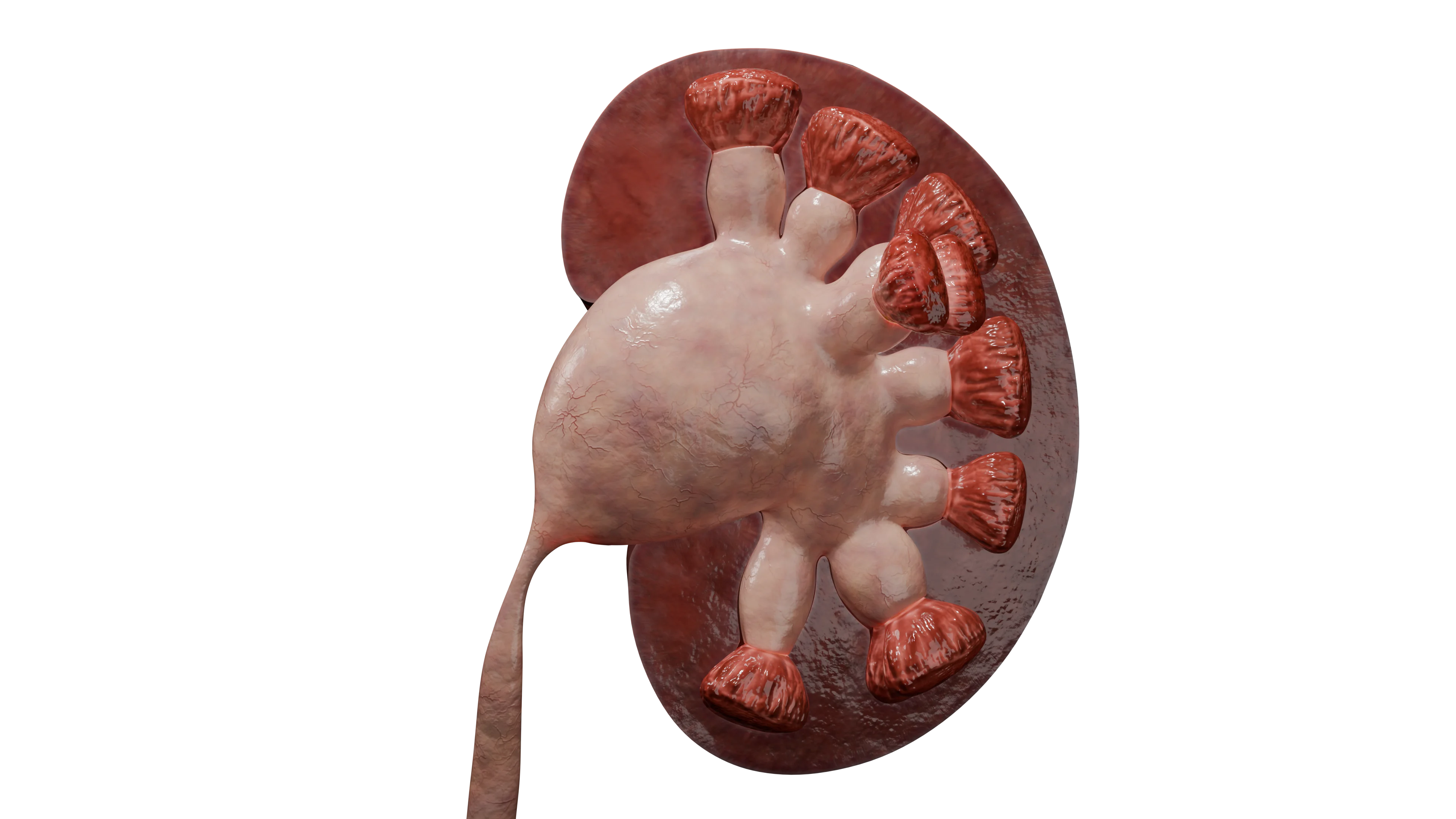
Renal Cyst: Etiology, Classification, Clinical Manifestations, Diagnosis, and Treatment
Egor G.Urologist, MD
10 min read·February 12, 2026
This article is for informational purposes only
The content on this website, including text, graphics, and other materials, is provided for informational purposes only. It is not intended as advice or guidance. Regarding your specific medical condition or treatment, please consult your healthcare provider.
A renal cyst is a thin-walled, fluid-filled lesion arising from the renal parenchyma and lined by the epithelium. Cysts are considered the most common benign renal lesion and occur in all age groups.
The clinical significance of renal cysts is determined by their impact on kidney function and associated symptoms. Asymptomatic cysts, incidentally detected during imaging studies, account for the majority of cases and require ongoing monitoring.
Symptomatic cysts may present with pain, hypertension, or urinary tract obstruction, necessitating active intervention.
Of particular concern is the potential malignant transformation of cysts, which requires careful monitoring even in asymptomatic patients.
Bosniak Classification of Renal Cysts
The Bosniak classification system, developed in 1986, provides a standardized approach to classifying renal cysts based on contrast-enhanced computed tomography (CT) findings. Key assessment criteria include the presence of septa, calcifications within the cyst walls or septa, and the degree of contrast enhancement.
Category I
Bosniak categories I and II are characterized as benign lesions with a minimal risk of malignancy. Category I includes simple cysts with thin walls and no septa, calcifications, or contrast enhancement.
Category II
Category II includes minimally complex cysts with a few thin septa or small calcifications that do not require active intervention. These lesions require dynamic monitoring with periodic ultrasound examinations.
Category IIF
A Bosniak category IIF cyst is neither definitively benign (as in Bosniak categories I or II) nor clearly suspicious for malignancy (as in Bosniak categories III or IV). Category IIF cysts exhibit certain “suspicious” features that are insufficient to warrant classification into a higher category but are sufficient to recommend active observation. Their defining characteristic is an uncertain malignant potential. The vast majority of these cysts are benign.
Category III
Categories III and IV include cysts with a high risk of malignant transformation. Category III comprises heterogeneous lesions with thickened, irregular walls, multiple septa, or irregular calcifications.
Category IV
Category IV includes cysts with definite solid components demonstrating contrast enhancement, which is pathognomonic of malignancy. These cases require surgical treatment with histopathologic confirmation.
A genetic approach identifies autosomal dominant polycystic kidney disease (ADPKD) as a distinct medical condition.
Epidemiology
Renal cysts are among the most common urologic conditions.
Their prevalence is directly associated with age. Renal cysts are rarely detected in individuals younger than 30 years; however, after age 50, they are found in approximately one in three individuals, and by age 70, in more than half of the population.
Regarding sex-related differences, renal cysts occur twice as frequently in men as in women.
Renal cysts are classified as congenital (resulting from developmental abnormalities of the renal tubules) or acquired (developing during person’s lifetime).
The primary pathogenetic mechanism underlying acquired cyst formation is obstruction of the renal tubular lumen due to inflammation, sclerosis, or ischemia. When the secretory activity of the epithelium above the site of obstruction is preserved, fluid accumulates, the tubular walls dilate, and an isolated cavity forms. Congenital forms (e.g., polycystic kidney disease) are associated with genetic defects that lead to uncontrolled formation of multiple cysts arising from nephron tubules.
Clinical Manifestations
Most simple renal cysts are asymptomatic and are detected incidentally during imaging studies performed for other conditions. The absence of clinical manifestations is characteristic of small, solitary lesions that do not compress surrounding tissues. The likelihood of developing symptoms increases with cyst size and proximity to vascular structures or the renal collecting system.
Less commonly, patients may develop hypertension secondary to parenchymal ischemia or impaired urinary drainage due to compression of the ureter or renal pelvis. Differential diagnosis is necessary when malignancy or an association with systemic diseases is suspected.
3D animation: renal cyst formation
Diagnosis of Renal Cysts
Ultrasonography is the primary modality for the initial diagnosis of renal cysts because it is noninvasive, widely available, and does not involve exposure to ionizing radiation. It allows detection of cysts and assessment of their location, size, and principal morphological characteristics. Ultrasonography serves as a basic tool for preliminary classification of cysts according to the Bosniak system, thereby guiding subsequent patient management.
Contrast-enhanced CT is used to further characterize complex cystic lesions identified by ultrasonography. This modality provides high spatial resolution and enables detailed assessment of cyst architecture, including the presence of thickened walls, septa, or solid components. CT is the gold standard for definitive Bosniak classification of renal cysts, which determines the need for continued observation or surgical intervention.
Magnetic resonance imaging (MRI) is used when ultrasonography and CT findings are inconclusive, particularly when malignancy is suspected. It provides excellent soft-tissue contrast and allows differentiation of hemorrhagic or proteinaceous cyst contents without exposure to ionizing radiation. MRI is particularly valuable in patients with contraindications to iodine-based contrast agents or when repeated follow-up imaging is required.
Comprehensive evaluation of contrast-enhanced CT findings and laboratory parameters helps minimize diagnostic errors. Particular attention is given to cysts with thickened walls or heterogeneous contents. Differentiation between simple renal cysts and polycystic kidney disease is based on family history and genetic testing results.
Find more scientifically accurate content on our social media
Subscribe and don’t miss out the latest resources
Treatment
Observation
Conservative management of renal cysts is indicated in patients with small, asymptomatic lesions. The primary criterion is a cyst diameter of less than 4 cm without compression of surrounding tissues or impairment of urinary drainage. An asymptomatic course is confirmed by the absence of pain, dysuria, and abnormalities in laboratory markers of kidney function.
Dynamic observation protocols include regular ultrasound examinations to monitor cyst characteristics. For Bosniak category I–II lesions, ultrasonography is recommended every 6–12 months to assess cyst size and the characteristics of its contents. If these parameters remain stable for two years, the interval between examinations may be extended to 24 months. Changes in the echogenicity of cyst contents or thickening of the cyst walls require reassessment of the Bosniak classification. Contrast-enhanced CT is indicated when malignancy is suspected.
Surgical Approaches to the Treatment of Renal Cysts
Absolute indications for surgical intervention in patients with renal cysts include persistent pain refractory to conservative treatment and impaired urinary drainage resulting in urinary tract obstruction. Additional indications include complications such as cyst infection, intracystic hemorrhage, or suspected malignancy. In these cases, surgical treatment becomes necessary to prevent life-threatening conditions.
Failure to intervene may result in progressive kidney failure and other irreversible changes. Relative indications include situations in which a cyst causes discomfort or functional impairment without posing an immediate threat. These include large cysts (greater than 5 cm) that compress surrounding tissues, as well as lesions demonstrating progressive growth during dynamic observation.
The decision to proceed with surgery is made on an individual basis, considering the patient’s age, comorbidities, and potential intervention risks. The patient’s preference for relief of symptoms affecting quality of life also plays an important role.
Several treatment options are available for renal cysts, each with specific indications. The choice of treatment depends on cyst size, type, location, symptoms, and suspicion of malignancy.
Percutaneous Cyst Aspiration
Procedure. Ultrasound-guided needle puncture through the skin, followed by aspiration of cyst contents, sometimes with instillation of a sclerosing agent (ethanol).
This technique is most effective for small cysts located on the posterior surface of the kidney.
Benefits. Minimally invasive, rapid procedure performed under local anesthesia.
Disadvantages. High risk of recurrence.
Laparoscopic Cyst Excision (Decortication)
Procedure. This technique is used to treat Bosniak category I and II cysts. Through 3–4 small abdominal incisions, the cyst is dissected, opened, and its walls are excised and submitted for histopathologic examination. The cyst cavity is left open.
Benefits. Gold standard for cyst treatment. Low risk of recurrence. The technique is suitable for cysts of any size and location. It provides good visualization and allows biopsy sampling.
Disadvantages. A surgical procedure requiring general anesthesia. Risks inherent to any surgical procedure include bleeding, infection, and injury to adjacent organs (bowel, blood vessels).
Endoscopic Cyst Fenestration (Marsupialization)
Procedure. A flexible endoscope is advanced into the kidney through the urethra, bladder, and ureter. The cyst wall protruding into the renal pelvis is incised using a laser, thereby performing cyst fenestration (marsupialization). This allows the cyst contents to drain freely into the renal collecting system. An internal stent is then placed for 7–14 days.
Benefits. Extremely minimally invasive, with no external incisions. Rapid recovery.
Disadvantages. Very limited indications: symptomatic parapelvic cysts immediately adjacent to the renal pelvis. The procedure is technically challenging and is not available at all medical centers. Moderate risk of recurrence. Risk of bleeding and infection.
Laparoscopic Resection
Procedure. This technique is used to treat Bosniak category III and IV cysts. A laparoscope (video camera) and specialized surgical instruments are introduced into the abdominal cavity through 3–4 small incisions (0.5–1 cm). The surgeon separates the kidney from the surrounding adipose tissue, isolates the renal vessels, and visualizes the cyst. A bulldog vascular clamp may be applied to the renal artery for 20–30 minutes to create a bloodless operative field for precise resection. The entire cyst is removed. The resection bed of the excised tumor (cyst) is sutured. The clamp is removed from the artery, and hemostasis is confirmed. The excised cyst is placed in a specialized retrieval bag and extracted through one of the incisions. All instruments are removed, and the incisions are closed with sutures.
Disadvantages. The most technically complex of the procedures described. High risk of intraoperative bleeding and injury to the renal collecting system and adjacent organs. Requires an exceptionally highly skilled surgeon.
FAQ
1. What is a renal cyst, and when does it pose a health risk?
A renal cyst is a thin-walled, fluid-filled lesion arising from the renal parenchyma and lined by the epithelium. Renal cysts pose a potential risk due to malignant transformation and may cause pain, hypertension, and impaired urinary drainage.
2. Can renal cysts disappear or resolve spontaneously?
No, a renal cyst cannot spontaneously resolve or disappear on its own. Its walls consist of cells that produce fluid; therefore, the cyst may only increase in size, remain stable, or develop complications.
3. At what size is surgical treatment indicated for a renal cyst?
Size is not the only criterion; however, lesions larger than 5 cm are more likely to cause symptoms. Surgery is recommended if a cyst of any size causes pain, impairs urinary drainage, contributes to hypertension, or exhibits features suggestive of malignancy according to the Bosniak classification.
4. Does the presence of a renal cyst affect blood pressure?
Yes, the presence of a renal cyst can directly affect blood pressure, causing a persistent elevation (secondary hypertension). This does not occur in all cases, but only under certain conditions; however, the association has been well established.
5. What is the difference between a simple renal cyst and polycystic kidney disease?
A simple renal cyst is a solitary, acquired lesion. Polycystic kidney disease is a systemic genetic disorder in which both kidneys are almost completely replaced by numerous small and large cysts, leading to a gradual decline in kidney function.
6. Can I participate in sports if I have been diagnosed with a renal cyst?
In patients with a simple asymptomatic renal cyst, participation in sports is not contraindicated and is even beneficial for cardiovascular health. Restrictions primarily apply to sports with a high risk of injury in patients with large cysts.
References
1.
VOKA 3D Anatomy & Pathology – Complete Anatomy and Pathology 3D Atlas. VOKA 3D Anatomy & Pathology.
Available from: https://catalog.voka.io/
2.
Al-Shukri SKh, Antonov AV, Radomsky YA, et al. K voprosu o vybore metoda lecheniya kist pochek [On the choice of treatment method for renal cysts]. Nefrologiya. 2005;9(1):75-8. [In Russian.]
Antonov AV. Zhidkostnye obrazovaniya zabryushinnogo prostranstva: diagnostika i lechenie [Fluid formations of the retroperitoneal space: diagnosis and treatment]. Urology Reports. 2012;(4):33-7. [In Russian.]
5.
Batyushin MM, Povilaytite PE. Klinicheskaya nefrologiya [Clinical nephrology]. Elista: Dzhangar; 2009. 656 p. [In Russian.]
Gromov AI. Nauchno-tekhnicheskiy progress luchevoy diagnostiki v urologii [Scientific and technical progress of radiation diagnostics in urology]. Russian Electronic Journal of Radiology. 2012;(2):43-7. [In Russian.]
8.
Dayneko VS, Ananyev AN, Nevirovich ES, et al. Struktura i chastota vstrechaemosti infektsii v kistakh u patsientov s autosomno-dominantnym polikistozom pochek v liste ozhidaniya transplantatsii pochek [Structure and incidence of cyst infection in patients with autosomal dominant polycystic kidney disease on the kidney transplant waiting list]. Vestnik Transplantologii i Iskusstvennykh Organov [Russian Journal of Transplantology and Artificial Organs]. 2018;(3):20-5. [In Russian.]
9.
Mukhtarov ShT, Akilov FA, Mirkhamidov DKh, et al. Otsenka effektivnosti retroperitoneoskopicheskoy kistektomii pochki [Evaluation of the effectiveness of retroperitoneoscopic renal cystectomy]. Vestnik Urologii [Herald of Urology]. 2019;(4):5-12. [In Russian.]
10.
Ryabov SI, Andrusev AM, Bagrov AYa, et al. Nefrologiya. Tom 2. Pochechnaya nedostatochnost’: rukovodstvo dlya vrachey [Nephrology. Vol. 2. Renal failure: guide for physicians]. S. Petersburg: SpetsLit; 2013. 232 p. [In Russian.]
11.
Shlomin VV, Grebenkina NYu, Bondarenko PB, et al. Otkrytoe khirurgicheskoe lechenie gigantskoy kisty pochki na fone arteriovenoznogo svishcha pravoy pochechnoy arterii [Open surgical treatment of a giant renal cyst against the background of an arteriovenous fistula of the right renal artery]. Angiologiya i Sosudistaya Khirurgiya [Angiology and Vascular Surgery]. 2017;(3):1-4. [In Russian.]
12.
Yusupov FA, Yuldashev AA, Abdykadyrov MSh, et al. Tserebral’nye i pochechnye kisty: sovremennoe sostoyanie problemy [Cerebral and renal cysts: current state of the problem]. Vestnik KRSU [Herald of KRSU]. 2022;(1):129-36. [In Russian.]
50 Franklin Street, STE 203 Boston, MA 02110 United States
Thank you!
Your message is sent! Our experts will contact you shortly. If you have any additional questions, please contact us at info@voka.io
Cookie Consent
We use cookies to enhance your browsing experience, analyze site traffic, and deliver content. Please choose whether you accept all cookies or wish to reject non-essential tracking.
Cookie Preferences
Manage your cookie preferences below:
Essential cookies enable basic functions and are necessary for the proper function of the website.
Name
Description
Duration
Geolocation Config
This cookie is used to store the consent settings based on the visitor's location.
30 days
Cookie Preferences
This cookie is used to store the user's cookie consent preferences.
30 days
Google reCAPTCHA helps protect websites from spam and abuse by verifying user interactions through challenges.
Name
Description
Duration
_GRECAPTCHA
Google reCAPTCHA sets a necessary cookie (_GRECAPTCHA) when executed for the purpose of providing its risk analysis.
179 days
Statistics cookies collect information anonymously. This information helps us understand how visitors use our website.
Google Analytics is a powerful tool that tracks and analyzes website traffic for informed marketing decisions.
Used to monitor number of Google Analytics server requests
10 minutes
__utmb
Used to distinguish new sessions and visits. This cookie is set when the GA.js javascript library is loaded and there is no existing __utmb cookie. The cookie is updated every time data is sent to the Google Analytics server.
30 minutes after last activity
__utmc
Used only with old Urchin versions of Google Analytics and not with GA.js. Was used to distinguish between new sessions and visits at the end of a session.
End of session (browser)
__utmz
Contains information about the traffic source or campaign that directed user to the website. The cookie is set when the GA.js javascript is loaded and updated when data is sent to the Google Anaytics server
6 months after last activity
__utmv
Contains custom information set by the web developer via the _setCustomVar method in Google Analytics. This cookie is updated every time new data is sent to the Google Analytics server.
2 years after last activity
__utmx
Used to determine whether a user is included in an A / B or Multivariate test.
18 months
_ga
ID used to identify users
2 years
_gali
Used by Google Analytics to determine which links on a page are being clicked
30 seconds
_ga_
ID used to identify users
2 years
_gid
ID used to identify users for 24 hours after last activity
24 hours
_gat
Used to monitor number of Google Analytics server requests when using Google Tag Manager
1 minute
_gac_
Contains information related to marketing campaigns of the user. These are shared with Google AdWords / Google Ads when the Google Ads and Google Analytics accounts are linked together.
90 days
__utma
ID used to identify users and sessions
2 years after last activity
Clarity is a web analytics service that tracks and reports website traffic.