Measles: Etiology, Clinical Manifestations, Diagnosis, Treatment, and Prevention
Yana Z.Infectious disease specialist, MD
19 min read·November 19, 2026
This article is for informational purposes only
The content on this website, including text, graphics, and other materials, is provided for informational purposes only. It is not intended as advice or guidance. Regarding your specific medical condition or treatment, please consult your healthcare provider.
Measles is one of the most contagious viral diseases. Measles spreads easily through coughing and sneezing. Transmission can also occur by breathing air in a room where an infected person has recently been. In some cases, the disease can lead to severe complications and death. Although measles is considered primarily a childhood disease, people of any age can become infected. Measles typically presents with high fever, cough, conjunctivitis (redness of the eyes), and a generalized rash.
Before widespread use of the measles vaccine, the disease claimed the lives of up to 7–8 million children worldwide each year. Between 2000 and 2024, measles vaccination prevented more than 59 million deaths.
Although measles is vaccine-preventable, its incidence is rising worldwide. The disease accounts for almost 50% of the 1.6 million deaths caused by vaccine-preventable diseases.
Historical Facts About Measles
Measles virus (MeV) is of zoonotic origin: it evolved from rinderpest virus. References to measles date back to the 7th century. The first description of the disease is attributed to the Persian physician Muhammad ibn Zakariya al-Razi (860–932), who published “The Book of Smallpox and Measles” and described measles as “more dreadful than smallpox”.
In 1757, the Scottish physician Francis Home demonstrated the infectious nature of measles. Measles was not clinically differentiated from scarlet fever until the 17th century, or from rubella until the late 19th century. It was not until 1918 that the British physician James Stratton Warrack described measles, scarlet fever, and rubella as distinct diseases.
In 1529, a measles outbreak claimed the lives of two-thirds of Cuba’s Indigenous population. In 1531, measles caused the deaths of half the population of Central American countries. In 1846, three-quarters of the inhabitants of the Faroe Islands contracted measles, and 1–2% died. Observations from this outbreak helped demonstrate that measles spreads through direct contact with infected individuals and establish the incubation period. In the 1850s, measles claimed the lives of 20% of Hawaii’s population. In 1875, it killed more than 40,000 inhabitants of Fiji (approximately one-third of the population).
Milestones in Measles Vaccine Development:
1954 — John Enders and Thomas Peebles, during a measles outbreak in Boston, Massachusetts, isolated MeV from the blood of 13-year-old David Edmonston and propagated it in cultures of human kidney and chick embryo tissue.
1963 — Maurice Hilleman developed the first single-dose live attenuated (weakened) measles vaccine based on the Edmonston-B strain, which was licensed in the United States. However, its use was associated with adverse reactions, including fever and rash. An inactivated measles vaccine was also available in the United States at that time. It was administered to at least 1 million people but was discontinued in favor of the live attenuated vaccine.
1965 — a vaccine based on the Schwarz strain was developed (it is no longer used in the United States).
1968 — an attenuated vaccine based on the Edmonston-Enders strain, also developed by Maurice Hilleman and his colleagues, was introduced in the United States.
1971 — the combined measles, mumps, and rubella (MMR) vaccine MMRII (Merck) was licensed.
1975 — the original vaccine based on the Edmonston-B strain was discontinued because of the relatively high frequency of adverse events.
2005 — a combined measles, mumps, rubella, and varicella vaccine was licensed.
2022 — the Priorix vaccine (GSK) was licensed.
Causative Agent and Routes of Transmission
Measles is caused by MeV, a nonsegmented, enveloped, negative-sense single-stranded RNA virus belonging to the genus Morbillivirus of the family Paramyxoviridae. Humans are the only reservoir of the virus; asymptomatic carriage has not been documented.
The viral RNA encodes two nonstructural and six structural proteins, including the phosphoprotein (P), hemagglutinin (H), and fusion protein (F). Human target cells express signaling lymphocytic activation molecule (CD150) and/or nectin-4, which is expressed on respiratory epithelial cells.
Only one antigenic type of MeV exists. Despite documented antigenic differences in the H protein, these differences do not appear to have epidemiological significance (no reduction in vaccine effectiveness has been observed).
The virus can remain infectious in the air and on surfaces for up to 2 hours. Each person with measles can transmit the virus to 14–18 susceptible individuals. MeV spreads from person to person during close contact, primarily through large respiratory droplets produced by coughing or sneezing. In enclosed spaces, transmission can also occur through aerosols for up to 2 hours after a person with measles has left the area. In countries where measles has been nearly eliminated, imported cases remain an important epidemiological factor.
MeV is rapidly inactivated by sunlight, acidic conditions, ether, and trypsin, as well as by alcohol-based hand antiseptics and surface disinfectants recommended by the World Health Organization (WHO).
Epidemiology
The incidence of measles varies worldwide and depends on the level of population immunization in a given region.
Before widespread vaccination, 3–4 million measles cases were reported annually in the United States. Immunization programs reduced the number of cases by more than 99%. However, according to estimates from the U.S. Centers for Disease Control and Prevention (CDC), MMR vaccination coverage among preschool-aged children in the United States has remained below the 95% target since 2020. In many areas, coverage has been substantially lower.
Worldwide, measles deaths increased by 43% in 2022 compared with 2021. The number of reported cases rose by 18% to approximately 9 million. A total of 136,000 people died, most of them children.
According to WHO estimates, in 2024:
about 84% of children received the first dose of measles vaccine, while only 76% received the second. Approximately 30 million children remained inadequately protected. Three-quarters of them lived in Africa and the Eastern Mediterranean, often in unstable, conflict-affected, or vulnerable regions;
the global number of measles cases was estimated at 11 million, 8% higher than the level before the SARS-CoV-2 pandemic;
major or disruptive measles outbreaks occurred in 59 countries;
the European Region recorded 127,350 measles cases, the highest number since 1997;
worldwide, 95,000 people died from measles complications, mostly unvaccinated or undervaccinated children under 5 years of age.
As a result of vaccination efforts, measles elimination was confirmed in 96 countries worldwide in 2025.
However, measles is once again spreading worldwide, including in high-income countries with high overall vaccination coverage, because unvaccinated individuals continue to contribute to transmission. Sporadic cases and outbreaks are reported annually across different continents and are associated with population movement from countries with unfavorable epidemiological conditions. Most reported measles cases worldwide occur in Africa.
Although measles has historically been considered a childhood disease, unvaccinated or partially vaccinated people of any age are at risk. In densely populated, less-developed countries, measles occurs more frequently in children younger than 2 years. In endemic regions with temperate climates, the disease is reported predominantly in late winter and spring. No association between measles incidence and sex or race has been described. Mortality is higher among females, particularly adolescent girls and young women.
Pathogenesis
Measles is a systemic infection. The initial target cells are alveolar macrophages and dendritic cells. After 2–3 days of replication in the respiratory tract, MeV spreads to the regional lymph nodes, causing primary viremia and dissemination to other organs.
Secondary viremia develops 5–7 days after infection. Infected lymphocytes and dendritic cells then migrate to the subepithelial layer and transmit the virus to epithelial cells. MeV is subsequently shed from the respiratory tract in droplets generated during coughing and sneezing.
Two envelope proteins play key roles in pathogenesis. The H protein binds the virus to receptors on target cells, whereas the F protein mediates fusion of the viral and target-cell membranes and allows the virus to enter the cell.
Infection begins when the H protein attaches to the cell and triggers a conformational change in the F protein. This change facilitates the release of viral particles. The viral genome is replicated and expressed in infected cells, which subsequently express H and F proteins on their surface. Fusion of adjacent infected cells then leads to the formation of multinucleated giant cells. Infected dendritic cells and macrophages also infect mononuclear cells, which disseminate the virus to nonlymphoid organs.
Interaction between MeV and CD150 receptors on dendritic cells leads to depletion of multiple B- and T-lymphocyte populations, loss of clonal diversity among memory B cells, and impaired antibody production.
Another study demonstrated a significant reduction in the number of recognized pathogen epitopes, resulting in a loss of antibody diversity compared with control groups.
Together, these effects lead to prolonged immune amnesia lasting several months or even years. This is associated with the loss of pre-existing specific antibodies to other infectious agents acquired through previous infection or vaccination and results in increased susceptibility to secondary bacterial and other infections. MeV also inhibits multiple stages of interferon induction and signaling cascades, further exacerbating immunosuppression.
In immunocompetent individuals, durable lifelong immunity develops after recovery.
Clinical Manifestations of Measles
The incubation period is 11–12 days, with a range of 7–21 days. People with measles are contagious from 4 days before through 4 days after rash onset. Viral load is highest in respiratory secretions during this period. Immunocompromised patients may remain contagious for as long as symptoms persist.
Prodromal Period
The prodromal period lasts 2–4 days, with a range of 1–7 days. It is characterized by malaise, decreased appetite, myalgia, photophobia, and sometimes periorbital edema and generalized lymphadenopathy. Other features include:
fever, usually above 38.3 °C and occasionally reaching 39–40 °C, which persists for 4–7 days;
the classic triad of cough, nasal obstruction, and nonpurulent conjunctivitis;
occasionally, a characteristic enanthem on the buccal mucosa opposite the second molars (Koplik spots: bluish-gray or white papules resembling “grains of sand” on a bright red mucosal background). Koplik spots usually appear 1–2 days before rash onset and may persist for up to 2 days after the rash appears. Although pathognomonic for measles, their absence does not exclude the diagnosis.
Koplik spots on the buccal mucosa in a patient with measles. Authors: Kondamudi NP, Tobin EH, Waymack JR. Measles. Source: StatPearls [7]
Exanthem period
The average time to rash onset is 14 days. A red macular or maculopapular rash first appears on the face at the hairline, along the sides of the neck, and behind the ears. Within 48 hours, the rash spreads downward to the trunk and extremities, including the palms and soles. It then gradually fades in the same order, beginning on the head and neck. The lesions are more densely distributed on the upper body, where they frequently become confluent.
Appearance of a child with measles. Source: DermNet [6]Generalized rash in a child with measles. Source: DermNet [6]
The rash may be accompanied by mild pruritus and lasts an average of 5–7 days. Initially, the lesions blanch with pressure. By day 3 or 4, most are replaced by copper-brown hyperpigmented macules.
In extensively affected areas, scaling may occur.
Epithelial desquamation after measles. Source: DermNet [6]:
Desquamation of the palms after measles
Desquamation of the soles after measles
Desquamation of the trunk after measles
Uncomplicated measles typically lasts 7–10 days, from the onset of late prodromal symptoms to normalization of body temperature and resolution of the rash. Cough may be the last symptom to resolve. Disease severity usually peaks on the first or second day after rash onset.
Measles generally has a more prolonged course in immunocompromised individuals. Severe complications, such as giant-cell pneumonia and encephalitis, are also more likely to develop. The rash may be absent in individuals with impaired cell-mediated immunity.
Modified and Atypical Measles
Breakthrough, or modified, measles is uncommon in people who have previously been vaccinated or received post-exposure prophylaxis. Approximately 3 in 100 people who have received two doses of MMR vaccine may still develop measles after intense or prolonged exposure to an infected person.
The incubation and contagious periods remain unchanged. The disease generally follows a milder course, with fewer complications and a lower likelihood of transmission. The rash may not follow its usual progression. It may begin on the trunk or extremities and spread centripetally, cluster in skin folds, or fail to involve the entire body. Cough, nasal obstruction, and conjunctivitis may occur independently of one another.
Atypical measles generally has a longer and more severe course than classic measles. It is characterized by prolonged high fever, an atypical rash that may be vesicular, petechial, or urticarial, and a high incidence of pneumonia.
Prognosis
The prognosis is generally favorable, although numerous complications can occur and the disease may be fatal. Measles is one of the leading causes of childhood blindness in developing countries, with an estimated 15,000–60,000 new cases annually.
According to the CDC, measles caused approximately 107,500 deaths worldwide in 2023. The highest mortality rates occur among infants aged 4–12 months and immunocompromised patients.
Risk factors for severe measles and complications:
protein-energy malnutrition;
immunosuppression;
pregnancy;
age younger than 5 years or older than 20 years;
vitamin A deficiency.
Complications
Measles complications are primarily associated with virus-mediated immunosuppression, which can lead to reactivation of latent infections or bacterial superinfection.
Acute complications:
otitis media (7–9%);
pneumonia (1–6%);
laryngotracheitis with stenosis;
reactivation of tuberculosis infection;
encephalitis;
encephalomyelitis;
diarrhea (8%);
sinusitis;
pericarditis;
myocarditis;
keratitis (primarily in children with vitamin A deficiency);
In addition to the complications listed above, measles can cause immune amnesia and may be fatal.
Pneumonia is a common complication, affecting approximately 1 in 20 unvaccinated individuals and potentially leading to hospitalization or death. It may be caused by MeV itself, as in giant cell pneumonia, or by secondary bacterial pathogens (Streptococcus pneumoniae, Haemophilus influenzae, and Staphylococcus aureus).
Neurologic complications of measles are rare but can cause severe functional impairment and carry a high risk of disability and death.
ADEM is an autoimmune demyelinating disease of the central nervous system, occurring in approximately 1 in 1,000 cases and associated with a mortality rate of 20%. It usually develops within 2 weeks after infection and can result in persistent cognitive and motor impairment, as well as visual impairment.
MIBE is a subacute, rapidly progressive encephalopathy with a mortality rate of 100%. It may develop in immunocompromised individuals, typically 1–9 months after measles infection.
SSPE is a rare degenerative disease of the central nervous system that may develop up to 20 years after recovery from measles. The mean latency period is approximately 5–10 years. It causes behavioral, intellectual, and motor impairment, personality changes, and seizures. Mortality reaches 100% within 1–3 years after onset. The condition most often occurs in children who had measles before 2 years of age. The risk of SSPE is 1 in 1,000 cases.
Pregnant women are at increased risk of maternal death, miscarriage, and intrauterine fetal death. Measles during pregnancy is also associated with giving birth to a small-for-gestational-age infant. The risk of perinatal transmission is low. Transmission occurs primarily at the peak of viremia, around the time of rash onset. Congenital measles is characterized by fever and rash within 10 days after birth. It can lead to serious complications, including pneumonia, encephalitis, and death, and is associated with an increased risk of SSPE.
Diagnosis of Measles
Clinical Case Definition
According to WHO:
fever;
generalized maculopapular rash;
cough, nasal obstruction, or conjunctivitis.
According to the CDC:
generalized rash lasting 3 or more days;
a body temperature of 38.3 °C or higher;
cough, nasal obstruction, or conjunctivitis.
Laboratory Tests
The most widely used methods for confirming measles are:
detection of MeV RNA in biological specimens by polymerase chain reaction (PCR);
detection of measles-specific IgM antibodies in blood using an enzyme-linked immunosorbent assay.
Polymerase chain reaction (PCR)
PCR is highly sensitive. In immunocompromised patients with impaired antibody production, PCR is the only available method for confirming measles. MeV RNA can be detected in oropharyngeal swabs (preferred), nasopharyngeal washings, blood, or urine for up to 10 days after rash onset. Sensitivity is highest during the first 3 days. Samples for PCR testing should be collected at the same time as blood samples.
A complete blood count may show leukopenia, particularly lymphopenia, and thrombocytopenia.
Serologic testing (antibody detection)
MeV-specific IgM is detectable in 77% of patients tested within 72 hours after rash onset and in 100% of patients tested 4–11 days after rash onset. MeV-specific IgM may persist for 30–60 days after recovery in most patients. False-positive results may occur in patients with rheumatologic diseases, rubella, parvovirus B19 infection, or Epstein–Barr virus infection. IgG becomes detectable 4 days after rash onset, although in most cases it appears after about 1 week.
Differential Diagnosis
Measles should be differentiated from:
dengue fever;
fixed erythema;
Kawasaki disease;
rubella;
scarlet fever;
serum sickness;
syphilis;
systemic lupus erythematosus;
parvovirus B19 infection;
toxic shock syndrome;
roseola infantum;
Zika virus infection;
infectious mononucleosis.
Treatment
There is currently no specific antiviral therapy for measles. Treatment is primarily symptomatic.
Supportive care includes antipyretics, prevention and correction of dehydration, and infection-control measures. Hospitalization may be indicated for patients with complications.
To reduce the risk of blindness, WHO and the American Academy of Pediatrics recommend vitamin A for all children with measles, regardless of country of residence, once daily for 2 consecutive days:
younger than 6 months: 50,000 IU;
6–11 months: 100,000 IU;
12 months and older: 200,000 IU.
For children with clinical signs of vitamin A deficiency, an additional third age-appropriate dose is recommended 2–4 weeks after the 2-day course. Measurement of serum vitamin A concentration before administration is not required. Vitamin A should not be used for post-exposure prophylaxis of measles.
Find more scientifically accurate content on our social media
Subscribe and don’t miss out the latest resources
Prevention and Vaccination
Routine vaccination
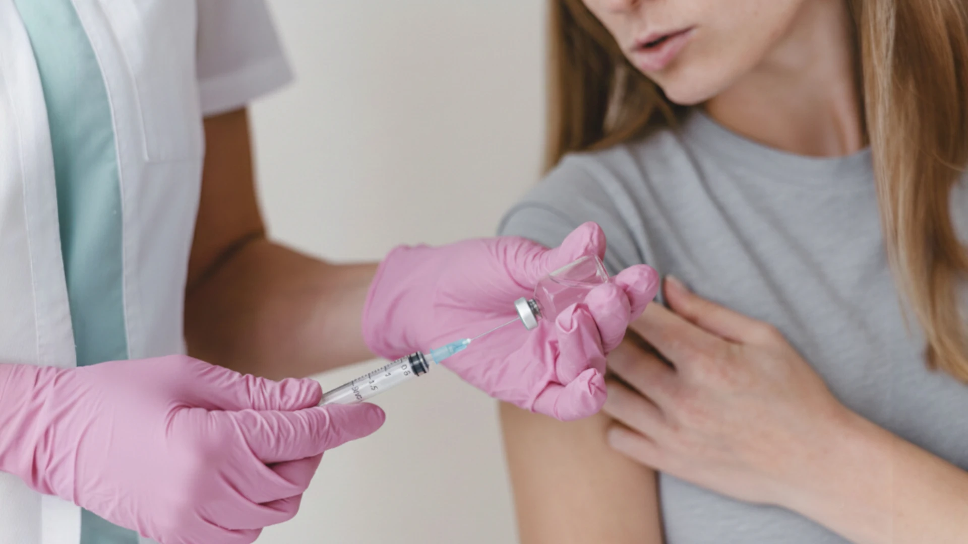
Population-wide vaccination is the most effective way to prevent measles. At least 95% of the population must be immune to prevent MeV transmission. Because MeV is highly contagious, even 5% of the population remaining unvaccinated can sustain periodic community outbreaks. Currently, first- and second-dose vaccine coverage in higher-income countries is estimated at 94% and 91%, respectively, compared with global coverage of 85% and 67%. In 2025, 84% of children worldwide (approximately 21 million) had received one dose, while 77% had received two doses.
The recommended two-dose vaccination schedule is as follows: the first dose is usually administered at 12–15 months of age, and the second at 4–6 years of age. During an outbreak or before travel to endemic areas, earlier vaccination at 6–11 months of age is acceptable.
The measles vaccine may be administered separately or combined with vaccines against mumps, rubella, and/or varicella.
A single dose of measles vaccine administered to children older than 12 months induces protective immunity in 95% of individuals. The second dose induces protective immunity in approximately 95% of the 5% who did not respond to the first dose. The vaccine induces both humoral and cell-mediated immunity. Vaccine-induced virus-neutralizing antibodies prevent the virus from entering target cells, while memory B and T cells provide long-term protection against subsequent infections. Although antibody titers gradually decline, routine revaccination later in life is not indicated.
3D Animation: Vaccination Against Measles, Rubella, and Mumps
Post-exposure Prophylaxis
In addition to routine vaccination, post-exposure prophylaxis (PEP) is used for secondary prevention of measles, particularly during outbreaks, and should be considered for susceptible contacts.
Human immunoglobulin (Ig) and measles-containing vaccine are used for measles PEP. The effectiveness of Ig ranges from 76% to 100%, while that of MMR vaccine ranges from 83.4% to 100%. Published evidence on the effectiveness of measles PEP is limited.
The timing of PEP is crucial. Administration of measles vaccine within 72 hours of exposure or Ig within 6 days of high-risk exposure may provide some protection against infection. It may also modify the clinical course of the disease and reduce the risk of complications.
Human Ig provides passive immunity and is indicated when vaccination is contraindicated:
immunocompromised patients;
infants aged 6–11 months;
infants younger than 6 months born to mothers without immunity to measles;
pregnant women.
Intramuscular Ig is administered at a dose of 0.5 mL/kg of body weight, up to a maximum of 15 mL. Intravenous Ig may be used in susceptible pregnant women and individuals with severe immunodeficiency, regardless of vaccination status. Ig is not indicated for individuals who received one dose of measles vaccine at 12 months of age or older, unless they have severe immunodeficiency.
Ig should not be used to control measles outbreaks. As an alternative to Ig, infants aged 6–11 months may receive the MMR vaccine. There are no recommendations for administering a third dose of MMR vaccine during measles outbreaks.
MMR vaccine and Ig should not be administered simultaneously because Ig may interfere with the immune response to the vaccine. After receiving intramuscular Ig, an exposed person should be vaccinated against measles according to the recommended schedule after 6 months. After intravenous Ig, vaccination should be delayed for 8 months. In either case, the person should be at least 12 months old at the time of vaccination and have no contraindications to vaccination.
Quarantine and Isolation
Timely contact tracing should be a priority. Patients with measles should be isolated for 4 days after rash onset, with the day of rash onset considered day 0. Immunocompromised patients should remain isolated for the entire duration of illness because of the potential for prolonged viral shedding.
In healthcare facilities, patients should preferably be placed in a single-occupancy, negative-pressure airborne infection isolation room (AIIR). Regardless of vaccination status or immunity, all healthcare personnel entering the room should wear an N95 respirator or a respirator providing equivalent protection.
Alternative strategies for the safer management of patients with suspected measles in clinics without an AIIR include:
examining the patient at the end of the day, after all other patients have been seen;
using a portable HEPA filtration system;
examining the patient in a room with a separate entrance, if available.
A room in which a patient with suspected measles has been examined should be disinfected and left unused for at least 2 hours.
FAQ
1. How does measles begin, and what are its first symptoms?
During the first 2–4 days, known as the prodromal period, measles may resemble a common cold. Typical symptoms include high fever, sometimes reaching 39–40 °C, cough, runny nose, and conjunctivitis. At this stage, characteristic small whitish spots known as Koplik spots may also appear on the buccal mucosa.
2. How long is the incubation period for measles?
The incubation period ranges from 7 to 21 days, with an average of 11–12 days. A person with measles is contagious from 4 days before rash onset through 4 days afterward.
3. How can measles be distinguished from chickenpox?
The two diseases have different rash morphologies. Measles causes a maculopapular rash that spreads in a characteristic top-to-bottom progression. Chickenpox causes a vesicular rash, including lesions on the scalp. New lesions continue to appear without following the same orderly pattern of spread.
4. Why is measles dangerous for children and adults?
Measles can cause severe bacterial complications, such as pneumonia, as well as serious neurologic complications, including measles encephalitis and subacute sclerosing panencephalitis. The disease tends to be most severe in children younger than 5 years, adults older than 20 years, pregnant women, and immunocompromised individuals.
5. How does the measles rash differ from the rashes of rubella and roseola infantum?
The rash of rubella is finer, does not usually become confluent, and does not show the characteristic pattern of progression seen in measles. In roseola infantum, which is caused by human herpesvirus 6 (HHV-6), the rash appears only after the fever has subsided. In measles, the rash appears while the fever is still present. Both conditions are included in the standard differential diagnosis of measles.
6. What is modified measles?
Modified measles is a form of the disease that may develop in previously vaccinated individuals or in people who have received post-exposure prophylaxis. The disease usually follows a milder course, with fewer complications. The rash may also lack the typical pattern of progression seen in classic measles.
7. Where is the risk of measles infection highest?
The risk is highest in regions with low vaccination coverage, including parts of Africa and the Eastern Mediterranean, and when traveling to countries experiencing ongoing outbreaks. MeV can remain infectious in the air and on surfaces for up to 2 hours after an infected person has left the room. Transmission can therefore occur even without direct contact.
8. What do measles IgM and IgG antibodies indicate in a blood test?
IgM antibodies appear shortly after rash onset and indicate acute measles infection. They may remain detectable for 30–60 days. IgG antibodies indicate previous infection or immunity acquired through vaccination. In immunocompromised patients with impaired antibody production, PCR is a more reliable method of confirming measles.
References
1.
VOKA 3D Anatomy & Pathology – Complete Anatomy and Pathology 3D Atlas. VOKA 3D Anatomy & Pathology.
Available from: https://catalog.voka.io
2.
World Health Organization. Measles — number of reported cases [Internet]. Geneva: World Health Organization.
Available from: https://www.who.int/data/gho/data/indicators/indicator-details/GHO/measles—number-of-reported-cases
3.
Immunize.org. Ask the experts about vaccines: MMR (Measles, mumps, and rubella) | Immunize.org [Internet]. Immunize.org. 2026.
Available from: https://www.immunize.org/ask-experts/topic/mmr/
4.
Burstain TL, Jacques D, Burstain JM. Association between first MMR vaccination before age 2 years and childhood autism in a U.S. EHR cohort of 2.5 million children. The Pediatric Infectious Disease Journal [Internet]. 2026 Jul 28.
Available from: https://doi.org/10.1097/inf.0000000000005349
5.
Au S, Saini S, Cruz WD, Venketaraman V. Measles: An updated literature review of the host response, pathogenesis, complications, prevention measures, and recent outbreaks. Current Issues in Molecular Biology [Internet]. 2026 Feb 13;48(2):206.
Available from: https://doi.org/10.3390/cimb48020206
50 Franklin Street, STE 203 Boston, MA 02110 United States
Thank you!
Your message is sent! Our experts will contact you shortly. If you have any additional questions, please contact us at info@voka.io
Cookie Consent
We use cookies to enhance your browsing experience, analyze site traffic, and deliver content. Please choose whether you accept all cookies or wish to reject non-essential tracking.
Cookie Preferences
Manage your cookie preferences below:
Essential cookies enable basic functions and are necessary for the proper function of the website.
Name
Description
Duration
Geolocation Config
This cookie is used to store the consent settings based on the visitor's location.
30 days
Cookie Preferences
This cookie is used to store the user's cookie consent preferences.
30 days
Google reCAPTCHA helps protect websites from spam and abuse by verifying user interactions through challenges.
Name
Description
Duration
_GRECAPTCHA
Google reCAPTCHA sets a necessary cookie (_GRECAPTCHA) when executed for the purpose of providing its risk analysis.
179 days
Statistics cookies collect information anonymously. This information helps us understand how visitors use our website.
Google Analytics is a powerful tool that tracks and analyzes website traffic for informed marketing decisions.
ID used to identify users for 24 hours after last activity
24 hours
_gat
Used to monitor number of Google Analytics server requests when using Google Tag Manager
1 minute
_gac_
Contains information related to marketing campaigns of the user. These are shared with Google AdWords / Google Ads when the Google Ads and Google Analytics accounts are linked together.
90 days
__utma
ID used to identify users and sessions
2 years after last activity
__utmt
Used to monitor number of Google Analytics server requests
10 minutes
__utmb
Used to distinguish new sessions and visits. This cookie is set when the GA.js javascript library is loaded and there is no existing __utmb cookie. The cookie is updated every time data is sent to the Google Analytics server.
30 minutes after last activity
__utmc
Used only with old Urchin versions of Google Analytics and not with GA.js. Was used to distinguish between new sessions and visits at the end of a session.
End of session (browser)
__utmz
Contains information about the traffic source or campaign that directed user to the website. The cookie is set when the GA.js javascript is loaded and updated when data is sent to the Google Anaytics server
6 months after last activity
__utmv
Contains custom information set by the web developer via the _setCustomVar method in Google Analytics. This cookie is updated every time new data is sent to the Google Analytics server.
2 years after last activity
__utmx
Used to determine whether a user is included in an A / B or Multivariate test.
18 months
_ga
ID used to identify users
2 years
_gali
Used by Google Analytics to determine which links on a page are being clicked
30 seconds
Clarity is a web analytics service that tracks and reports website traffic.