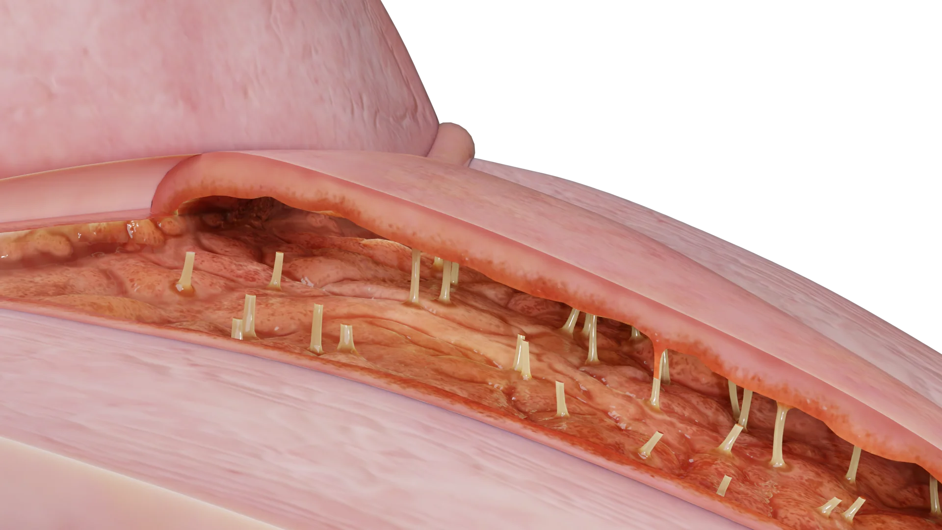
Vulvitis: Predisposing Factors, Clinical Manifestations, Diagnosis, and Treatment
Vulvitis refers to vulvar inflammation affecting the labia, clitoris, mons pubis, and vestibule of the vagina. Clinical manifestations, diagnosis, and treatment.
Anesthesia
Pain management and sedation techniques
Angiology
Arterial and venous pathologies
Cardiology
Acquired and congenital heart diseases
Dentistry
Diseases of teeth, gums, and the oral cavity
Dermatology
Disorders of the skin and subcutaneous tissue
Endocrinology
Disorders of the glands and hormonal imbalance
Gastroenterology
Stomach, intestinal, and digestive diseases
Gynecology
Diseases of female reproductive organs
Hematology
Hematopoiesis and blood-related disorders
Hepatology
Liver, gallbladder, and biliary tract diseases
Histology
Microscopic tissue and cell structures
Infectious diseases
Bacterial, viral, and parasitic infections
Neurology
Brain, spinal cord, and peripheral nerve disorders
Obstetrics
Pregnancy complications and abnormal fetal positions
Oncology
Cancer types, benign and malignant tumors
Ophthalmology
Conditions affecting the eyes and vision
Orthopedics
Bone, joint, and soft tissue disorders
Otorhinolaryngology
Ear, nose, and throat diseases
Pediatrics
Child health, development, and clinical conditions
Physiology
Biological processes within organs and systems
Pulmonology
Lung and respiratory tract diseases
Traumatology
Acute injuries and musculoskeletal trauma
Urology
Urinary tract and male reproductive disorders

This article is for informational purposes only
The content on this website, including text, graphics, and other materials, is provided for informational purposes only. It is not intended as advice or guidance. Regarding your specific medical condition or treatment, please consult your healthcare provider.
Uterine fibroids (also known as leiomyomas or myomas) are benign hyperplastic lesions of smooth muscle cells in the uterus or cervix.
The exact pathophysiology of uterine fibroids is unclear. Various studies indicate that the myoma initiating cell originates from one of the smooth muscle cells of the uterus (myometrium). This cell typically deviates from the normal signaling pathways of cell division.
As an estrogen-dependent tumor, uterine fibroids may also affect the estrogen and progesterone receptors, causing them to function abnormally compared to healthy myometrium.
A genetic pathology has also been identified, associated with mutations in genes regulating the growth of smooth muscle cells (MED12, HMGA2).
As it becomes larger, the myoma compresses the surrounding tissues (myometrium and connective tissue). This leads to a progressively growing pseudocapsule around the formation, which is rich in collagen fibers, nerve fibers, and blood vessels.
So far, cases of uterine fibroids have not been identified in girls before puberty. The condition is more likely to develop later in life with prevalence of up to 80 % during reproductive age. However, postmenopausal women are rarely affected.
Risk factors include:
At the same time, the odds of uterine fibroids are lower in women with
There is still no clear association between smoking and uterine fibroids, necessitating further research.
FIGO (International Federation of Gynecology and Obstetrics) Classification of Uterine Fibroids:
3D Models of Uterine Fibroids according to FIGO Classification:
 Type 0 — Pedunculated intracavitary
 Type 1 — < 50 % intramural
 Type 2 — ≥ 50 % intramural
 Type 3 — 100 % intramural, contacts endometrium but does not protrude into uterine
 Type 4 — 100 % intramural
 Type 5 — < 50 % protrudes into cavity of lesser pelvis
 Type 6 — ≥ 50 % above uterine subserous layer
 Type 7 — Subserous pedunculated, 100 % in cavity of lesser pelvis
Symptoms and management strategy typically depend on myoma localization. For this purpose, the FIGO classification system is used to categorize myomas into the following types:
A uterine myoma may be absolutely asymptomatic. In this case it may be detected accidentally during a routine imaging study.
General symptoms of uterine leiomyomas include:
These symptoms are commonly produced by submucous fibroids.
Less common symptoms include:
Myomatous nodes that deform the uterine cavity or grow on a peduncle inside the cavity may cause infertility. Such fibroids are generally recommended for surgical removal irrespective of their size or other symptoms.
Besides anemia and infertility, myomatous nodes may undergo degenerative changes and cause torsion of a subserosal node pedicle, leading to acute abdominal pain.
When choosing a treatment option for uterine fibroids, the following aspects should be considered: the patient’s age, symptoms (if any), their willingness to preserve fertility, and the experience of the healthcare professional. The exact technique is typically dictated by the location and size of the myoma.
Watchful waiting is preferred in women with asymptomatic fibroids. According to current guidelines, such patients do not require routine instrumental follow‑up.
Drug therapy aims to reduce bleeding and pain.
Find more scientifically accurate content on our social media


1. What is a uterine fibroid and what causes it?
2. What complications can uterine fibroids cause?
3. Can a woman with fibroids get pregnant?
4. Is a uterine fibroid a form of cancer?
5. How fast do uterine fibroids grow?
6. How is surgery for fibroid removal performed?
7. What is contradicted if you have uterine fibroids?
8. Why do uterine fibroids grow?
References
1.
VOKA 3D Anatomy & Pathology – Complete Anatomy and Pathology 3D Atlas [Internet]. VOKA 3D Anatomy & Pathology.
Available from: https://catalog.voka.io/
2.
American College of Obstetricians and Gynecologists. Uterine fibroids: diagnosis and treatment (Practice Bulletin No. 228). Obstet Gynecol. 2021;137(6):e100-e115. doi: 10.1097/AOG.0000000000004401.
3.
European Society of Human Reproduction and Embryology. Management of uterine fibroids (Guideline). Hum Reprod Open. 2023;2023(3):hoad028. doi: 10.1093/hropen/hoad028.
4.
NICE. Overview | Heavy menstrual bleeding: assessment and management | Guidance | NICE [Internet]. 2018.
Available from: https://www.nice.org.uk/guidance/ng88
5.
Bulun SE, et al. Uterine fibroids: mechanisms and pathogenesis. Semin Reprod Med. 2021;39(1-2):3–10. doi: 10.1055/s-0041-1730892.
6.
Stewart EA, et al. Uterine fibroids: from molecular pathogenesis to therapy. Nat Rev Dis Primers. 2022;8(1):43. doi: 10.1038/s41572-022-00373-9.
7.
Munro MG, et al. FIGO classification system for uterine fibroids. Int J Gynaecol Obstet. 2021;153(2):241-244. doi: 10.1002/ijgo.13761.
8.
Van den Bosch T, et al. Sonographic classification and reporting system for uterine fibroids. Ultrasound Obstet Gynecol. 59(3), pp. 409–416, 2022. doi: 10.1002/uog.24794.
9.
Donnez J, Dolmans MM. Uterine fibroid management: from the present to the future. Hum Reprod Update. 2023;29(6):715–739. doi: 10.1093/humupd/dmad012.
10.
Al-Hendy A, et al. Treatment of uterine fibroids: current and future options. Womens Health (Lond). 2022;18:17455057221111360. doi: 10.1177/17455057221111360.
11.
Laughlin-Tommaso SK, et al. Long-term outcomes after uterine artery embolization: focus on patient-centered outcomes. Fertil Steril. 114(5), pp. 944–951, 2020. doi: 10.1016/j.fertnstert.2020.07.026.
12.
Tropeano G, et al. Focused ultrasound surgery for fibroids: a systematic review and meta-analysis. J Minim Invasive Gynecol. 30(1), pp. 5–15, 2023. doi: 10.1016/j.jmig.2022.09.003.
Make VOKA your preferred source
See more VOKA articles in Google Search
Table of Contents


Summarize article with AI
Choose your preferable AI assistant:
Link successfully copied to clipboard
Thank you!
Your message is sent!
Our experts will contact you shortly. If you have any additional questions, please contact us at info@voka.io