Vulvitis: Predisposing Factors, Clinical Manifestations, Diagnosis, and Treatment
Vulvitis refers to vulvar inflammation affecting the labia, clitoris, mons pubis, and vestibule of the vagina. Clinical manifestations, diagnosis, and treatment.
Anesthesia
Pain management and sedation techniques
Angiology
Arterial and venous pathologies
Cardiology
Acquired and congenital heart diseases
Dentistry
Diseases of teeth, gums, and the oral cavity
Dermatology
Disorders of the skin and subcutaneous tissue
Endocrinology
Disorders of the glands and hormonal imbalance
Gastroenterology
Stomach, intestinal, and digestive diseases
Gynecology
Diseases of female reproductive organs
Hepatology
Liver, gallbladder, and biliary tract diseases
Neurology
Brain, spinal cord, and peripheral nerve disorders
Obstetrics
Pregnancy complications and abnormal fetal positions
Oncology
Cancer types, benign and malignant tumors
Ophthalmology
Conditions affecting the eyes and vision
Otorhinolaryngology
Ear, nose, and throat diseases
Pediatrics
Child health, development, and clinical conditions
Pulmonology
Lung and respiratory tract diseases
Traumatology
Acute injuries and musculoskeletal trauma
Urology
Urinary tract and male reproductive disorders

This article is for informational purposes only
The content on this website, including text, graphics, and other materials, is provided for informational purposes only. It is not intended as advice or guidance. Regarding your specific medical condition or treatment, please consult your healthcare provider.
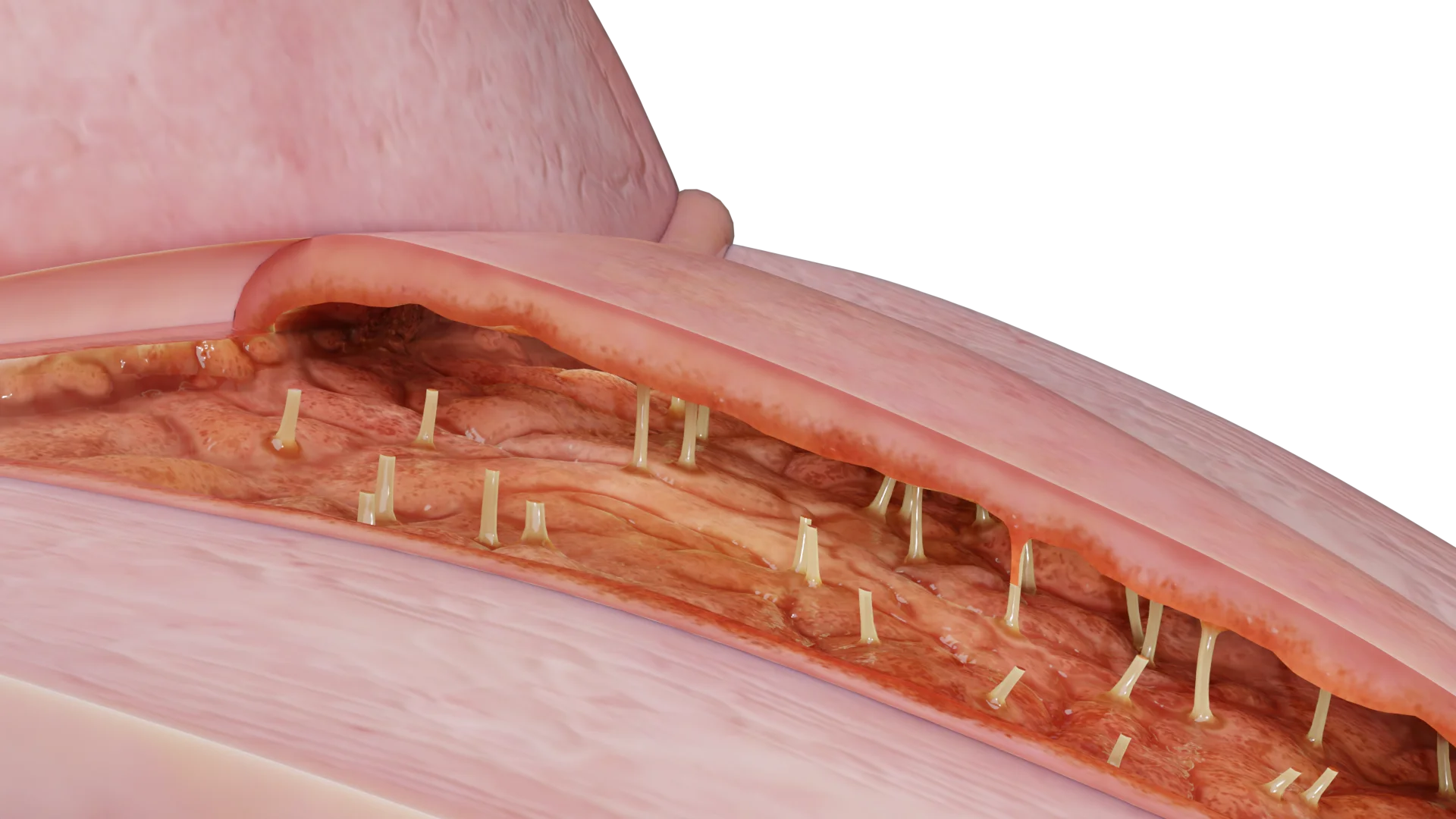
Uterine myoma (or leiomyoma) is a benign hyperplastic lesion of the smooth muscle cells of the uterus or cervix.
The exact pathophysiology of uterine myoma development remains unclear. Studies show that the first myoma cell develops from a single uterine smooth muscle cell (myometrium) that is characterized by deviation from normal cell division signaling pathways.
Myoma is an estrogen-dependent tumor, with the potential to alter estrogen and progesterone receptors compared to the normal surrounding myometrium.
A genetic pathology associated with a mutation in genes regulating smooth muscle cell growth (MED12, HMGA2) has also been identified.
During its growth, myoma squeezes the surrounding structures (myometrium and connective tissue), causing progressive formation of a pseudocapsule rich in collagen fibers, neurofibers, and blood vessels.
No cases of uterine myoma have been described in pre-pubertal girls. The probability of the disease increases with age and may reach 80% during the reproductive years, with a decreasing incidence at menopause.
Risk factors for developing myoma include:
The risk of developing uterine myoma is reduced in women with:
The effect of smoking on myoma development remains unclear, and more research is needed.
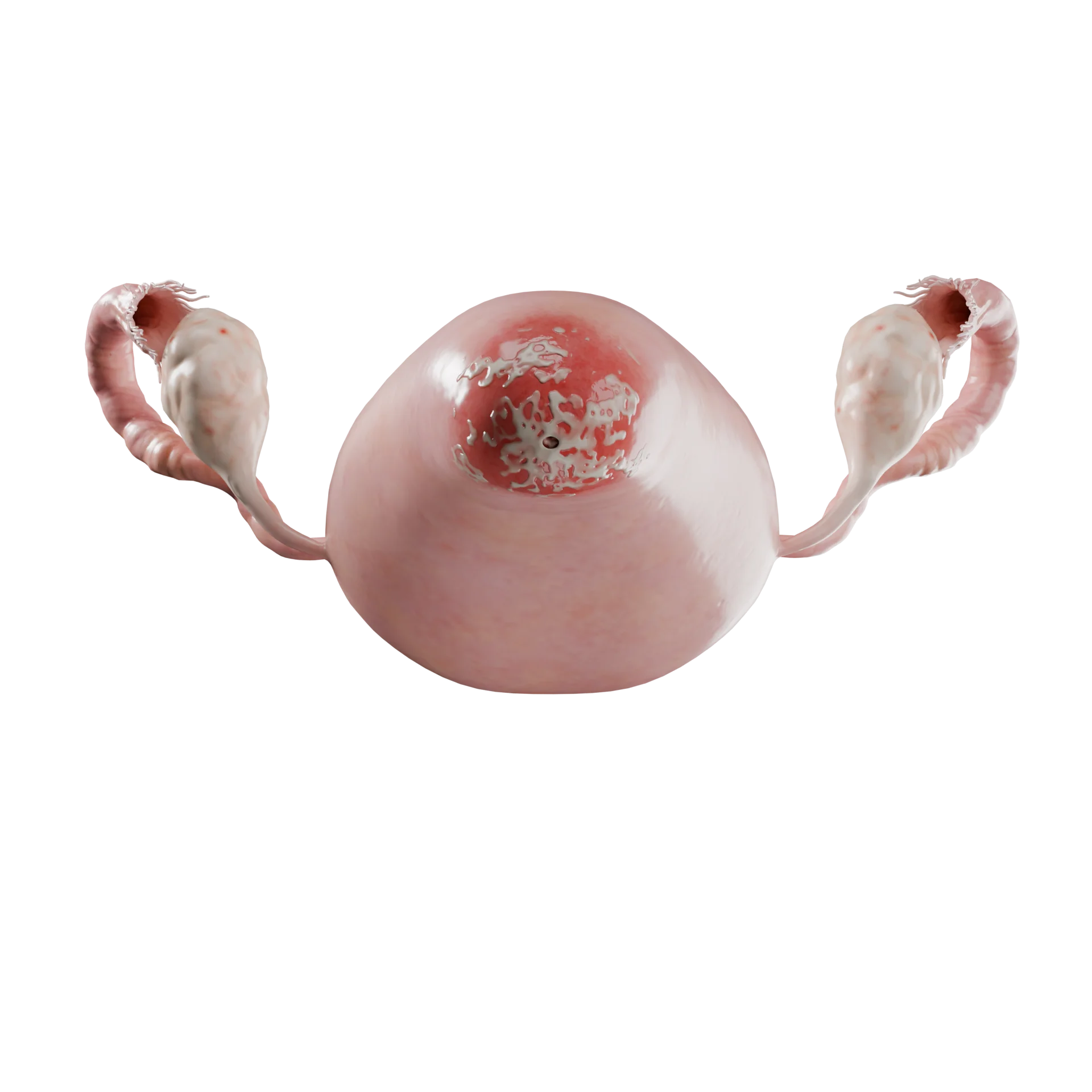
FIGO classification of uterine myomas:
3D models of uterine myoma types according to the FIGO classification:
 Type 0 – the pedicle node is located in the uterine cavity
 Type 1 – less than 50% of the node is located intramurally
 Type 2 – more than 50% of the node is located in the muscle layer
 Type 3 – nodule located in the myometrium, the edge is adjacent to the endometrium, but does not extend into the uterine cavity
 Type 4 – myoma located entirely in the myometrial thickness
 Type 5 – less than 50% of the node protrudes into the pelvic cavity
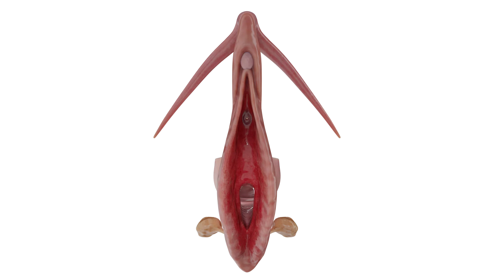
 Type 6 – more than 50% of the node is located above the serous layer of the uterus
 Type 7 – subserosal nodule on a pedicle, completely located in the pelvic cavity
The localization of myoma affects not only the presence of symptoms, but also the tactics of treatment. For this purpose, myomatous nodes are divided into:
Uterine myoma can exist completely asymptomatic and be an incidental finding with any imaging modality.
Common symptoms of uterine leiomyoma include:
These symptoms are characteristic of myomas with a submucosal component.
Less common symptoms include:
Myomatous nodes can be a cause of infertility, especially those that deform the uterine cavity or are located on a pedicle in the cavity.Such myomas are to be treated surgically regardless of size and the presence of other symptoms.
In addition to anemia and infertility, myomatous nodes can be complicated by degenerative processes and torsion of the stem of the subserosal node with the development of a clinical picture of acute abdomen.
When choosing uterine myoma treatment options, it is important to consider the patient’s age, present symptoms, desire to preserve fertility, and the experience of the physician. The location and size of the myoma will determine the treatment options available.
Surveillance: this is the preferred method for women with asymptomatic myomas. Current recommendations do not require periodic surveillance using imaging techniques for female patients.
Drug treatment is primarily aimed at reducing the severity of bleeding and pain symptoms.
Find more scientifically accurate content on our social media


1. What is a uterine myoma and what causes it?
2. What are the dangers of uterine myoma?
3. Can I get pregnant with a uterine myoma?
4. Is uterine myoma a cancer or not?
5. How fast does a uterine myoma grow?
6. How is uterine myoma surgery performed?
7. What should not be done with uterine myoma?
8. Why does a uterine myoma grow?
List of Sources
1.
VOKA Catalog.
https://catalog.voka.io/
2.
ACOG (American College of Obstetricians and Gynecologists). (2021). *Uterine Fibroids: Diagnosis and Treatment* (Practice Bulletin No. 228). *Obstetrics & Gynecology, 137*(6), e100-e115. [DOI: 10.1097/AOG.0000000000004401].
3.
ESHRE (European Society of Human Reproduction and Embryology). (2023). *Management of uterine fibroids* (Guideline). *Human Reproduction Open, 2023*(3). [DOI: 10.1093/hropen/hoad028].
4.
NICE (National Institute for Health and Care Excellence). (2021). *Heavy menstrual bleeding: assessment and management* (NG88).
https://www.nice.org.uk/guidance/ng88
5.
Bulun, S. E., et al. (2021). *Uterine fibroids: mechanisms and pathogenesis*. *Seminars in Reproductive Medicine, 39*(1-02), 3-10. [DOI: 10.1055/s-0041-1730892].
6.
Stewart, E. A., et al. (2022). *Uterine fibroids: from molecular pathogenesis to therapy*. *Nature Reviews Disease Primers, 8*(1), 43. [DOI: 10.1038/s41572-022-00373-9].
7.
Munro, M. G., et al. (2021). *FIGO classification system for uterine fibroids*. *International Journal of Gynecology & Obstetrics, 153*(2), 241-244. [DOI: 10.1002/ijgo.13761].
8.
Van den Bosch, T., et al. (2022). *Sonographic classification and reporting system for uterine fibroids*. *Ultrasound in Obstetrics & Gynecology, 59*(3), 409-416. [DOI: 10.1002/uog.24794].
9.
Donnez, J., & Dolmans, M. M. (2023). *Uterine fibroid management: from the present to the future*. *Human Reproduction Update, 29*(6), 715-739. [DOI: 10.1093/humupd/dmad012].
10.
Al-Hendy, A., et al. (2022). *Treatment of uterine fibroids: current and future options*. *Women’s Health, 18*, 174550572211113. [DOI: 10.1177/17455057221111360].
11.
Laughlin-Tommaso, S. K., et al. (2020). *Long-term outcomes after uterine artery embolization: focus on patient-centered outcomes*. *Fertility and Sterility, 114*(5), 944-951. [DOI: 10.1016/j.fertnstert.2020.07.026].
12.
Tropeano, G., et al. (2023). *Focused ultrasound surgery for fibroids: a systematic review and meta-analysis*. *Journal of Minimally Invasive Gynecology, 30*(1), 5-15. [DOI: 10.1016/j.jmig.2022.09.003].
Loading test 6 questions
Table of Contents


Summarize article with AI
Choose your preferable AI assistant:
Link successfully copied to clipboard
Thank you!
Your message is sent!
Our experts will contact you shortly. If you have any additional questions, please contact us at info@voka.io