Breast Cancer: Causes, Symptoms, Classification, Diagnosis, Treatment and Prognosis
Svetlana D.Surgical oncologist, MD
38 min read·December 11, 2025
This article is for informational purposes only
The content on this website, including text, graphics, and other materials, is provided for informational purposes only. It is not intended as advice or guidance. Regarding your specific medical condition or treatment, please consult your healthcare provider.
Breast cancer is the most commonly diagnosed malignancy and the second most common cause of death from malignancy in women. Among men, this tumor is extremely rare (0.5–1% of total cases).
According to WHO, globally in 2022, breast cancer was diagnosed in 2.3 million women and caused 670,000 deaths.
This malignancy is reported in all countries of the world, with a significantly higher incidence in countries with a high Human Development Index (HDI) compared to countries with a low HDI (1:12 vs. 1:27), but a lower mortality rate (1:71 vs. 1:48).
The risk factors for breast cancer currently include:
Female sex;
Age: the disease is more often detected in more mature age (perimenopausal and menopausal period);
Family history and presence of genetic mutations — carrying a mutation in the BRCA1 and BRCA2 genes is most important;
Early onset of menarche (before age 12);
First childbirth after age 30 or nulliparity;
Late menopause (after age 55);
Obesity;
Alcohol and tobacco abuse;
History of exposure to ionizing radiation.
Pathogenesis
The pathogenesis of breast cancer is not fully understood. A combination of genetic and environmental risk factors, dyshormonal disorders, play a role in oncogenesis.
Currently, a high probability of breast cancer development in patients with mutations in BRCA1, BRCA2, PALB-2 genes has been proved. In addition, the association of breast cancer with mutations in STK11, CHEK2, CDH1, and PTEN genes has been identified. The vast majority of mutations (up to 90–95%) are sporadic; only in 5–10% of patients they are inherited.
Dyshormonal disorders such as imbalance of progesterone and estrogen levels, thyroid disorders, obesity, and other endocrine diseases lead to hyperplasia of glandular epithelium and in combination with other risk factors cause the development of tumor pathology.
Classification of breast cancer
By histologic structure
Depending on the histological structure, the following types of malignant breast tumors are distinguished:
Epithelial tumors
Invasive carcinoma unspecified;
Pleomorphic carcinoma;
Invasive lobular carcinoma
Tubular carcinoma;
Cribriform carcinoma;
Mucinous carcinoma;
Medullary carcinoma;
Invasive micropapillary carcinoma;
Metaplastic carcinoma unspecified;
Squamous cell carcinoma;
Spindle cell carcinoma;
Mixed metaplastic carcinoma;
Myoepithelial carcinoma;
Invasive papillary carcinoma;
Acinic cell carcinoma;
Mucosa-epidermoid carcinoma;
Polymorphic carcinoma.
Non-invasive tumors
Ductal carcinoma in situ;
Classic lobular carcinoma in situ;
Pleomorphic lobular carcinoma in situ.
Mesenchymal tumors
Liposarcoma;
Angiosarcoma;
Rhabdomyosarcoma;
Osteosarcoma;
Leiomyosarcoma.
Fibroepithelial tumors
Phyllodes tumor with malignization.
Nipple tumors
Paget’s disease of the nipple.
Breast tumors in men:
Invasive carcinoma;
Carcinoma in situ.
In addition, the inflammatory breast cancer is separately distinguished.
By receptor status and proliferative activity
Depending on the presence of estrogen, progesterone, HER2/neu receptors in the tumor and the rate of cell division, the following subtypes of breast cancer are distinguished:
Luminal A. This type includes tumors that have receptors for estrogen and progesterone or estrogen alone, are HER2-negative, and have a Ki67 proliferative activity index of less than 20%.
Luminal B. This type includes tumors with a Ki-67 index greater than 20%, retaining estrogen and progesterone receptors, HER2 status can be either positive or negative.
HER2-positive. This type includes HER2-positive tumors that lack receptors for estrogen and progesterone.
Triple-negative. Estrogen and progesterone-negative, HER2-negative.
Luminal A and luminal B subtypes of breast cancer develop from the epithelial cells of the ducts and lobules, clinically, tend to have a more favorable prognosis compared to other subtypes.
Triple-negative subtype is characterized by the most aggressive course, early metastasis, but at the same time it is the most sensitive to chemotherapy. That, if timely treatment is started, helps to achieve a good response to therapy and persistent remission in numerous patients.
TNM classification and staging of breast cancer
TNM classification is used to evaluate the primary tumor (category T), the status of regional lymph nodes (category N), the presence of distant metastases (category M) and further staging of the tumor process prevalence based on the criteria obtained.
There is a clinical classification (cTNM), where categories are determined based on clinical findings, and a pathologic classification (pTNM), where categories are established based on histologic findings.
Primary tumor (T)
Tx — primary tumor could not be evaluated;
T0 — primary tumor is undetectable;
Tis (DCIS) — ductal carcinoma in situ;
Tis (LCIS) — lobular carcinoma in situ;
Tis (Paget) — Paget’s disease of the nipple in situ;
T1 — tumor up to 2 cm in diameter:
T1mi — tumor up to 1 mm in diameter;
T1a — tumor 1 to 5 mm in diameter;
T1b — tumor 5 to 10 mm in diameter;
T1c — tumor 10 to 20 mm in diameter.
T2 — tumor is 2 to 5 cm in diameter;
T3 — tumor is more than 5 cm in diameter;
T4 — tumor of any size with direct spread to the chest wall and/or skin (but not isolated to the dermis):
T4a — spread to the chest wall (but not isolated invasion of the pectoral muscles);
The cT and pT categories correspond to each other.
Regional lymph nodes (N)
Clinical classification (cN):
cNx — regional lymph nodes could not be evaluated;
cN0 — metastases in regional lymph nodes are not determined;
cN1 — metastases in ipsilateral axillary lymph nodes of levels I-II:
cN1mi — micrometastases (larger than 0.2 mm but less than 2.0 mm).
cN2 — metastases in ipsilateral axillary lymph nodes of level I–II fused to each other or isolated metastasis in an intramammary lymph node on the side of the lesion:
cN2a — metastases in ipsilateral axillary lymph nodes of level I–II fused to each other;
cN2b — isolated metastasis in the ipsilateral intramammary lymph node.
сN3 – наличие метастазов в ипсилатеральных подключичных лимфоузлах (лимфоузлы III уровня), или наличие интрамаммарных ипсилатеральных метастазов и метастазов в подмышечных лимфоузлах I-II уровня, или наличие метастазов в ипсилатеральных надключичных лимфоузлах:
cN3a — presence of metastases in ipsilateral subclavian lymph nodes;
cN3b — presence of metastases in ipsilateral intramammary and axillary lymph nodes of level I-II;
cN3c — presence of metastases in ipsilateral supraclavicular lymph nodes.
Pathologic Classification (pN):
pNx — regional lymph nodes could not be evaluated;
pN0 — metastases in regional lymph nodes are not determined:
pN0(i+) — morphological examination reveals only isolated tumor cells (ITCs) — individual tumor cells or their clusters no larger than 0.2 mm;
pN0(mol+) — metastases were not detected by morphological examination, ITCs were detected by non-morphological methods (PCR).
pN1 – micrometastases or metastases in 1–3 ipsilateral axillary lymph nodes and/or metastases in intramammary lymph nodes according to sentinel lymph node biopsy but not clinically detectable:
pN1mi — micrometastases (larger than 0.2 mm but less than 2.0 mm);
pN1a — metastases in 1–3 ipsilateral axillary lymph nodes (at least one of them more than 2.0 mm in diameter);
pN1b — metastases (excluding ITCs) to sentinel intramammary lymph nodes;
pN1c — combination of pN1a and pN1b criteria.
pN2 — metastases in 4–9 axillary lymph nodes or clinically detectable metastases in intramammary lymph nodes in the absence of metastases in axillary lymph nodes:
pN2a — metastases in 4–9 axillary lymph nodes (at least one of which is greater than 2.0 mm);
pN2b — clinically detectable metastases in intramammary lymph nodes in the absence of metastases in axillary lymph nodes.
pN3 — metastases in 10 or more axillary lymph nodes, or metastases in subclavian lymph nodes, or clinically detectable metastases in ipsilateral intramammary lymph nodes in the presence of metastases in axillary lymph nodes of levels I–II, or more than 3 metastases in axillary lymph nodes in the presence of metastases in sentinel intramammary lymph nodes (but not clinically detectable), or metastases in supraclavicular lymph nodes:
pN3a — metastasis to 10 or more axillary lymph nodes (at least one of which is greater than 2.0 mm in diameter), or metastasis to subclavian lymph nodes;
pN3b — clinically detectable metastases in ipsilateral intramammary lymph nodes in the presence of metastases in axillary lymph nodes of levels I-II, or more than 3 metastases in axillary lymph nodes in the presence of metastases in sentinel intramammary lymph nodes (but not clinically detectable);
pN3c — metastasis to supraclavicular lymph nodes.
Distant metastases (category M)
M0 — no distant metastases were detected;
M1 — distant metastases are identified.
The cM and rM categories correspond to each other.
Breast cancer staging
Stage
Т
N
М
0
Tis
N0
M0
IA
T1 (including T1mi)
N0
M0
IB
T0, T1 (including T1mi)
N1mi
M0
IIA
T0, T1 (including T1mi) T2
N1
N0
M0
IIB
T2 T3
N1 N0
M0
IIIA
T0, T1 (including T1mi), T2 T3
N2
N1, N2
M0
IIIB
T4
N0, N1, N2
M0
IIIC
any T
N3
M0
IV
any T
any N
M1
Clinical presentation of breast cancer
Symptoms and localization of breast cancer
At the initial stages, when the size of the primary tumor reaches no more than 1–2 cm, the disease is usually asymptomatic and is detected only through screening methods of examination (ultrasound, mammography).
The presence of a palpable mass is the most common symptom of breast cancer. The tumor is characterized by a round shape, dense consistency, and is usually painless on palpation. The size of the mass can vary from a few millimeters to tens of centimeters. Giant tumor size is typical for malignant phyllodes tumor, breast sarcomas, and locally advanced breast cancer. Small tumor sizes are typical for lobular breast cancer, Paget’s disease.
Tumors can be solitary or multiple, localized in one breast or affect both.
In the early stages, the neoplasm is easily displaced relative to the surrounding tissues; the skin covering the mass is not changed. When the tumor process spreads, the so-called “lemon peel” symptom occurs — changes in the skin (swelling, thickening, more pronounced pores — the appearance of the skin resembles a lemon peel, hence the name of the symptom) and underlying tissues due to lymphostasis caused by blockage of lymphatic pathways by tumor masses.
The following symptoms are also diagnosed with breast cancer:
“Tethering” sign — when you try to take the skin over the tumor into a fold, a depression appears;
Umbilication — retraction of the skin over the tumor;
Skin ulceration above the tumor is possible when it invades with the development of bleeding of varying degrees of intensity, as well as infection of the wound with the development of purulent-septic complications.
Palpable mass of dense consistency in the axillary region, supraclavicular region indicates the spread of the tumor process to the regional lymph nodes. Metastases in lymph nodes can be single or multiple, can be located separately or form tumor conglomerates.
The clinical presentation of breast cancer is diverse and depends on many factors such as morphological type of tumor, tumor localization, tumor stage, and physiological features of the patient.
Clinical forms of breast cancer
The following breast cancer variants are of greatest clinical significance:
Non-invasive ductal cancer, or ductal carcinoma in situ (DCIS)
It is the most common variant of non-invasive breast cancer. DCIS develops from the ductal lobular unit epithelium and may spread along the ductal system down to the nipple, but either the basal membrane or the myoepithelial cell layer remains intact. DCIS may be a precursor to invasive ductal carcinoma, making its detection and treatment of great prognostic value.
DCIS is mostly asymptomatic and is detected by screening mammography (70–90% of all cases).
The symptoms typical for ductal carcinoma in situ include:
Presence of bloody, sometimes brown discharge from the nipple;
Palpable tumor mass in the retroareolar region (occurs in 1–5% of cases).
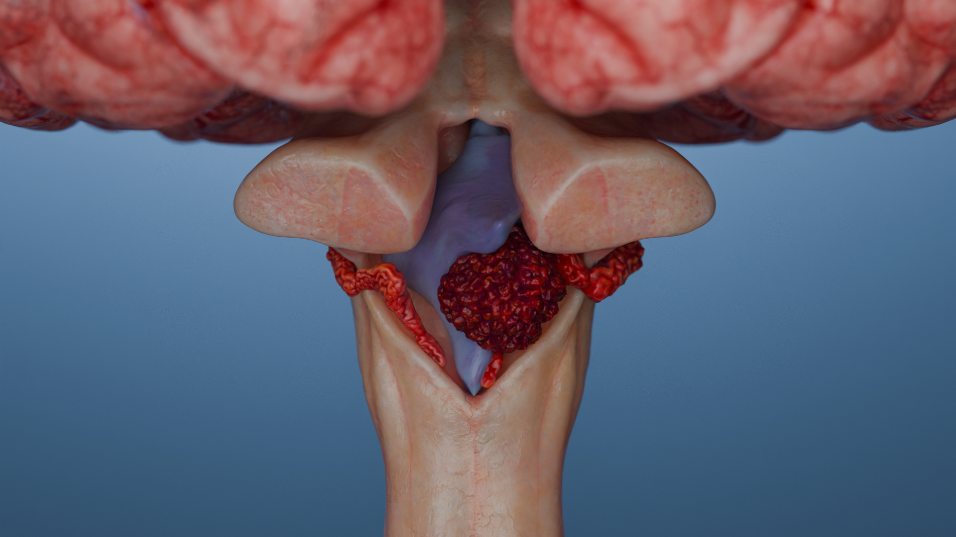
3D animation: ductal carcinoma in situ
Invasive ductal cancer
Invasive ductal cancer accounts for 50 to 75% of all invasive breast cancers.
It is clinically manifested by the presence of a palpable tumor mass in the breast of dense consistency, usually painless. When the tumor process spreads, the skin symptoms described above are joined.
Non-invasive lobular cancer
Non-invasive lobular cancer, or lobular carcinoma in situ, is often multicenter and can be localized as multiple foci in either one breast (up to 70%) or both (in 20–60% of patients).
Clinically, it is usually asymptomatic and is detected by screening mammography.
3D animation: lobular cancer in situ
Invasive lobular cancer
Invasive lobular cancer accounts for about 10–15% of all invasive breast cancers.
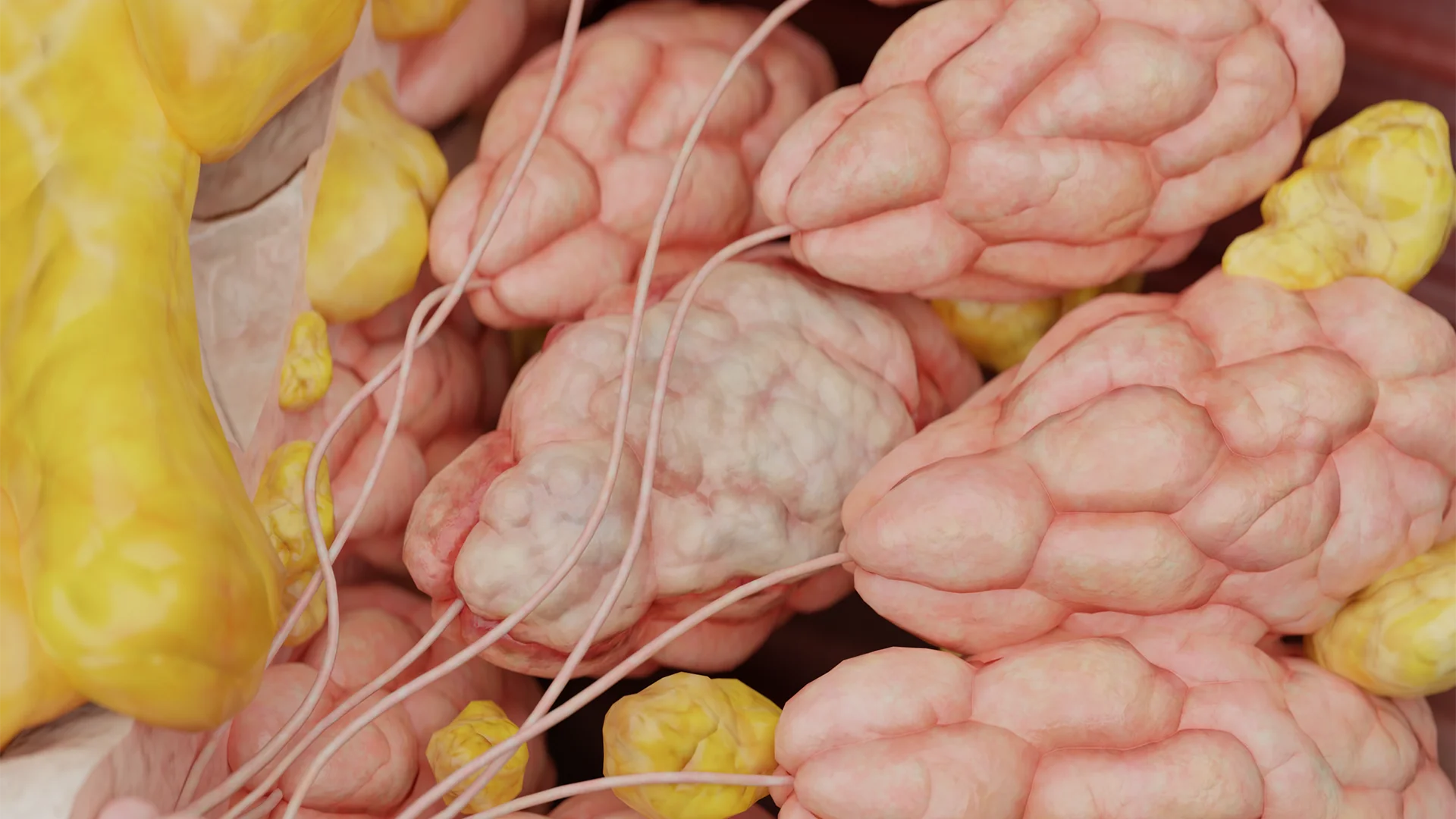
Invasive lobular cancer T2: 3D model
It may be asymptomatic for a long time due to the small size of the tumor, and the first symptoms may be enlargement of axillary lymph nodes due to metastatic lesions. Invasive lobular cancer is the most common cause of multicenter breast cancer (either one or both breasts may be affected).
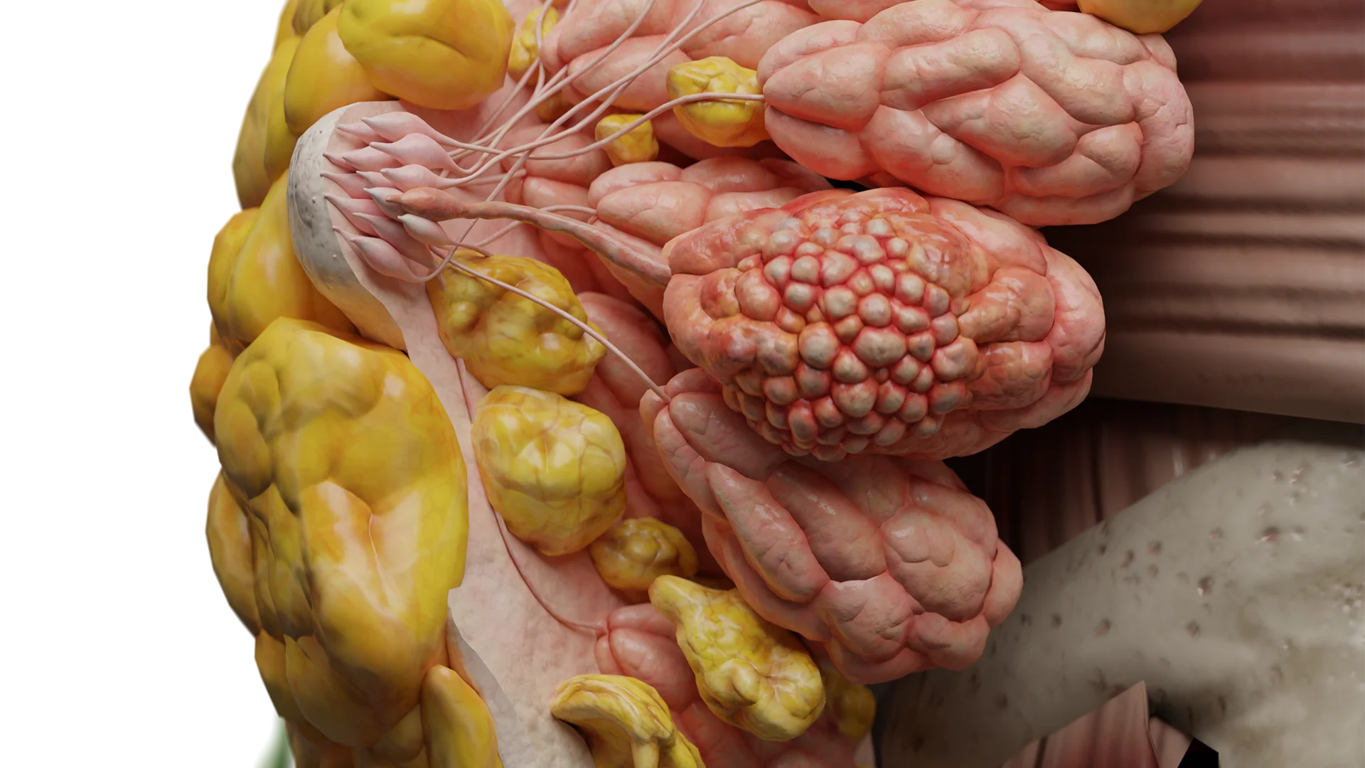
3D animation: invasive lobular cancer
Paget’s disease of the nipple
Paget’s disease of the nipple is a relatively rare form of breast cancer in which the skin of the areolar area and nipple is affected, usually combined with ductal carcinoma in situ or invasive ductal carcinoma (in 80–90%), but can be isolated.
Presence of palpable dense tumor in the retroareolar area.
3D animation: Paget’s disease of the nipple
Inflammatory breast cancer
Inflammatory breast cancer is a relatively rare variant of breast cancer characterized by a rapid aggressive course with whole breast involvement and early metastasis.
Unlike other forms of breast cancer in the inflammatory variant, usually there is no palpable tumor.
The main symptoms of inflammatory breast cancer are:
Swelling, redness of the skin of the breast;
Breast deformity;
Nipple retraction;
Prominent lemon peel symptom.
This variant of breast cancer requires differential diagnosis with infectious breast lesions.
3D animation: inflammatory breast cancer
Breast sarcomas
Breast sarcomas are rare (about 1%) mesenchymal tumors of the breast characterized by an aggressive course, rapid tumor growth, and early, predominantly hematogenous, metastasis.
Breast sarcoma: 3D model
The development of breast sarcoma is due to both genetic developmental anomalies (Li-Fraumeni syndrome, neurofibromatosis type 1, familial adenomatous polyposis) and the impact of external factors (prolonged contact with arsenic compounds, vinyl chloride, alkylating agents, the consequences of radiation therapy to the chest area in the treatment of other malignant neoplasms).
The main symptom of breast sarcoma is the presence of a rapidly growing dense tumor of the breast, which can reach giant size with the formation of areas of tumor necrosis, skin ulceration, invading surrounding tissues with the development of pain syndrome and erosive bleeding (in the absence of timely surgical treatment).
3D animation: breast sarcoma
Malignant phyllodes tumor
Malignant phyllodes tumor is a rare fibroepithelial tumor characterized by rapid growth, aggressive course and early, mainly hematogenous, metastasis. It can be either a primary tumor or develop from a phyllodes fibroadenoma of the breast.
Malignant phyllodes tumor, as well as breast sarcomas, due to low sensitivity of the tumor to chemoradiation treatment requires aggressive surgical treatment (mastectomy) followed by regular follow-up.
3D animation: malignant phyllodes tumor of the breast
Diagnosis of breast cancer
History and physical examination
When taking a medical history, please note that identifying breast cancer in immediate blood relatives is crucial for recognizing familial forms of the disease. Mandatory genetic counseling is indicated for such patients and their relatives.
The initial examination includes examination and palpation of the breast and areas of regional metastasis (axillary, supraclavicular areas).
Examination begins in a standing position:
Symmetry of the breasts, mobility when raising and lowering the arms is assessed;
Skin changes are checked: edema, hyperemia, skin retraction, ulceration;
Nipple retraction, nipple discharge are evaluated.
Palpation:
In standing and supine position, starting from the upper quadrants, clockwise, both breasts are palpated to assess the presence of discharge from the nipple when pressing.
Next, the axillary and supraclavicular lymph nodes are palpated.
Breast examination and palpation is recommended for all women as a self-examination monthly on the same day of the menstrual cycle (optimally in the 1st phase of the cycle, on day 7–10).
Mammography
Mammography is the primary method of diagnosing breast cancer, especially for non-invasive forms of breast cancer. Radiologic signs:
For intraductal cancer in situ, the typical radiologic feature is the presence of grouped microcalcifications. When performing ductography, the main signs are ductal deformation and the symptom of “amputation” (abrupt interruption of the duct due to tumor obstruction).
The main radiological sign of infiltrating breast cancer is the presence of a mass of irregular shape without clear boundaries, heterogeneous structure, high density (above the breast tissue), with the presence of streaking and clusters of microcalcifications in the tumor and adjacent areas, skin edema.
Ultrasound
Ultrasound, along with mammography, is among the main methods of imaging breast tumors.
The main ultrasound signs of malignant breast neoplasm are:
Presence of a hypoechoic mass with indistinct irregular boundaries;
Heterogenous structure that may give a dorsal acoustic shadow;
Vertical dimension is usually equal to the horizontal dimension;
Presence of blood flow on Doppler study.
Magnetic Resonance Imaging (MRI)
Magnetic resonance imaging (MRI) is used as a method of clarifying diagnosis.
MRI is recommended as an annual screening method for patients at high risk of developing breast cancer (carriers of genetic mutations, particularly BRCA1 and BRCA2).
In addition, MRI allows visualization of intraductal breast tumors without calcifications, which is challenging for ultrasound and mammography.
Pathognomonic symptoms of breast cancer on MRI are:
Indistinct boundaries of the tumor;
Pointed edges of the tumor;
Uneven contrast accumulation.
Fine-needle aspiration biopsy
Fine-needle aspiration (FNA) biopsy is mainly used to verify metastases in regional lymph nodes. FNA biopsy of breast tumors is currently less preferred than core needle biopsy due to the need to determine the morphological type of the tumor prior to specific treatment and is rarely performed.
Core needle biopsy
Core needle biopsy of a breast tumor allows to determine the morphological type of the tumor, which allows planning the tactics of special treatment individually with the greatest predictable effect.
Core needle biopsy with determination of the receptor status of the tumor (presence of receptors to estrogen and progesterone, HER2/neu, and Ki-67) is performed to decide on neoadjuvant chemotherapy and is mandatory before starting specific treatment.
Basic principles of breast cancer treatment
Treatment of breast cancer is based on the determination of the morphological type of the tumor, its sensitivity to this or that type of therapy, the prevalence of the tumor process. It includes surgery, chemotherapy, hormone therapy, targeted therapy, radiation therapy.
Surgical therapy
Surgical treatment is one of the main stages of therapy of malignant breast neoplasms. It can be performed in the first stage of special treatment or after neoadjuvant chemotherapy courses.
Surgical treatment is indicated for all patients with resectable tumor process in the absence of distant metastases. Patients with advanced breast cancer may undergo palliative mastectomy (if there is a threat of bleeding, for sanitary reasons).
Currently, the gold standard for surgical treatment of breast cancer is curative resection (lumpectomy) with biopsy of the sentinel lymph node in the presence of clinically undetectable metastases or with axillary lymph node dissection in the presence of clinically detectable metastases.
When performing breast-conserving surgery, intraoperative morphological examination of the resection margins for the presence of tumor is mandatory. In case of a positive margin, re-dissection is indicated until a negative response is obtained or mastectomy is performed (when a negative margin is technically impossible to achieve).
In patients with multicenter lesions, diffuse microcalcifications in the breast, and inflammatory breast cancer, mastectomy with sentinel lymph node biopsy or axillary lymph node dissection is indicated.
In the presence of mutation carriers in BRCA1 and BRCA2 genes in patients with diagnosed breast cancer, it is reasonable to consider performing bilateral mastectomy due to the high risk of cancer development in the second breast.
Reconstructive breast surgeries can be performed both simultaneously with tumor removal and delayed after completion of specialized treatment. Currently, there is no evidence of an association between the risk of disease recurrence and the timing of reconstructive surgery, therefore, one-stage mammoplasty can be recommended to improve the quality of life of female patients. Delayed mammoplasty may be recommended when implants are needed and radiation therapy is planned in the postoperative period, as this increases the risk of a (fibro)capsular contracture.
Chemotherapy treatment
Chemotherapy can be performed both preoperatively (neoadjuvant therapy) and postoperatively (adjuvant therapy) to achieve better treatment outcomes.
Adjuvant chemotherapy
Administration of adjuvant chemotherapy (ACT) after curative surgical treatment is determined by the morphological type of the tumor and the extent of the tumor spread.
In luminal A type of tumor, in addition to hormone therapy, chemotherapy is prescribed in case of widespread tumor process (pT>=3, pN>=2), low grade of tumor differentiation (G3), patients younger than 35 years old with marked lymphovascular invasion and high risk of tumor recurrence. The standard regimen of ACT includes 4 courses of AC (doxorubicin+cyclophosphamide) and 6 courses of CMF (cyclophosphamide+metotrexate+fluorouracil).
In luminal B type and HER2-negative status, hormone therapy and ACT with anthracyclines (doxorubicin) and taxanes (paclitaxel, docetaxel) are indicated.
In luminal B type and HER2-positive status, ACT (anthracyclines, taxanes) as well as hormone therapy and anti-HER2/neu therapy are also indicated.
In non-luminal HER2-positive breast cancer, courses of chemotherapy with anthracyclines and taxanes are indicated in addition to anti-HER2/neu therapy.
In triple-negative breast cancer, courses of chemotherapy with anthracyclines and taxanes are also indicated. It is possible to use platinum preparations in patients with BRCA1 mutation.
Neoadjuvant chemotherapy
Neoadjuvant chemotherapy (NACT) in the primary resectable tumor process allows for improving the prognosis, creating conditions for performing breast-conserving surgeries, and, in some cases, achieving complete pathomorphological regression of the tumor; in the primary non-resectable process, in the case of a good tumor response to treatment, it allows for making the tumor resectable.
NACT courses are carried out until the process stabilizes or until the tumor regresses completely. Typically, 6–8 courses are prescribed, with evaluation of efficacy after every 2 courses. If there is no effect after the first two courses, it is possible to change the NACT regimen or perform surgical treatment.
Patients with luminal type A breast cancer in some cases may be prescribed hormone therapy as neoadjuvant treatment for a period of 4–8 months, with constant monitoring of treatment efficacy.
Hormone therapy
Adjuvant hormone therapy is indicated for all patients with hormone-dependent tumor types (luminal A, luminal B) and can be administered immediately after surgical treatment (in the absence of indications for chemotherapy) or after courses of adjuvant chemotherapy. Hormone therapy may be combined with radiation therapy and the administration of targeted drugs. The choice of drugs and the duration of hormone therapy depend on the age of the patient (premenopausal or postmenopausal) and the presence of comorbidities or factors of poor prognosis.
Factors of poor prognosis include:
Age less than 35 years old;
T3–4 and/or involvement of 4 or more axillary lymph nodes;
Tumors of low grade of differentiation (G3);
HER2/neu-positive status;
High Ki-67;
Prominent lymphovascular invasion.
The first-line hormone therapy drug is tamoxifen, which can be prescribed in both premenopausal and postmenopausal periods. The minimum duration of administration is 5 years, and in the presence of at least one factor of poor prognosis is 10 years (or administration of tamoxifen for 5 years, followed by a switch to aromatase inhibitors for 5 years, provided that ovarian function is blocked/failed).
Tamoxifen use is associated with an increased risk of thromboembolic complications and endometrial hyperplasia, up to the development of endometrial cancer. Patients receiving tamoxifen should undergo transvaginal pelvic ultrasound to assess the thickness and structure of the endometrium before starting therapy and every three months throughout treatment.
In premenopausal patients with a high risk of tumor recurrence, bilateral ovariectomy or administration of gonadotropin-releasing hormone (GnRH) analogues is indicated along with tamoxifen administration
Aromatase inhibitors are contraindicated in preserved ovarian function and may be administered to patients in the menopausal period or after ovarian function has been blocked/failed. The latter condition is achieved either by performing bilateral ovariectomy or by administering GnRH under control of FSH and estradiol levels for the entire period of hormone therapy. Aromatase inhibitors may be started 6–8 weeks after the first administration of GnRH, provided menopausal levels of FSH and estradiol are achieved.
Taking aromatase inhibitors increases the risk of osteoporosis. In this regard, such patients are shown the prescription of vitamin D in a dose of 800 to 2000 IU per day, calcium in a dose of 1300 mg per day with periodic monitoring of bone mineral density (densitometry).
Menopausal patients may be recommended to administer zoledronic acid 4 mg intravenously 2 times a year for 3–5 years to prevent osteoporosis and reduce the risk of tumor progression.
Radiotherapy
Radiation therapy is performed in addition to breast-conserving surgeries or in case of a primary tumor size of more than 5 cm regardless of the extent of surgery performed, as well as in case of 4 or more metastases in axillary lymph nodes.
Radiation therapy in patients after adjuvant chemotherapy courses should be started within 3–4 weeks after the end of treatment, but no later than 6 months after surgery. Patients who are not indicated for adjuvant chemotherapy should begin radiation therapy within the first 8 weeks after surgical treatment.
Radiation therapy regimens are determined individually, based on the morphological type of the tumor, the extent of the process, the presence of adverse prognostic factors.
Targeted therapy
Targeted therapy is indicated for HER2/neu-positive patients. Trastuzumab is prescribed at a dose of 6 mg/kg once every 3 weeks (first dose 8 mg/kg) or 2 mg/kg (first dose 4 mg/kg) every week for 1 year. Trastuzumab may be administered subcutaneously at a dose of 600 mg once every 3 weeks regardless of the patient’s weight.
Due to the cardiotoxicity of the drug, patients receiving targeted therapy should undergo cardiac ultrasound with monitoring of the heart contractile function every 3 months.
A contraindication to trastuzumab administration is a decrease in myocardial contractility (left ventricular ejection fraction less than 50%).
Administration of CDK4/6 inhibitors (amebaciclib, palbociclib, ribociclib) in combination with antiestrogen hormone therapy may be recommended for the treatment of patients with hormone-receptor-positive advanced or metastatic breast cancer that has progressed after endocrine therapy or patients at high risk for tumor progression.
Administration of adjuvant PaRP inhibitors (olaparib) is approved in the European Union as adjuvant therapy for patients with early HER2/neu-negative high-risk breast cancer with the presence of BRCA gene mutations. Olaparib can be used as monotherapy or in combination with hormone therapy if these patients have previously received neoadjuvant or adjuvant chemotherapy.
Treatment of breast sarcomas
Treatment of breast sarcomas is based primarily on curative surgical treatment. The preferred extent of surgery is mastectomy, due to the rapid growth and aggressive nature of the disease course. Lymphogenic metastasis is not typical, therefore lymph node dissection in clinically undetectable metastases in lymph nodes is not reasonable.
Radiation therapy is ineffective due to radioresistance of mesenchymal tumors and is performed on the area of primary tumor focus in order to reduce the risk of tumor recurrence in case of doubt that the performed surgery was curative.
The standard of chemotherapy is the administration of regimens containing anthracycline drugs in combination with cisplatin. The following polychemotherapy regimens are possible: CYVADIK (cyclophosphan + vincristine + doxorubicin + dacarbazine), AP (doxorubicin + cisplatin), PC (cisplatin + cyclophosphan).
Hormone therapy is not used due to the absence of estrogen and progesterone receptors in the tumor.
The principles of treatment of malignant phyllodes tumor are similar to those of breast sarcomas.
Find more scientifically accurate content on our social media
Subscribe and don’t miss out the latest resources
Prognosis
Survival prognosis in breast cancer depends on the morphological type of the tumor, the extent of the process (stage) at the time of diagnosis, and the presence of adverse prognostic factors.
Patients after completion of special treatment are to be followed up and evaluated every 3 months during the 1st year, every 6 months during the 2nd through 5th year, and annually thereafter for life.
The average 5-year progression-free survival rate is 80–98% for stage I, 51–91% for stage II, 10–50% for stage III, and less than 10% for stage IV.
FAQ
1. How does breast cancer manifest?
The most common sign of early stage breast cancer is the presence of a palpable mass (lump). In the early stages, the tumor is usually painless on palpation. Other symptoms of breast cancer may include skin changes (such as a “lemon peel symptom,” stretching or redness), breast deformity, nipple retraction, or nipple discharge, especially bloody discharge.
2. What are the causes of breast cancer?
The causes of breast cancer are not fully understood, but a combination of hereditary (mutations of BRCA1, BRCA2 genes, etc.), environmental factors and dyshormonal disorders play a role in carcinogenesis. Risk factors include female gender, age (perimenopause), early menarche, nulliparity, obesity, alcohol abuse, and hormone replacement therapy.
3. Can breast cancer be caused by psychosomatics?
Breast cancer is not a disease with a proven psychosomatic nature. Current scientific evidence and clinical guidelines do not consider psychological factors (such as stress) as a direct cause of breast cancer.
4. Does breast cancer occur in men and what are the symptoms?
Yes, breast cancer in men happens, but it is extremely rare (0.5–1% of the total number of cases). The symptoms of breast cancer in men are similar to those of women: it is usually an invasive carcinoma that manifests as a lump.
5. What is breast carcinoma and what are the stages of its progression?
Carcinoma is a malignant tumor made of epithelial cells; it is the vast majority of breast cancers. The classification distinguishes breast cancer stages (0 to IV) according to the TNM system (tumor size, lymph node involvement, metastasis) as well as molecular subtypes (e.g., luminal A/B, HER2-positive, triple-negative).
6. How fast does breast cancer spread?
The rate at which breast cancer develops depends on the type of cancer. Some forms (e.g., inflammatory, sarcomas, triple-negative cancers) are characterized by a rapid, aggressive course and early metastasis. Luminal A subtype tends to have a slower course.
7. Is breast cancer treatable and what is the prognosis?
Yes, breast cancer is treatable. The current approach, based on clinical guidelines, is comprehensive and may include surgery, chemotherapy, radiation, hormonal and targeted therapies. Prognosis and survival depend on the histologic subtype of the tumor and the stage at which treatment was initiated.
8. How long does one live with breast cancer?
Breast cancer life expectancy and overall survival rates are directly related to the stage of the disease at the time of diagnosis. The statistical five-year survival rate for women in stage I is 80–98%; in stage II, 51–91%; in stage III, 10–50%; and in stage IV (presence of distant metastases), less than 10%.
9. Where does breast cancer metastasize to?
Breast cancer most often metastasizes lymphogenously to regional lymph nodes (axillary, supraclavicular, intramammary). Hematogenous (through the blood) metastasis is typical for aggressive forms (e.g., sarcomas) and at advanced stages.
References
1.
VOKA Catalogue. [Electronic resource].
https://catalog.voka.io/
2.
NCCN Clinical Practice Guidelines in Oncology. Breast Cancer, version 4.2025 – April 17, 2025. [Internet].
Clinical protocol “Algorithms of diagnosis and treatment of malignant neoplasms” of the Ministry of Health of the Republic of Belarus dated 06.07.2018. [Source in Russian] (Клинический протокол “Алгоритмы диагностики и лечения злокачественных новообразований” МЗ РБ от 06.07.2018г.)
7.
A. N. Sencha. Breast ultrasound. Step by step. From simple to complex. (А. Н. Сенча. Ультразвуковое исследование молочных желез. Шаг за шагом. От простого к сложному.) \[Book in Russian] 2nd edition.
8.
A. N. Sencha, Yu. V. Bikeev. Ultrasound examination of mammary glands. Atlas. (Сенча А. Н., Бикеев Ю. В. Ультразвуковое исследование молочных желез. Атлас.) \[Book in Russian]
9.
S. K. Ternovoy, A. B. Abduraimov. Radiation mammology. (С. К. Терновой, А. Б. Абдураимов. Лучевая маммология.) \[Book in Russian]
10.
World Health Organization. Breast Cancer. [Internet].