Vulvitis: Predisposing Factors, Clinical Manifestations, Diagnosis, and Treatment
Vulvitis refers to vulvar inflammation affecting the labia, clitoris, mons pubis, and vestibule of the vagina. Clinical manifestations, diagnosis, and treatment.
Anesthesia
Pain management and sedation techniques
Angiology
Arterial and venous pathologies
Cardiology
Acquired and congenital heart diseases
Dentistry
Diseases of teeth, gums, and the oral cavity
Dermatology
Disorders of the skin and subcutaneous tissue
Endocrinology
Disorders of the glands and hormonal imbalance
Gastroenterology
Stomach, intestinal, and digestive diseases
Gynecology
Diseases of female reproductive organs
Hepatology
Liver, gallbladder, and biliary tract diseases
Neurology
Brain, spinal cord, and peripheral nerve disorders
Obstetrics
Pregnancy complications and abnormal fetal positions
Oncology
Cancer types, benign and malignant tumors
Ophthalmology
Conditions affecting the eyes and vision
Otorhinolaryngology
Ear, nose, and throat diseases
Pediatrics
Child health, development, and clinical conditions
Physiology
Biological processes within organs and systems
Pulmonology
Lung and respiratory tract diseases
Traumatology
Acute injuries and musculoskeletal trauma
Urology
Urinary tract and male reproductive disorders

This article is for informational purposes only
The content on this website, including text, graphics, and other materials, is provided for informational purposes only. It is not intended as advice or guidance. Regarding your specific medical condition or treatment, please consult your healthcare provider.
Endometriosis is a chronic inflammatory gynecologic disease characterized by the presence of endometrial glands and stroma outside the uterine cavity and myometrium. The exact prevalence of endometriosis is unknown. However, it is thought to affect approximately 10% of women of reproductive age and up to 50% of women with infertility.
The pathogenesis of endometriosis is complex and involves many factors and processes that occur simultaneously. There are many interactions of the immune system, hormones, genes, local and stem cells – all of which influence the development of endometriosis and its further progression.
Many theories have been studied in recent years, but there is no single theory that can explain all aspects of endometriosis. Currently, there are several hypotheses that explain its development:
Superficial endometriosis – superficial peritoneal foci of endometrial tissue with < 5 mm peritoneal invasion. This is the most common type of the disease.
According to the ENZIaN classification, three stages are distinguished:
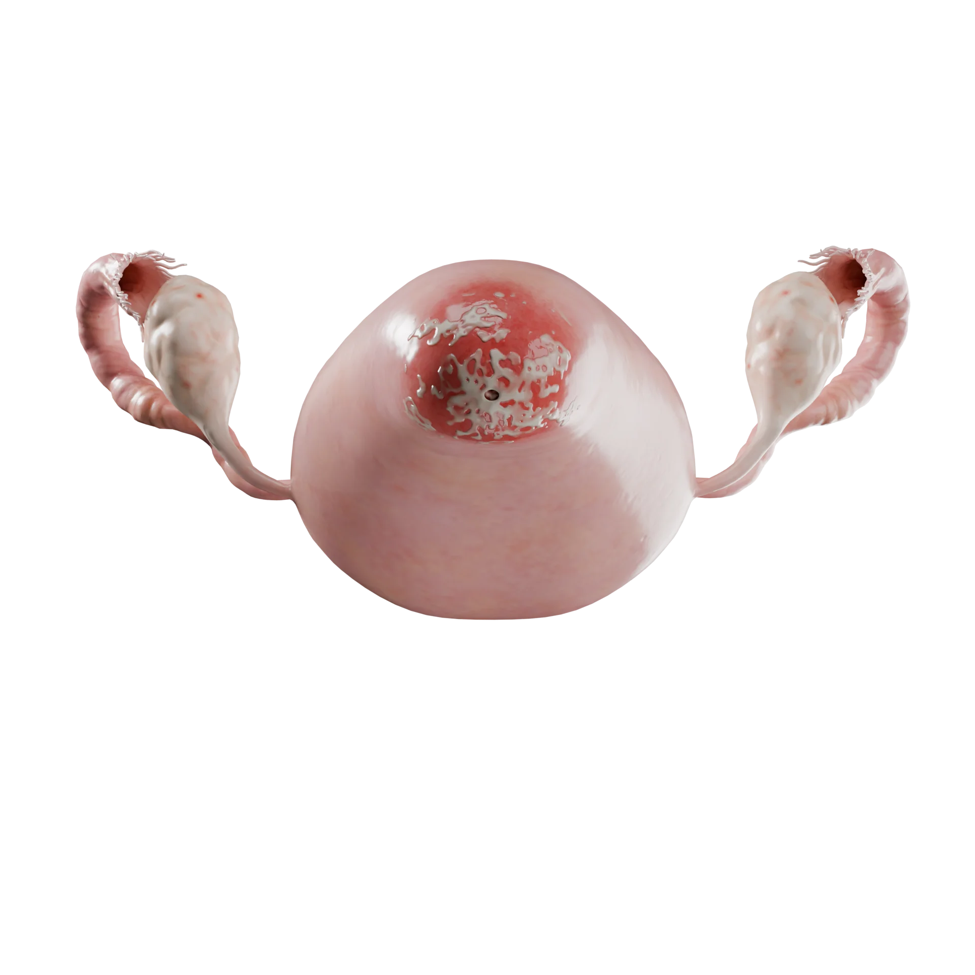
Endometriomas are thick-walled, cavitary ovarian lesions containing viscous protein and hemorrhagic products. They are often bilateral (in 50% of cases). According to the classification of Adamyan L.V., the following stages are distinguished:
3D Models of the stages of ovarian endometriosis:
 Stage 1 ovarian endometriosis
 Stage 2 ovarian endometriosis.
 Stage 3 ovarian endometriosis.
 Stage 4 ovarian endometriosis.
According to the ENZIaN classification, three stages are distinguished:
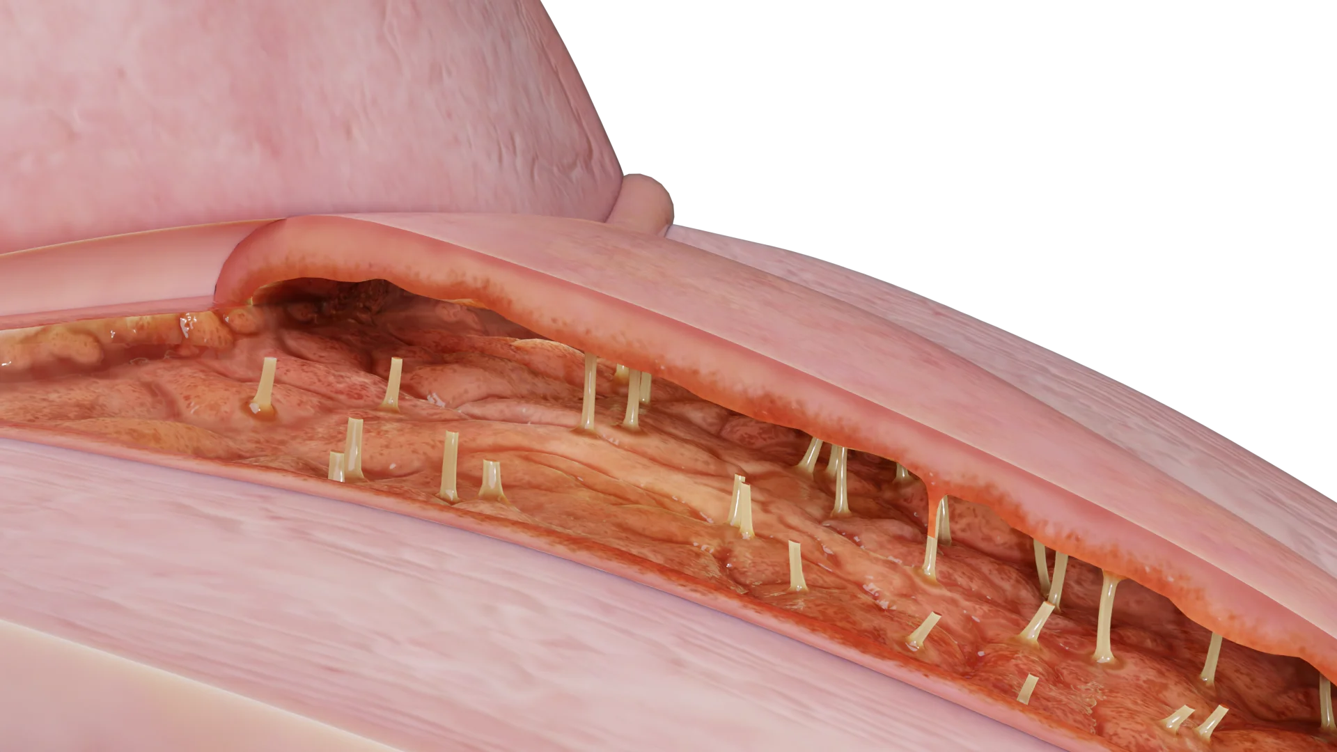
Deep infiltrative endometriosis are foci consisting of fibromuscular hyperplasia surrounding the gland on the peritoneum. These lesions are more than 5 mm deep. The ENZIaN classification provides a more detailed understanding of the location of the pathologic foci. This classification is based on the location of the infiltrate, the depth of its invasion into the pelvic cavity, as well as infiltration into adjacent abdominal organs and violation of their functions. The designation is made using the Latin alphabet and Arabic numerals, where:
The American Society for Reproductive Medicine has developed its own classification. This system rates the stages of endometriosis according to a point scale, which is determined according to surgical assessment of the size, location, severity of endometriotic lesions and the occurrence of adhesions. Thus, women with endometriosis are categorized into four stages: I (1-5 points), II (6-15 points), III (16-40 points), and IV (>40 points).
Endometriosis can be accompanied by various symptoms such as:
Non-gynecologic symptoms:
Pain is a major symptom for many women with endometriosis. The perception of pain can vary individually in intensity, location, time of onset, and duration. In addition, the quality of pain and the associated sympathetic and parasympathetic responses can sometimes differ.
The more symptoms present, the more likely the diagnosis. In a prospective study by Forman and colleagues that only severe dysmenorrhea was a predictor of endometriosis in women who underwent laparoscopy for infertility. This is also supported by other studies that suggest that increased severity of dysmenorrhea may indicate the presence of endometriosis.
However, there is no convincing correlation between the stage of the disease and the severity of symptoms, making the prognosis for each individual patient much more difficult. The growth, frequency and progression of endometrioid lesions, cysts and nodules, remain incompletely understood. This is due to the lack of understanding of pathophysiology, lack of standardized clinical indicators.
Studies suggest that endometriosis may progress in about one-third of women within six to twelve months, while similar forms of endometriosis have been observed to be non-progressive or even regressive. However, these reports should be interpreted with caution because they are few in number and do not take into account the biological activity of individual lesions.
Late diagnosis of endometriosis is a hallmark of the disease. Numerous studies have demonstrated a significant period of time between the onset of the first symptoms and a definitive diagnosis. These studies rely on data that use surgical confirmation as the gold standard.
Imaging modalities such as:
Standard transvaginal ultrasound remains the first-line diagnostic method due to its ability for real-time evaluation in addition to reproducibility, accessibility, cost, and non-invasiveness.
The International Consensus on Deep Endometriosis Analysis (IDEA) has developed a systematic sonographic approach to improve the detection of endometriosis on pelvic ultrasound by evaluating four components: uterus and appendages, deep infiltrative endometriosis, sliding sign, and soft markers. Thus, the components of this specialized ultrasound examination exceed those of the “standard” ultrasound examination.
Superficial peritoneal endometriosis (SPE) has traditionally been described as undetectable by any imaging modality because the size of the foci in the peritoneum isless than 5 mm. Modern equipment and specialist skills allow visualization of SPE lesions at the utero-sacral ligament (USL), parametrium and Douglas space (POD). SPE lesions appear as avascular hypoechogenic areas with irregular borders, less than 5 mm deep. In addition, ovarian motility and local soreness (SST) are two commonly assessed soft markers that are associated with the presence of SPEs.
The sensitivity and specificity of transvaginal ultrasound for detecting endometriomas approaches 90%. Endometriomas have different appearances depending on the degree of viscous proteinaceous material, blood products, and blood degradation. As free fluid is reabsorbed into the cyst, protein and iron concentrations increase. Cyclic bleeding will contribute to the variety of echogenicity, but typically, as bleeding becomes chronic, endometriomas produce a lot of hemorrhagic debris, taking on the appearance of classic frosted glass.
However, early in their formation, the sonographic characteristics of endometriomas may be indistinguishable from hemorrhagic ovarian cysts. They may be unicameral or multicameral (usually less than 5 chambers), and 50% of endometriomas are bilateral. Typically, an endometrioma is a homogeneous cyst with low internal echo, with a wall without solid areas or internal vascularization.
Atypical endometriomas may occur in 50% of patients, more commonly in the postmenopausal age group. Features include:
During pregnancy, endometrioma may undergo decidualization and mimic malignancy due to the presence of vascularized solid areas.
Lesions appear as hypoechogenic thickening of the wall of the lesions or as hypo- or isoechogenic solid nodules that may vary in size and have smooth or irregular contours. The intestinal form of DIE occurs in approximately 8-12% of patients with endometriosis. Rectal and rectosigmoid endometriosis are considered severe forms of DIE, and these forms account for 70-93% of intestinal endometriosis cases.
It is recommended to always include renal ultrasound to evaluate hydronephrosis to assess the urinary tract involvement. Ureteral dilation > 6 mm and detection of nodules > 17 mm in patients scheduled for surgery due to DIE was associated with ureteral endometriosis in 100% of cases.
It is worth noting that the sensitivity of ultrasound varies considerably depending on the location of the DIE.
The uterine slip sign is a real-time dynamic TV-US sign. There are two separate stages:
The sliding sign is considered positive if smooth sliding occurs between the posterior wall of the uterus/cervix and the anterior wall of the sigmoid/rectum.
If there is no sliding, it is usually due to the formation of adhesions or nodules that cause fibrosis between the two structures.
Preoperative knowledge of POD obliteration is important because it allows for appropriate surgical planning and patient counseling involving colorectal surgeons.
Pelvic organ mobility can also be detected on MRI, either directly (using a cine loop) or indirectly (identification of bowel distortion). Direct mobility assessment on MRI has been reported, with absent MRI slip sign correlating well with absent TV-US slip sign and organ fixation detectable on laparoscopy.
Although superficial peritoneal lesions are difficult to visualize with TV-US, there are some soft markers that can help determine the presence or absence of superficial endometriosis.
Ovarian mobility and local soreness (SST) are two commonly evaluated soft markers that are associated with the presence of SPE. In addition, studies suggest that SST may be a marker of endometriosis of the peritoneal lateral pelvic wall.
Thus, in the absence of hard markers of TV-US such as endometrioma/deep endometriosis/obliterated POD, soft markers may provide insight into associated superficial lesions, aiding in the management of chronic pelvic pain.
Ovarian immobility in preoperative TV-US is also significantly associated with the need for complex laparoscopic pelvic lateral wall surgery, including ureterolysis and tuboovariolysis. Therefore, ovarian immobility in TV-US should be considered not only a red flag of increased risk of endometriosis/pelvic lateral wall adhesion, but also a necessity for complex surgery and advanced laparoscopic skills.
These techniques include TV-US-guided rectal contrast injection, sonovaginography, and bowel preparation before TV-US (diet for 1-3 days, oral laxative the day before the examination, rectal enema). These techniques are mainly used as additional information for surgical planning, in particular to determine the number of affected intestinal layers and the distance from the lesion to the anal verge.
MRI for endometriosis is complementary to ultrasound. MRI can be used for diagnosis but is most often required for preoperative determination of the extent of disease, both for surgical planning and patient counseling. However, if conservative treatment is planned, dynamic ultrasound scans are usually performed at 6-12 months. MRI can detect endometrioid lesions in the small bowel, sigmoid colon and/or cecum, as well as endometriosis of the abdominal wall or diaphragm.
Laparoscopic identification of endometrioid lesions with histologic diagnosis has been described as the gold standard for diagnosis in the past. However, advances in the quality and availability of imaging techniques for some forms of endometriosis, surgical risk, limited access to highly skilled surgeons, and financial implications have relegated this method of diagnosis to last place, but laparoscopy still remains the most reliable diagnostic method.
It is worth mentioning that serum CA-125 determination has no diagnostic value. An elevated CA-125 concentration (i.e. 35 IU/mL or more) can be detected in endometriosis, but endometriosis can also be present despite normal CA-125 values (less than 35 IU/mL).
Find more scientifically accurate content on our social media


The choice of treatment will depend on the severity of the symptoms, the extent and location of the disease, the desire to become pregnant and the age of the patient. There are medical and surgical treatments, as well as a combination of both.
Pharmacologic therapy for endometriosis is aimed at improving symptoms or preventing recurrence postoperatively.
Surgical treatment is indicated when symptoms persist or when the side effects of drug therapy outweigh its therapeutic effect. Patients with anatomical changes in pelvic structures, adhesions, bowel or urinary tract obstruction are also indicated for surgical treatment.
Conservative surgery consists of coagulation of endometrioid foci and restoration of normal pelvic anatomy. When ectopic foci are excised, there is a significant reduction in pelvic pain and improvement in fertility.
Despite this, the risk of symptom recurrence after surgery remains high.
Ablation of endometriosis foci is applicable in women with superficial endometriosis. The evidence in favor of ablation over excision is based on studies involving women with heterogeneous endometriosis.
Some of these studies excluded women with deep endometriosis, in whom ablation is not usually used. The excisional approach is likely to be more appropriate for deep foci, as it is impossible to know whether the entire nidus has been destroyed by ablation.
When intervening in women with ovarian endometrioma, cystectomy is preferred over drainage and coagulation because it reduces the risk of recurrence and pain.
Alternatively, CO2 laser vaporization may be performed. Both techniques have similar recurrence rates in the first year after surgery, but the early postoperative recurrence rate may be lower after cystectomy.
When performing surgery for ovarian endometrioma, extreme care should be taken to minimize damage to healthy ovarian tissue.
The final surgical treatment includes hysterectomy with or without ovarian removal, which depends on the age of the patient.
Hysterectomy with bilateral salpingo-oophorectomy and excision of all foci of endometriosis showed effective cure in 90% of cases.
1. What is uterine endometriosis in women?
2. What causes endometriosis?
3. What are the first symptoms of endometriosis?
4. How is endometriosis diagnosed?
5. What are the dangers of endometriosis?
6. Can I get pregnant with endometriosis?
7. Will endometriosis disappear at menopause?
List of Sources
1.
VOKA Catalog.
https://catalog.voka.io/
2.
Becker CM, Bokor A, Heikinheimo O, Horne A, Jansen F, Kiesel L, King K, Kvaskoff M, Nap A, Petersen K, Saridogan E, Tomassetti C, van Hanegem N, Vulliemoz N, Vermeulen N; ESHRE Endometriosis Guideline Group. ESHRE guideline: endometriosis. Hum Reprod Open. 2022 Feb 26;2022(2):hoac009. doi: 10.1093/hropen/hoac009. PMID: 35350465; PMCID: PMC8951218.
3.
Practice bulletin no. 114: management of endometriosis. Obstet Gynecol. 2010 Jul;116(1):223-236. doi: 10.1097/AOG.0b013e3181e8b073. PMID: 20567196.
4.
Practice Committee of the American Society for Reproductive Medicine. Endometriosis and infertility: a committee opinion. Fertil Steril. 2012 Sep;98(3):591-8. doi: 10.1016/j.fertnstert.2012.05.031. Epub 2012 Jun 15. PMID: 22704630.
5.
International Working Group of AAGL, ESGE, ESHRE and WES, Carla Tomassetti, Neil P Johnson, John Petrozza, Mauricio S Abrao, Jon I Einarsson, Andrew W Horne, Ted T M Lee, Stacey Missmer, Nathalie Vermeulen, Krina T Zondervan, Grigoris Grimbizis, Rudy Leon De Wilde, An international terminology for endometriosis, 2021,, Human Reproduction Open, Volume 2021, Issue 4, 2021, hoab029.
https://doi.org/10.1093/hropen/hoab029
6.
International Federation of Gynecology and Obstetrics (FIGO). (2021). FIGO Recommendations on Endometriosis.International Journal of Gynecology & Obstetrics, 155(1), 1-6. DOI: [10.1002/ijgo.13860].
7.
Revised American Society for Reproductive Medicine (rASRM). (1997). Revised classification of endometriosis. Fertility and Sterility, 67(5), 817-821. DOI:[10.1016/S0015-0282(97)81391-X].
8.
Haas, D., et al. (2021). The ENZIAN classification for deep endometriosis.Archives of Gynecology and Obstetrics, 303(1), 1-7. DOI: [10.1007/s00404-020-05839-1].
9.
Adamson GD, Pasta DJ. Endometriosis fertility index: the new, validated endometriosis staging system. Fertil Steril. 2010 Oct;94(5):1609-15. doi: 10.1016/j.fertnstert.2009.09.035. PMID: 19931076.
10.
Zondervan KT, Becker CM, Missmer SA. Endometriosis. N Engl J Med. 2020 Mar 26;382(13):1244-1256. doi: 10.1056/NEJMra1810764. PMID: 32212520.
11.
Vannuccini, S., & Petraglia, F.. Recent advances in understanding the pathogenesis of endometriosis, 2019. F1000Research, 8, F1000 Faculty Rev-345. DOI: [10.12688/f1000research.14842.1].
12.
Nisenblat, V., et al. Combination of non-invasive tests for the diagnosis of endometriosis. Cochrane Database of Systematic Reviews, 2016, 7, CD012281. DOI: [10.1002/14651858.CD012281].
13.
Dunselman, G. A. J., et al. ESHRE guideline: Management of women with endometriosis. Human Reproduction, 2014. 29(3), 400-412. DOI: [10.1093/humrep/det457].
14.
Johnson, N. P., et al. . Surgical treatment for endometriosis-associated infertility. Cochrane Database of Systematic Reviews, 2013, 1, CD003678. DOI: [10.1002/14651858.CD003678.pub3].
Loading test 6 questions
Table of Contents


Summarize article with AI
Choose your preferable AI assistant:
Link successfully copied to clipboard
Thank you!
Your message is sent!
Our experts will contact you shortly. If you have any additional questions, please contact us at info@voka.io