The content on this website, including text, graphics, and other materials, is provided for informational purposes only. It is not intended as advice or guidance. Regarding your specific medical condition or treatment, please consult your healthcare provider.
Traumatic subdural hematoma is a disease characterized by the accumulation of blood in the subdural space beneath the dura mater, associated with head and brain trauma.
The frequency of acute subdural hematomas is approximately 20% in severe traumatic brain injury. According to the literature, the mortality rate from this condition ranges from 50 to 90%, with only 30% of patients achieving full neurological recovery after a traumatic hemorrhage.
Classification of traumatic subdural hemorrhages
Traumatic subdural hematomas are classified as follows:
Acute: detected during the examination of the patient within the first 24 to 72 hours after the hemorrhage onset;
Chronic: detected more than 14 days after the hemorrhage event.
Etiology of traumatic subdural hematomas
The etiological factors in the development of traumatic subdural hematomas include the same causes that underlie the occurrence of traumatic brain injury:
Road traffic accidents;
Falls;
Violent injuries;
Sports injuries.
Notably, up to 55% of the frequency of subdural hematoma development is attributed to falls.
Pathogenesis of traumatic subdural hematomas
The pathogenesis of traumatic subdural hematomas is characterized by a сoup-contrecoup mechanism, attributing this type of traumatic brain injury to a focal category.
Formation mechanisms
Coup mechanism (direct impact). During the application of a traumatic agent to the rigid bones of the skull vault, brain tissue is injured, forming contusion foci, and ruptures of pial arterial or venous (most commonly bridging) vessels. Due to vessel injury and ongoing bleeding, blood accumulates in the subdural space.
Cavitation mechanism (counter-impact). This mechanism explains the formation of subdural hematomas on the side opposite the application of the mechanical factor, associated with the cavitation theory.
Rotational mechanism. Modern literature also highlights the rotational injury theory, which suggests angular acceleration and shear stresses lead to predominant damage to bridging veins stretched between the superior sagittal sinus and the surface of the cerebral hemispheres.
Age-related characteristics
The development of traumatic subdural hematomas is further influenced by age-related characteristics: in elderly patients, due to brain atrophy, the subdural space and the tension on bridging veins increase, heightening the risk of rupture even from relatively minor trauma.
Complications
Ischemia progression. Due to ongoing bleeding, accumulation of blood in the subdural space, and increased intracranial pressure, the perfusion pressure in brain tissue decreases, leading to worsening brain damage, development of secondary ischemic foci, and deteriorating patient prognosis.
Dislocation syndrome. Axial dislocation caused by acute subdural hemorrhages may trigger temporal-sartorial herniation syndrome due to the displacement of the temporal lobe hook into the notch area of the tentorium cerebelli, compressing the brainstem, the oculomotor nerve, and the posterior cerebral artery on the affected side, potentially leading to ischemic changes in these structures and rapid deterioration of the patient’s condition.
Clinical presentation
Symptoms of traumatic subdural hematomas
The clinical symptoms of traumatic subdural hematomas comprise non-focal, focal, and meningeal symptoms:
Non-focal symptoms include intense headaches, nausea, vomiting, dizziness, consciousness impairment at varying levels — from mild stupor to deep coma.
Focal symptoms are most commonly represented by contralateral hemiparesis or hemiplegia, sensory disruptions, and aphasia involving the dominant hemisphere. Seizure activity may include generalized and focal seizures.
Meningeal syndrome is manifested by neck muscle rigidity, Kernig’s and Brudzinski’s signs, associated with irritation of the meninges by blood clots.
Seizure syndrome manifests with convulsive fits due to cortical brain damage and pathological nerve signal activity.
Autonomous manifestations (bradycardia, hypertension, and respiratory disturbances) indicate elevated intracranial pressure and may point to the onset of dislocation syndrome or the damage sustained.
Development of the pathological process
Acute subdural hematomas typically progress through three phases:
Initial loss of consciousness at the time of injury (10–20 minutes) followed by recovery to a stupor level.
“Lucid interval” is a temporary improvement in condition.
Abrupt deterioration of condition with depression of consciousness up to coma.
However, this progression is observed in no more than 20–30% of patients. More often, the lucid interval is absent or diminished, and symptoms escalate swiftly, depending on the rate of intracerebral hematoma formation.
Subacute subdural hematomas progress less evidently. The lucid interval may last 1–2 weeks. Gradually, headaches intensify, drowsiness and lethargy increase, and signs of intracranial hypertension escalate. Neurological focal symptoms appear later and are induced by brain compression and displacement.
Diagnosis of acute subdural hematomas
After admission to the emergency department, the diagnostic algorithm for acute subdural hematomas consists of the following steps:
1. Assessment of consciousness level using the GCS (Glasgow Coma Scale)
This step allows for determining the patient’s level of consciousness and estimating the severity of brain structure damage, suggesting possible treatment options (surgical or non-surgical).
2. Medical history collection
Medical history collection includes the timing and mechanism of the injury and the presence of consciousness loss in the patient after the injury.
This step allows understanding of symptom development dynamics and inferring possible damages considering the injury mechanism. For certain injury mechanisms (e.g., fall from height), it’s essential to consider the possibility of combined head and body injuries, which may complicate the treatment prognosis.
3. Visual examination of the patient
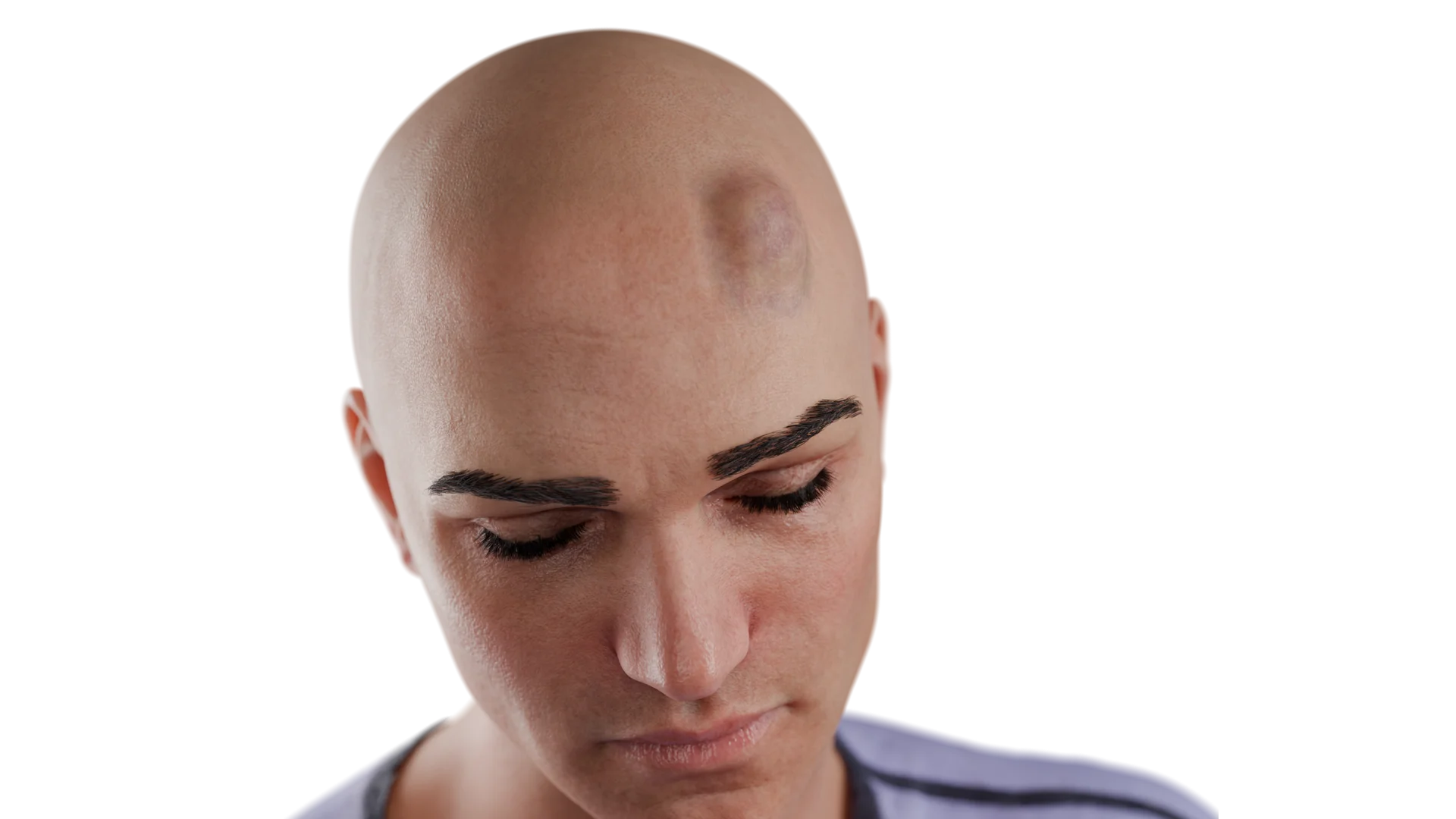
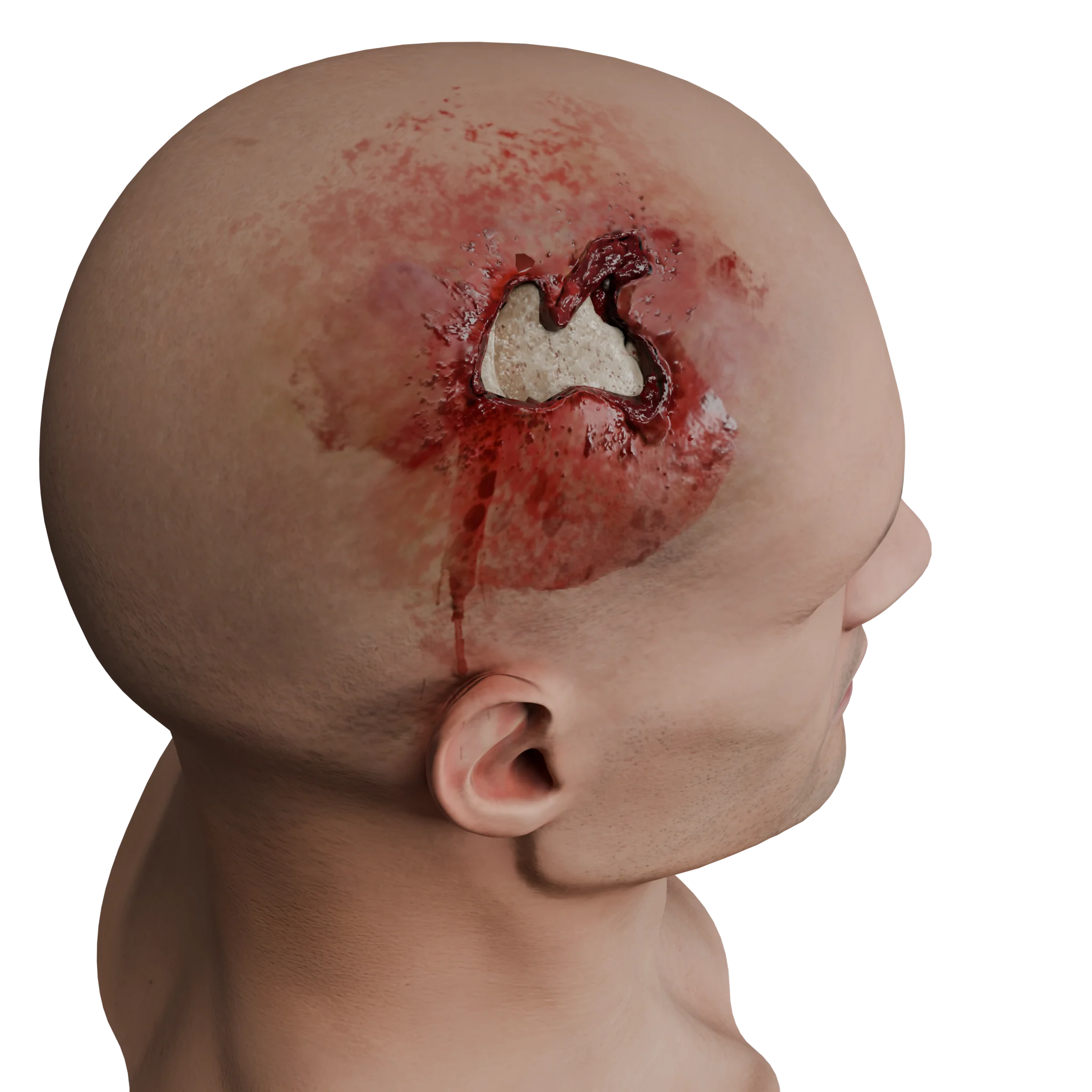
The head area is examined, checking for concomitant soft tissue damage in areas of the body other than the head.
This step helps highlight the location of soft tissue in head injuries, allowing inference of the brain contusion’s possible location. Additionally, this step is essential for patient care, treating soft tissue bruises and performing primary surgical treatment if needed.
4. Neurological examination of the patient
Assessment of non-focal, focal, and meningeal symptoms is performed.
This stage enables preliminary topical diagnosis of brain injury to determine indications for neuroimaging studies. If the patient presents with non-focal, focal, or meningeal symptoms, neuroimaging is indicated.
5. Neuroimaging studies
Brain CT
Computed tomography of the brain is the “gold standard” in diagnosing traumatic brain injury, including subdural hematomas.
The advantages of CT of the brain compared to other methods (including MRI) are:
Examination completion speed;
Ability to clearly evaluate the localization, extent, and nature of brain damage;
Ability to assess associated bone structure damage;
Capability (if necessary) to quickly scan other parts of the body to exclude concurrent injury.
A CT image of acute subdural hematoma is characterized by the following:
Localization of a crescent-shaped clot above the cerebral hemisphere.
In the hyperacute phase of hemorrhage, hyperintense blood clots are present, separated by an isointense signal, explained by the presence of coagulated clots and liquid blood fraction.
In the acute phase of hemorrhage, the subdural hematoma is represented by a hyperintense (50-60 HU) homogeneous signal forming a crescent over the entire cerebral hemisphere.
In the subacute phase of hemorrhage, subdural hematoma is represented by a hyper-isointense signal on CT scans (35-45 HU).
Brain MRI
Magnetic resonance imaging is used for the diagnosis of subdural hematomas. Visualization of hematomas is directly dependent on the timeline of occurrence and the stage of hemoglobin degradation.
Hyperacute subdural hematoma
T1: isointense to gray matter;
T2: iso-hyperintense to gray matter;
FLAIR: hyperintense to CSF.
Acute subdural hematoma
T1: iso-hypointense to gray matter;
T2: hypointense to gray matter;
FLAIR: hyperintense to CSF.
Subacute subdural hematoma
T1: generally hyperintense due to the presence of methemoglobin.
T2: variable appearance, usually hyperintense.
FLAIR: hyperintense.
6. Laboratory diagnostics
Laboratory diagnosis includes complete blood count, urinalysis, coagulation profile, blood biochemistry, and blood group + rhesus factor.
These tests do not confirm or suspect a subdural hematoma, but they allow for the assessment of the patient’s overall condition, accompanying coagulation system disorders, and preparation for potential surgery.
Find more scientifically accurate content on our social media
Subscribe and don’t miss out the latest resources
Treatment of traumatic subdural hematomas
Conservative treatment of acute and subacute subdural hematomas
Conservative treatment of acute and subacute subdural hematomas is possible with small hematomas that do not cause mass effect or shift of the brain’s median structures.
In such cases, conservative treatment primarily consists of symptomatic therapy, which includes:
Pain relief (NSAIDs, narcotic analgesics);
Antiemetic drugs;
Anticonvulsants (if seizures manifest).
Osmotic diuretics are prescribed in the presence of cerebral edema.
Prescription of glucocorticosteroids is contraindicated in traumatic brain injury due to worsening outcomes.
For moderate and severe traumatic brain injury (including acute subdural hematomas), a single dose of 1000 mg of tranexamic acid reduces the risk of adverse outcomes in studies.
Surgical treatment of acute subdural hematomas
Regardless of the patient’s consciousness level, surgical treatment of acute subdural hematomas is indicated in the following cases:
Presence of hematoma thickness of 10 mm or more.
Presence of displacement of median structures of 5 mm or more.
Hematoma volume over 35 ml.
With a hematoma of less than 10 mm, hematoma volume less than 35 ml, or displacement of median structures less than 5 mm, and a progressive decline in the level of consciousness (by 2 or more points on the GCS), or if monitoring indicates an increase in ICP over 20 mmHg. Surgery is also indicated for the patient.
In certain cases, with hematomas over 35 ml in volume, a hematoma thickness over 10 mm, without progressive deterioration in the patient’s condition, and absence of displacement syndrome, conservative treatment is possible with subsequent neuroimaging in 24-48 hours.
Craniotomy
The volume of surgical intervention involves craniotomy (usually in the frontal/parietal/temporal region)
With the removal of the subdural hematoma;
Searching for the source of bleeding (usually either parasagittal veins or cortical arteries);
Stopping the bleeding from the source.
Subsequently, the dura mater is tightly sutured, the bone flap is returned to place, and the wound is closed layer by layer.
Decompressive craniectomy
In certain cases (with hematoma volume over 140 cm³, displacement syndrome more than 13 mm, and consciousness level of the patient below 7 on the GCS), decompressive craniectomy is indicated for the patient.
Surgical procedure:
A wide trepanation of the skull in the frontal/parietal/temporal region is performed by removing the squama of the temporal bone to the base of the middle cranial fossa.
After opening the dura mater and removing the subdural hematoma with bleeding source control, plastic surgery of the dura mater is performed.
The bone flap is sent for conservation. The wound is sutured in layers.
Surgical treatment of subacute subdural hematomas
Surgical treatment of subacute subdural hematomas, regardless of the patient’s level of consciousness, is indicated in the following cases:
Presence of hematoma thickness of 10 mm or more.
Shift of median structures of 5 mm or more.
Hematoma volume over 35 ml.
Surgical intervention for subacute subdural hematomas includes:
Performing craniotomy in the frontal/parietal/temporal region, with removal of subdural content. During exploration, a source of bleeding is typically not found in the subdural space due to the injury’s age. After clot removal, tight suturing of the dura mater is performed, followed by the bonding of the bone flap in place.
Use of two drill holes to wash out the subdural space in the frontal and parietal regions, aiming to drain the subdural space and remove the hematoma through irrigation with saline.
FAQ
1. What is a subdural hematoma?
A subdural hematoma is a collection of blood between the dura mater and the surface of the brain, resulting from vessel damage, most often bridging veins, during a traumatic brain injury.
2. What are the types of subdural hematomas?
Subdural hematomas are categorized into three types: acute, manifesting within the first 72 hours post-injury, subacute, detectable between the 4th and 10th days, and chronic, diagnosed two weeks or more post-injury.
3. How dangerous is a subdural hematoma?
It is among the most threatening forms of traumatic brain injury: mortality in acute forms reaches 50–90%. Prognosis depends on the hematoma’s volume, the patient’s age, the level of consciousness upon admission, and the presence of accompanying injuries.
4. What are the main causes of subdural hematomas?
The main causes of subdural hematomas include traffic accidents, falls (especially in the elderly), sports and domestic injuries, and violent head injuries.
5. How does a subdural hematoma manifest?
The clinical presentation of a subdural hematoma consists of non-focal symptoms such as headache, nausea, vomiting, and consciousness depression, as well as focal disturbances including paralysis, speech impairments, and sensory changes.
6. What is the “lucid interval” in acute subdural hematoma?
It is a temporary improvement in the patient’s condition after the initial loss of consciousness. This is followed by a sharp deterioration up to coma. The typical “lucid interval” occurs in only 20–30% of patients.
7. How is a subdural hematoma diagnosed?
The gold standard is computed tomography (CT). For subacute subdural hematomas, MRI is highly informative.
8. Is surgery always required for acute and subacute subdural hematomas?
Surgery is not always necessary. In cases where the hematoma is small, does not cause clinical manifestations, and there is no displacement of midline structures, conservative symptomatic treatment with patient monitoring may be possible.
9. What surgeries are performed?
The range of surgical interventions includes craniotomy with hematoma evacuation and hemostasis, as well as decompressive craniectomy, which is performed for massive hematomas and significant brain displacement. As a treatment option for subacute forms, the creation of a burr hole and drainage of the subdural space can be applied.
10. What is the prognosis for acute and subacute subdural hematomas?
Better outcomes are observed with small hematomas, early diagnosis, and subacute forms of progression. An unfavorable prognosis is noted in older patients with significant displacement and deep coma in acute subdural hematomas.
References
1.
VOKA 3D Anatomy & Pathology – Complete Anatomy and Pathology 3D Atlas [Internet]. VOKA 3D Anatomy & Pathology.
Available from: https://catalog.voka.io/
2.
Bullock M.R., Chesnut R., Ghajar J., et al. Surgical management of acute subdural hematomas // Neurosurgery. – 2006. – Vol. 58, Suppl. 3. – P. S16–S24.
3.
Yang W., Huang J. Chronic subdural hematoma: epidemiology and natural history // Neurosurg Clin N Am. – 2017. – Vol. 28(2). – P. 205–210.
4.
Morgalla M., Will B.E., Roser F., Tatagiba M. Do recurrent subdural hematomas grow faster? // Acta Neurochir (Wien). – 2008. – Vol. 150. – P. 1059–1065.
5.
Alvis-Miranda H.R., Rubiano A.M., Moscote-Salazar L.R., et al. Acute subdural hematoma: a concise review // Neuroimmunol Neuroinflamm. – 2014. – Vol. 1(2). – P. 68–76.
6.
Weigel R., Schmiedek P., Krauss J.K. Outcome of contemporary surgery for chronic subdural haematoma: evidence based review // J Neurol Neurosurg Psychiatry. – 2003. – Vol. 74(7). – P. 937–943.
7.
Stippler M., Zhao Y., Kim Y.S., et al. Extended Glasgow Outcome Scale: an international study of interrater reliability // J Neurotrauma. – 2016. – Vol. 33(5). – P. 409–414.
8.
Kolias A.G., Chari A., Santarius T., Hutchinson P.J. Chronic subdural haematoma: modern management and emerging therapies // Nat Rev Neurol. – 2014. – Vol. 10. – P. 570–578.
9.
Ducruet A.F., Grobelny B.T., Zacharia B.E., et al. The surgical management of chronic subdural hematoma // Neurosurg Rev. – 2012. – Vol. 35(2). – P. 155–169.
10.
Park S.H., Kang D.H., Park J., et al. Fibrinolysis in chronic subdural hematomas: comparison of urokinase and tissue plasminogen activator // Neurosurgery. – 2008. – Vol. 63(4). – P. 905–910.
11.
Santarius T., Lawton R., Kirkpatrick P.J., et al. Use of drains versus no drains after burr-hole evacuation of chronic subdural haematoma: a randomised controlled trial // Lancet. – 2009. – Vol. 374(9695). – P. 1067–1073.
12.
Brennan P.M., Kolias A.G., Joannides A.J., et al. The management and outcome for patients with chronic subdural hematoma: a prospective, multicenter, observational cohort study in the United Kingdom // J Neurosurg. – 2017. – Vol. 127(4). – P. 732–739.
13.
Edlmann E., Giorgi-Coll S., Whitfield P.C., et al. Pathophysiology of chronic subdural haematoma: inflammation, angiogenesis and implications for pharmacotherapy // J Neuroinflammation. – 2017. – Vol. 14. – P. 108.
14.
Hutchinson P.J., Kolias A.G., Timofeev I.S., et al. Trial of decompressive craniectomy for traumatic intracranial hypertension // N Engl J Med. – 2016. – Vol. 375. – P. 1119–1130.
15.
Honeybul S., Ho K.M. Long-term complications of decompressive craniectomy for head injury // J Neurotrauma. – 2011. – Vol. 28(6). – P. 929–935.
16.
Osuka K., Watanabe Y., Usuda N., et al. Vasogenic edema and angiogenesis in chronic subdural hematoma // J Neurosurg. – 2012. – Vol. 117. – P. 1119–1126.
17.
Bartley A., Tajsic T., Barone D.G. A pragmatic approach to the management of chronic subdural haematoma in older patients // Drugs Aging. – 2020. – Vol. 37. – P. 1–11.
18.
Won S.Y., Konczalla J., Dubinski D., et al. A systematic review of epileptic seizures in adults with subdural haematomas // Seizure. – 2017. – Vol. 45. – P. 28–35.
19.
Miranda L.B., Braxton E., Hobbs J., Quigley M.R. Chronic subdural hematoma in the elderly: not a benign disease // J Neurosurg. – 2011. – Vol. 114. – P. 72–76.