Scapular Anatomy Quiz
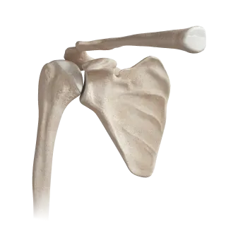
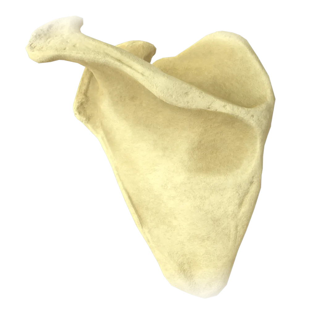
The quiz covers the structure of the scapula, its surfaces, borders, angles, processes, muscle attachments, innervation, and blood supply.
1/20
bold
text
1. To which division of the skeleton does the scapula belong?
-
Skeleton of the free upper limb
The scapula (scapula), together with the clavicle, is part of the pectoral girdle (cingulum pectorale). The skeleton of the free upper limb begins with the humerus.
-
Skeleton of the pectoral girdle
The scapula (scapula), together with the clavicle, is part of the pectoral girdle (cingulum pectorale). The skeleton of the free upper limb begins with the humerus.
-
Thoracic skeleton
The scapula (scapula), together with the clavicle, is part of the pectoral girdle (cingulum pectorale). The skeleton of the free upper limb begins with the humerus.
-
Skeleton of the vertebral column
The scapula (scapula), together with the clavicle, is part of the pectoral girdle (cingulum pectorale). The skeleton of the free upper limb begins with the humerus.
-
I find it difficult to answer
The scapula (scapula), together with the clavicle, is part of the pectoral girdle (cingulum pectorale). The skeleton of the free upper limb begins with the humerus.
2. How many angles does the scapula comprise?
-
Two
The scapula comprises three angles: superior (angulus superior), inferior (angulus inferior), and lateral (angulus lateralis), where the glenoid cavity is located.
-
Four
The scapula comprises three angles: superior (angulus superior), inferior (angulus inferior), and lateral (angulus lateralis), where the glenoid cavity is located.
-
Three
The scapula comprises three angles: superior (angulus superior), inferior (angulus inferior), and lateral (angulus lateralis), where the glenoid cavity is located.
-
One
The scapula comprises three angles: superior (angulus superior), inferior (angulus inferior), and lateral (angulus lateralis), where the glenoid cavity is located.
-
I find it difficult to answer
The scapula comprises three angles: superior (angulus superior), inferior (angulus inferior), and lateral (angulus lateralis), where the glenoid cavity is located.
3. What is the articular surface of the scapula that articulates with the head of the humerus called?
-
Subscapular fossa
The glenoid cavity (cavitas glenoidalis) is located at the lateral angle of the scapula and forms the shoulder (glenohumeral) joint with the head of the humerus.
-
Glenoid cavity
The glenoid cavity (cavitas glenoidalis) is located at the lateral angle of the scapula and forms the shoulder (glenohumeral) joint with the head of the humerus.
-
Acromion
The glenoid cavity (cavitas glenoidalis) is located at the lateral angle of the scapula and forms the shoulder (glenohumeral) joint with the head of the humerus.
-
Coracoid process
The glenoid cavity (cavitas glenoidalis) is located at the lateral angle of the scapula and forms the shoulder (glenohumeral) joint with the head of the humerus.
-
I find it difficult to answer
The glenoid cavity (cavitas glenoidalis) is located at the lateral angle of the scapula and forms the shoulder (glenohumeral) joint with the head of the humerus.
4. Which process of the scapula is the site of origin of the short head of the biceps brachii and the coracobrachialis muscle?
-
Acromion
The coracoid process (processus coracoideus) serves as the site of origin of the short head of the m. biceps brachii and m. coracobrachialis, as well as the attachment of the m. pectoralis minor.
-
Coracoid process
The coracoid process (processus coracoideus) serves as the site of origin of the short head of the m. biceps brachii and m. coracobrachialis, as well as the attachment of the m. pectoralis minor.
-
Scapular spine
The coracoid process (processus coracoideus) serves as the site of origin of the short head of the m. biceps brachii and m. coracobrachialis, as well as the attachment of the m. pectoralis minor.
-
Articular process
The coracoid process (processus coracoideus) serves as the site of origin of the short head of the m. biceps brachii and m. coracobrachialis, as well as the attachment of the m. pectoralis minor.
-
I find it difficult to answer
The coracoid process (processus coracoideus) serves as the site of origin of the short head of the m. biceps brachii and m. coracobrachialis, as well as the attachment of the m. pectoralis minor.
5. Which muscle attaches to the medial border of the scapula?
-
M. serratus anterior
M. rhomboideus major attaches to the medial (vertebral) border of the scapula from the level of the spine to the inferior angle. M. serratus anterior attaches to the costal surface of the medial border.
-
M. rhomboideus major
M. rhomboideus major attaches to the medial (vertebral) border of the scapula from the level of the spine to the inferior angle. M. serratus anterior attaches to the costal surface of the medial border.
-
M. trapezius
M. rhomboideus major attaches to the medial (vertebral) border of the scapula from the level of the spine to the inferior angle. M. serratus anterior attaches to the costal surface of the medial border.
-
M. infraspinatus
M. rhomboideus major attaches to the medial (vertebral) border of the scapula from the level of the spine to the inferior angle. M. serratus anterior attaches to the costal surface of the medial border.
-
I find it difficult to answer
M. rhomboideus major attaches to the medial (vertebral) border of the scapula from the level of the spine to the inferior angle. M. serratus anterior attaches to the costal surface of the medial border.
6. The following is located on the costal (anterior) surface of the scapula:
-
Infraspinous fossa
The costal surface of the scapula is represented by the subscapular fossa (fossa subscapularis), from which the m. subscapularis.
-
Supraspinous fossa
The costal surface of the scapula is represented by the subscapular fossa (fossa subscapularis), from which the m. subscapularis.
-
Subscapular fossa
The costal surface of the scapula is represented by the subscapular fossa (fossa subscapularis), from which the m. subscapularis.
-
Scapular notch
The costal surface of the scapula is represented by the subscapular fossa (fossa subscapularis), from which the m. subscapularis.
-
I find it difficult to answer
The costal surface of the scapula is represented by the subscapular fossa (fossa subscapularis), from which the m. subscapularis.
7. Which nerve passes through the scapular notch (incisura scapulae), which is converted into a foramen by the superior transverse scapular ligament?
-
Axillary nerve
N. suprascapularis passes through the scapular notch (beneath the lig. transversum scapulae superius), whereas the suprascapular artery passes above the ligament. The nerve innervates the mm. supraspinatus et infraspinatus.
-
N. suprascapularis
N. suprascapularis passes through the scapular notch (beneath the lig. transversum scapulae superius), whereas the suprascapular artery passes above the ligament. The nerve innervates the mm. supraspinatus et infraspinatus.
-
N. thoracicus longus
N. suprascapularis passes through the scapular notch (beneath the lig. transversum scapulae superius), whereas the suprascapular artery passes above the ligament. The nerve innervates the mm. supraspinatus et infraspinatus.
-
N. subscapularis
N. suprascapularis passes through the scapular notch (beneath the lig. transversum scapulae superius), whereas the suprascapular artery passes above the ligament. The nerve innervates the mm. supraspinatus et infraspinatus.
-
I find it difficult to answer
N. suprascapularis passes through the scapular notch (beneath the lig. transversum scapulae superius), whereas the suprascapular artery passes above the ligament. The nerve innervates the mm. supraspinatus et infraspinatus.
8. Which muscle originates in the supraspinous fossa of the scapula?
-
M. infraspinatus
M. supraspinatus originates in the fossa supraspinata, located superior to the spine of the scapula. M. infraspinatus occupies the infraspinous fossa (fossa infraspinata).
-
M. supraspinatus
M. supraspinatus originates in the fossa supraspinata, located superior to the spine of the scapula. M. infraspinatus occupies the infraspinous fossa (fossa infraspinata).
-
M. teres minor
M. supraspinatus originates in the fossa supraspinata, located superior to the spine of the scapula. M. infraspinatus occupies the infraspinous fossa (fossa infraspinata).
-
M. deltoideus
M. supraspinatus originates in the fossa supraspinata, located superior to the spine of the scapula. M. infraspinatus occupies the infraspinous fossa (fossa infraspinata).
-
I find it difficult to answer
M. supraspinatus originates in the fossa supraspinata, located superior to the spine of the scapula. M. infraspinatus occupies the infraspinous fossa (fossa infraspinata).
9. Which structure separates the supraspinous fossa from the infraspinous fossa of the scapula?
-
Medial border
The spine of the scapula (spina scapulae) is a horizontal bony ridge that divides the posterior surface of the scapula into the supraspinous (fossa supraspinata) and infraspinous (fossa infraspinata) fossae.
-
Scapular notch
The spine of the scapula (spina scapulae) is a horizontal bony ridge that divides the posterior surface of the scapula into the supraspinous (fossa supraspinata) and infraspinous (fossa infraspinata) fossae.
-
Scapular spine
The spine of the scapula (spina scapulae) is a horizontal bony ridge that divides the posterior surface of the scapula into the supraspinous (fossa supraspinata) and infraspinous (fossa infraspinata) fossae.
-
Coracoid process
The spine of the scapula (spina scapulae) is a horizontal bony ridge that divides the posterior surface of the scapula into the supraspinous (fossa supraspinata) and infraspinous (fossa infraspinata) fossae.
-
I find it difficult to answer
The spine of the scapula (spina scapulae) is a horizontal bony ridge that divides the posterior surface of the scapula into the supraspinous (fossa supraspinata) and infraspinous (fossa infraspinata) fossae.
10. What is the lateral expansion of the spine of the scapula that articulates with the clavicle called?
-
Coracoid process
The acromion (acromion) is a flattened process that forms the lateral continuation of the spine of the scapula. It forms the acromioclavicular joint with the clavicle and serves as the attachment site for some fibers of the m. deltoideus and m. trapezius.
-
Infraglenoid tubercle
The acromion (acromion) is a flattened process that forms the lateral continuation of the spine of the scapula. It forms the acromioclavicular joint with the clavicle and serves as the attachment site for some fibers of the m. deltoideus and m. trapezius.
-
Acromion
The acromion (acromion) is a flattened process that forms the lateral continuation of the spine of the scapula. It forms the acromioclavicular joint with the clavicle and serves as the attachment site for some fibers of the m. deltoideus and m. trapezius.
-
Lateral angle
The acromion (acromion) is a flattened process that forms the lateral continuation of the spine of the scapula. It forms the acromioclavicular joint with the clavicle and serves as the attachment site for some fibers of the m. deltoideus and m. trapezius.
-
I find it difficult to answer
The acromion (acromion) is a flattened process that forms the lateral continuation of the spine of the scapula. It forms the acromioclavicular joint with the clavicle and serves as the attachment site for some fibers of the m. deltoideus and m. trapezius.
11. Which muscle attaches to the tuberculum supraglenoidale of the scapula?
-
Short head of the m. biceps brachii
Long head of the m. biceps brachii originates from the supraglenoid tubercle (tuberculum supraglenoidale), located superior to the glenoid cavity. The short head originates from the coracoid process.
-
Long head of the m. biceps brachii
Long head of the m. biceps brachii originates from the supraglenoid tubercle (tuberculum supraglenoidale), located superior to the glenoid cavity. The short head originates from the coracoid process.
-
Coracobrachialis
Long head of the m. biceps brachii originates from the supraglenoid tubercle (tuberculum supraglenoidale), located superior to the glenoid cavity. The short head originates from the coracoid process.
-
M. subscapularis
Long head of the m. biceps brachii originates from the supraglenoid tubercle (tuberculum supraglenoidale), located superior to the glenoid cavity. The short head originates from the coracoid process.
-
I find it difficult to answer
Long head of the m. biceps brachii originates from the supraglenoid tubercle (tuberculum supraglenoidale), located superior to the glenoid cavity. The short head originates from the coracoid process.
12. To which tubercle of the scapula does the long head of the triceps brachii attach?
-
Supraglenoid tubercle
Long head of the m. triceps brachii originates from the infraglenoid tubercle (tuberculum infraglenoidale), located inferior to the glenoid cavity of the scapula.
-
Infraglenoid tubercle
Long head of the m. triceps brachii originates from the infraglenoid tubercle (tuberculum infraglenoidale), located inferior to the glenoid cavity of the scapula.
-
Coracoid process
Long head of the m. triceps brachii originates from the infraglenoid tubercle (tuberculum infraglenoidale), located inferior to the glenoid cavity of the scapula.
-
Inferior angle
Long head of the m. triceps brachii originates from the infraglenoid tubercle (tuberculum infraglenoidale), located inferior to the glenoid cavity of the scapula.
-
I find it difficult to answer
Long head of the m. triceps brachii originates from the infraglenoid tubercle (tuberculum infraglenoidale), located inferior to the glenoid cavity of the scapula.
13. Which nerve innervates the m. serratus anterior, which attaches to the medial border of the scapula?
-
N. thoracodorsalis
M. serratus anterior is innervated by the long thoracic nerve (n. thoracicus longus, n. thoracicus lateralis), which originates from the C5–C7 roots of the brachial plexus.
-
N. thoracicus longus
M. serratus anterior is innervated by the long thoracic nerve (n. thoracicus longus, n. thoracicus lateralis), which originates from the C5–C7 roots of the brachial plexus.
-
N. subscapularis
M. serratus anterior is innervated by the long thoracic nerve (n. thoracicus longus, n. thoracicus lateralis), which originates from the C5–C7 roots of the brachial plexus.
-
N. suprascapularis
M. serratus anterior is innervated by the long thoracic nerve (n. thoracicus longus, n. thoracicus lateralis), which originates from the C5–C7 roots of the brachial plexus.
-
I find it difficult to answer
M. serratus anterior is innervated by the long thoracic nerve (n. thoracicus longus, n. thoracicus lateralis), which originates from the C5–C7 roots of the brachial plexus.
14. The blood supply to the supraspinous and infraspinous fossae of the scapula is derived mainly from:
-
A. circumflexa humeri posterior
The suprascapular artery (a. suprascapularis) supplies the supraspinous fossa and partly the infraspinous fossa, whereas the circumflex scapular artery (a. circumflexa scapulae) supplies mainly the infraspinous fossa, forming the scapular arterial anastomosis.
-
A. suprascapularis and a. circumflexa scapulae
The suprascapular artery (a. suprascapularis) supplies the supraspinous fossa and partly the infraspinous fossa, whereas the circumflex scapular artery (a. circumflexa scapulae) supplies mainly the infraspinous fossa, forming the scapular arterial anastomosis.
-
A. thoracoacromialis
The suprascapular artery (a. suprascapularis) supplies the supraspinous fossa and partly the infraspinous fossa, whereas the circumflex scapular artery (a. circumflexa scapulae) supplies mainly the infraspinous fossa, forming the scapular arterial anastomosis.
-
A. subscapularis
The suprascapular artery (a. suprascapularis) supplies the supraspinous fossa and partly the infraspinous fossa, whereas the circumflex scapular artery (a. circumflexa scapulae) supplies mainly the infraspinous fossa, forming the scapular arterial anastomosis.
-
I find it difficult to answer
The suprascapular artery (a. suprascapularis) supplies the supraspinous fossa and partly the infraspinous fossa, whereas the circumflex scapular artery (a. circumflexa scapulae) supplies mainly the infraspinous fossa, forming the scapular arterial anastomosis.
15. From which vessel does the circumflex scapular artery (a. circumflexa scapulae) arise?
-
Directly from the a. axillaris
A. circumflexa scapulae is a branch of the a. subscapularis, which in turn arises from the axillary artery. After passing through the triangular space, it reaches the infraspinous fossa.
-
A. subscapularis
A. circumflexa scapulae is a branch of the a. subscapularis, which in turn arises from the axillary artery. After passing through the triangular space, it reaches the infraspinous fossa.
-
A. suprascapularis
A. circumflexa scapulae is a branch of the a. subscapularis, which in turn arises from the axillary artery. After passing through the triangular space, it reaches the infraspinous fossa.
-
A. thoracica lateralis
A. circumflexa scapulae is a branch of the a. subscapularis, which in turn arises from the axillary artery. After passing through the triangular space, it reaches the infraspinous fossa.
-
I find it difficult to answer
A. circumflexa scapulae is a branch of the a. subscapularis, which in turn arises from the axillary artery. After passing through the triangular space, it reaches the infraspinous fossa.
16. What type of bone is the scapula classified as according to its shape?
-
Long bone
The scapula is classified as a flat bone (ossa plana). It consists of two compact plates with a thin layer of cancellous bone between them and primarily performs protective and supportive functions.
-
Flat bone
The scapula is classified as a flat bone (ossa plana). It consists of two compact plates with a thin layer of cancellous bone between them and primarily performs protective and supportive functions.
-
Short cancellous bone
The scapula is classified as a flat bone (ossa plana). It consists of two compact plates with a thin layer of cancellous bone between them and primarily performs protective and supportive functions.
-
Sesamoid bone
The scapula is classified as a flat bone (ossa plana). It consists of two compact plates with a thin layer of cancellous bone between them and primarily performs protective and supportive functions.
-
I find it difficult to answer
The scapula is classified as a flat bone (ossa plana). It consists of two compact plates with a thin layer of cancellous bone between them and primarily performs protective and supportive functions.
17. At the level of which ribs is the scapula projected when standing with the arms lowered?
-
Ribs I–IV
In the functional position of the body, the scapula lies at the level of ribs II–VII. Its superior angle corresponds to the level of rib II, and its inferior angle to the level of rib VII (occasionally rib VIII).
-
Ribs III–VII
In the functional position of the body, the scapula lies at the level of ribs II–VII. Its superior angle corresponds to the level of rib II, and its inferior angle to the level of rib VII (occasionally rib VIII).
-
Ribs II–VI
In the functional position of the body, the scapula lies at the level of ribs II–VII. Its superior angle corresponds to the level of rib II, and its inferior angle to the level of rib VII (occasionally rib VIII).
-
Ribs V–IX
In the functional position of the body, the scapula lies at the level of ribs II–VII. Its superior angle corresponds to the level of rib II, and its inferior angle to the level of rib VII (occasionally rib VIII).
-
I find it difficult to answer
In the functional position of the body, the scapula lies at the level of ribs II–VII. Its superior angle corresponds to the level of rib II, and its inferior angle to the level of rib VII (occasionally rib VIII).
18. Which muscle originates in the infraspinous fossa of the scapula and is innervated by the n. suprascapularis?
-
M. teres minor
M. infraspinatus originates in the fossa infraspinata and is innervated by the n. suprascapularis (C5–C6). M. teres minor, which also partially occupies the infraspinous fossa, is innervated by the n. axillaris branch from.
-
M. infraspinatus
M. infraspinatus originates in the fossa infraspinata and is innervated by the n. suprascapularis (C5–C6). M. teres minor, which also partially occupies the infraspinous fossa, is innervated by the n. axillaris branch from.
-
M. subscapularis
M. infraspinatus originates in the fossa infraspinata and is innervated by the n. suprascapularis (C5–C6). M. teres minor, which also partially occupies the infraspinous fossa, is innervated by the n. axillaris branch from.
-
M. teres major
M. infraspinatus originates in the fossa infraspinata and is innervated by the n. suprascapularis (C5–C6). M. teres minor, which also partially occupies the infraspinous fossa, is innervated by the n. axillaris branch from.
-
I find it difficult to answer
M. infraspinatus originates in the fossa infraspinata and is innervated by the n. suprascapularis (C5–C6). M. teres minor, which also partially occupies the infraspinous fossa, is innervated by the n. axillaris branch from.
19. Which ligament connects the coracoid process to the acromion of the scapula?
-
Lig. transversum scapulae superius
The coracoacromial ligament (lig. coracoacromiale) extends between the processus coracoideus and the acromion. It forms the arch of the glenohumeral joint, limiting excessive abduction of the arm.
-
Lig. coracoacromiale
The coracoacromial ligament (lig. coracoacromiale) extends between the processus coracoideus and the acromion. It forms the arch of the glenohumeral joint, limiting excessive abduction of the arm.
-
Lig. coracoclaviculare
The coracoacromial ligament (lig. coracoacromiale) extends between the processus coracoideus and the acromion. It forms the arch of the glenohumeral joint, limiting excessive abduction of the arm.
-
Lig. glenohumerale
The coracoacromial ligament (lig. coracoacromiale) extends between the processus coracoideus and the acromion. It forms the arch of the glenohumeral joint, limiting excessive abduction of the arm.
-
I find it difficult to answer
The coracoacromial ligament (lig. coracoacromiale) extends between the processus coracoideus and the acromion. It forms the arch of the glenohumeral joint, limiting excessive abduction of the arm.
20. Which nerve innervates the m. subscapularis, which originates from the fossa subscapularis?
-
N. thoracicus longus
M. subscapularis is innervated by the subscapular nerve (n. subscapularis), which originates from the posterior cord (fasciculus posterior) of the brachial plexus (C5–C7). The nerve reaches the muscle directly through the subscapular fossa.
-
N. thoracodorsalis
M. subscapularis is innervated by the subscapular nerve (n. subscapularis), which originates from the posterior cord (fasciculus posterior) of the brachial plexus (C5–C7). The nerve reaches the muscle directly through the subscapular fossa.
-
N. subscapularis
M. subscapularis is innervated by the subscapular nerve (n. subscapularis), which originates from the posterior cord (fasciculus posterior) of the brachial plexus (C5–C7). The nerve reaches the muscle directly through the subscapular fossa.
-
Axillary nerve
M. subscapularis is innervated by the subscapular nerve (n. subscapularis), which originates from the posterior cord (fasciculus posterior) of the brachial plexus (C5–C7). The nerve reaches the muscle directly through the subscapular fossa.
-
I find it difficult to answer
M. subscapularis is innervated by the subscapular nerve (n. subscapularis), which originates from the posterior cord (fasciculus posterior) of the brachial plexus (C5–C7). The nerve reaches the muscle directly through the subscapular fossa.
Retake this quiz?
Your current progress will be reset.
