1/20
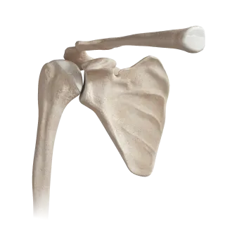
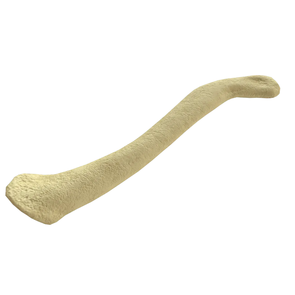
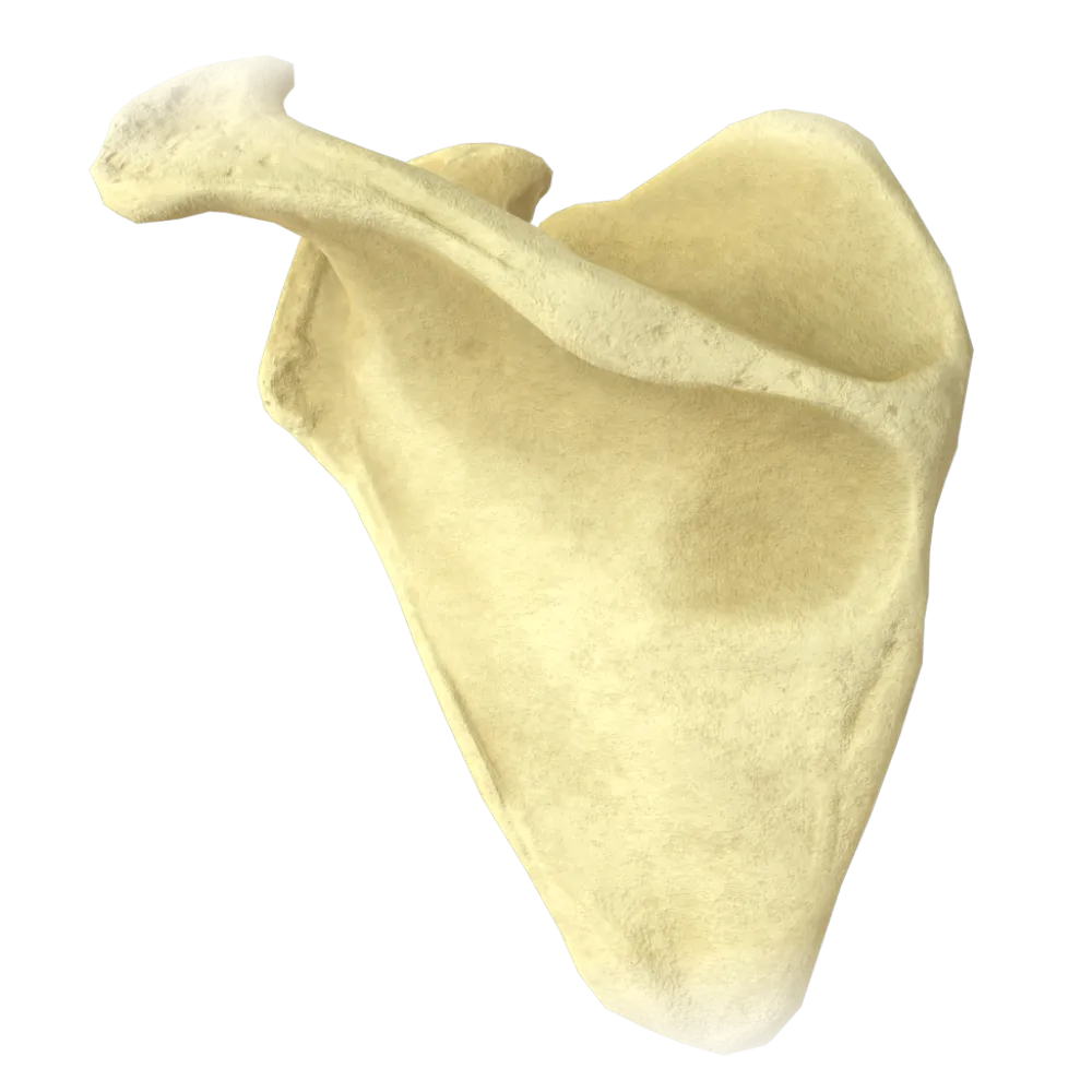
Pectoral Girdle Anatomy Quiz
Pectoral girdle normal anatomy quiz: bones, joints, muscles, vessels, and nerves.
1. Which bone forms the acromioclavicular joint together with the clavicle?
-
Scapula (acromion)
The acromioclavicular joint is formed by the acromion of the scapula and the acromial end of the clavicle. It is a flat joint with an articular disc.
-
Humerus (head)
The acromioclavicular joint is formed by the acromion of the scapula and the acromial end of the clavicle. It is a flat joint with an articular disc.
-
Sternum (manubrium)
The acromioclavicular joint is formed by the acromion of the scapula and the acromial end of the clavicle. It is a flat joint with an articular disc.
-
Ribs (first rib)
The acromioclavicular joint is formed by the acromion of the scapula and the acromial end of the clavicle. It is a flat joint with an articular disc.
-
I find it difficult to answer
The acromioclavicular joint is formed by the acromion of the scapula and the acromial end of the clavicle. It is a flat joint with an articular disc.
2. Based on the shape of its articular surfaces, what type of joint is the sternoclavicular joint?
-
Ball-and-socket
The sternoclavicular joint is a saddle-shaped (sellar) joint based on the shape of articular surfaces. Despite this, due to the intra-articular disc, it functions as a ball-and-socket joint, permitting movement in three axes.
-
Ellipsoidal
The sternoclavicular joint is a saddle-shaped (sellar) joint based on the shape of articular surfaces. Despite this, due to the intra-articular disc, it functions as a ball-and-socket joint, permitting movement in three axes.
-
Saddle-shaped
The sternoclavicular joint is a saddle-shaped (sellar) joint based on the shape of articular surfaces. Despite this, due to the intra-articular disc, it functions as a ball-and-socket joint, permitting movement in three axes.
-
Flat
The sternoclavicular joint is a saddle-shaped (sellar) joint based on the shape of articular surfaces. Despite this, due to the intra-articular disc, it functions as a ball-and-socket joint, permitting movement in three axes.
-
I find it difficult to answer
The sternoclavicular joint is a saddle-shaped (sellar) joint based on the shape of articular surfaces. Despite this, due to the intra-articular disc, it functions as a ball-and-socket joint, permitting movement in three axes.
3. Which ligament prevents upward displacement of the clavicle in the acromioclavicular joint?
-
Lig. coracoacromiale
Lig. coracoclaviculare, consisting of lig. trapezoideum and lig. conoideum, prevents the upward displacement of the clavicle by attaching to the coracoid process of the scapula.
-
Lig. coracoclaviculare
Lig. coracoclaviculare, consisting of lig. trapezoideum and lig. conoideum, prevents the upward displacement of the clavicle by attaching to the coracoid process of the scapula.
-
Lig. costoclaviculare
Lig. coracoclaviculare, consisting of lig. trapezoideum and lig. conoideum, prevents the upward displacement of the clavicle by attaching to the coracoid process of the scapula.
-
Lig. interclaviculare
Lig. coracoclaviculare, consisting of lig. trapezoideum and lig. conoideum, prevents the upward displacement of the clavicle by attaching to the coracoid process of the scapula.
-
I find it difficult to answer
Lig. coracoclaviculare, consisting of lig. trapezoideum and lig. conoideum, prevents the upward displacement of the clavicle by attaching to the coracoid process of the scapula.
4. What is the glenoid cavity of the scapula?
-
Infraspinous fossa on the posterior surface of the scapula
The glenoid cavity is the articular socket of the scapula, located at the lateral angle, articulating with the head of the humerus to form the shoulder joint.
-
Glenoid cavity for the head of the humerus
The glenoid cavity is the articular socket of the scapula, located at the lateral angle, articulating with the head of the humerus to form the shoulder joint.
-
Subscapular fossa on the costal surface of the scapula
The glenoid cavity is the articular socket of the scapula, located at the lateral angle, articulating with the head of the humerus to form the shoulder joint.
-
Supraspinous fossa above the spine of the scapula
The glenoid cavity is the articular socket of the scapula, located at the lateral angle, articulating with the head of the humerus to form the shoulder joint.
-
I find it difficult to answer
The glenoid cavity is the articular socket of the scapula, located at the lateral angle, articulating with the head of the humerus to form the shoulder joint.
5. Which muscle originates from the coracoid process of the scapula together with the short head of the biceps brachii?
-
M. subscapularis
M. coracobrachialis (coracobrachialis muscle) originates from the coracoid process of the scapula together with the short head of the m. biceps brachii. It is Innervated by the musculocutaneous nerve.
-
M. coracobrachialis
M. coracobrachialis (coracobrachialis muscle) originates from the coracoid process of the scapula together with the short head of the m. biceps brachii. It is Innervated by the musculocutaneous nerve.
-
M. pectoralis minor
M. coracobrachialis (coracobrachialis muscle) originates from the coracoid process of the scapula together with the short head of the m. biceps brachii. It is Innervated by the musculocutaneous nerve.
-
M. serratus anterior
M. coracobrachialis (coracobrachialis muscle) originates from the coracoid process of the scapula together with the short head of the m. biceps brachii. It is Innervated by the musculocutaneous nerve.
-
I find it difficult to answer
M. coracobrachialis (coracobrachialis muscle) originates from the coracoid process of the scapula together with the short head of the m. biceps brachii. It is Innervated by the musculocutaneous nerve.
6. Which nerve innervates the m. trapezius?
-
N. thoracicus longus
M. trapezius is innervated by the accessory nerve (n. accessorius, XI pair of cranial nerves), as well as by branches of the cervical plexus (C3–C4), providing proprioceptive innervation.
-
N. accessorius (XI cranial nerve pair) and cervical plexus
M. trapezius is innervated by the accessory nerve (n. accessorius, XI pair of cranial nerves), as well as by branches of the cervical plexus (C3–C4), providing proprioceptive innervation.
-
N. suprascapularis
M. trapezius is innervated by the accessory nerve (n. accessorius, XI pair of cranial nerves), as well as by branches of the cervical plexus (C3–C4), providing proprioceptive innervation.
-
Axillary nerve
M. trapezius is innervated by the accessory nerve (n. accessorius, XI pair of cranial nerves), as well as by branches of the cervical plexus (C3–C4), providing proprioceptive innervation.
-
I find it difficult to answer
M. trapezius is innervated by the accessory nerve (n. accessorius, XI pair of cranial nerves), as well as by branches of the cervical plexus (C3–C4), providing proprioceptive innervation.
7. Which artery is the primary source of blood supply to the muscles of the scapular region through the suprascapular and circumflex scapular arteries?
-
A. axillaris
A. subclavia gives rise to a. suprascapularis (via truncus thyrocervicalis) and a. transversae cervicis. A. circumflexa scapulae — branch of a. subscapularis from a. axillaris. The main trunk supplying blood vessels to the scapula is a. subclavia.
-
A. subclavia
A. subclavia gives rise to a. suprascapularis (via truncus thyrocervicalis) and a. transversae cervicis. A. circumflexa scapulae — branch of a. subscapularis from a. axillaris. The main trunk supplying blood vessels to the scapula is a. subclavia.
-
A. brachialis
A. subclavia gives rise to a. suprascapularis (via truncus thyrocervicalis) and a. transversae cervicis. A. circumflexa scapulae — branch of a. subscapularis from a. axillaris. The main trunk supplying blood vessels to the scapula is a. subclavia.
-
A. thoracoacromialis
A. subclavia gives rise to a. suprascapularis (via truncus thyrocervicalis) and a. transversae cervicis. A. circumflexa scapulae — branch of a. subscapularis from a. axillaris. The main trunk supplying blood vessels to the scapula is a. subclavia.
-
I find it difficult to answer
A. subclavia gives rise to a. suprascapularis (via truncus thyrocervicalis) and a. transversae cervicis. A. circumflexa scapulae — branch of a. subscapularis from a. axillaris. The main trunk supplying blood vessels to the scapula is a. subclavia.
8. Which muscle moves the scapula away from the spine and rotates its inferior angle laterally?
-
M. rhomboideus major
M. serratus anterior is attached to the medial edge and inferior angle of the scapula. When contracted, it pulls the scapula forward and laterally, and also rotates its inferior angle outward, which is necessary for raising the arm above the horizontal plane. It is innervated by n. long thoracic.
-
M. levator scapulae
M. serratus anterior is attached to the medial edge and inferior angle of the scapula. When contracted, it pulls the scapula forward and laterally, and also rotates its inferior angle outward, which is necessary for raising the arm above the horizontal plane. It is innervated by n. long thoracic.
-
M. serratus anterior
M. serratus anterior is attached to the medial edge and inferior angle of the scapula. When contracted, it pulls the scapula forward and laterally, and also rotates its inferior angle outward, which is necessary for raising the arm above the horizontal plane. It is innervated by n. long thoracic.
-
M. trapezius (middle part)
M. serratus anterior is attached to the medial edge and inferior angle of the scapula. When contracted, it pulls the scapula forward and laterally, and also rotates its inferior angle outward, which is necessary for raising the arm above the horizontal plane. It is innervated by n. long thoracic.
-
I find it difficult to answer
M. serratus anterior is attached to the medial edge and inferior angle of the scapula. When contracted, it pulls the scapula forward and laterally, and also rotates its inferior angle outward, which is necessary for raising the arm above the horizontal plane. It is innervated by n. long thoracic.
9. Where is the lig. transversum scapulae superius located?
-
Above the acromion between its process and the coracoid process
Lig transversum scapulae superius bridges over the incisure scapulae, converting it into a foramen for the passage of n. suprascapularis. Through the ligament passes a. suprascapularis (above the ligament).
-
Above the scapular notch, converting it into a foramen
Lig transversum scapulae superius bridges over the incisure scapulae, converting it into a foramen for the passage of n. suprascapularis. Through the ligament passes a. suprascapularis (above the ligament).
-
Along the medial edge of the scapula
Lig transversum scapulae superius bridges over the incisure scapulae, converting it into a foramen for the passage of n. suprascapularis. Through the ligament passes a. suprascapularis (above the ligament).
-
Between the clavicle and the first rib
Lig transversum scapulae superius bridges over the incisure scapulae, converting it into a foramen for the passage of n. suprascapularis. Through the ligament passes a. suprascapularis (above the ligament).
-
I find it difficult to answer
Lig transversum scapulae superius bridges over the incisure scapulae, converting it into a foramen for the passage of n. suprascapularis. Through the ligament passes a. suprascapularis (above the ligament).
10. What is the insertion point of m. deltoideus on the humerus?
-
Crest of the greater tubercle
M. deltoideus is attached to the deltoid tuberosity on the lateral surface of the humeral body. It is innervated by n. axillaris (C5–C6).
-
Deltoid tuberosity of the humerus
M. deltoideus is attached to the deltoid tuberosity on the lateral surface of the humeral body. It is innervated by n. axillaris (C5–C6).
-
Surgical neck of the humerus
M. deltoideus is attached to the deltoid tuberosity on the lateral surface of the humeral body. It is innervated by n. axillaris (C5–C6).
-
Lesser tubercle of the humerus
M. deltoideus is attached to the deltoid tuberosity on the lateral surface of the humeral body. It is innervated by n. axillaris (C5–C6).
-
I find it difficult to answer
M. deltoideus is attached to the deltoid tuberosity on the lateral surface of the humeral body. It is innervated by n. axillaris (C5–C6).
11. Which nerve passes through the quadrilateral foramen together with the posterior circumflex humeral artery?
-
N. radialis
N. axillaris and a. circumflexa humeri posterior pass through the quadrilateral foramen, which is bounded: medially – by the long head of m. triceps, laterally – by the surgical neck of the arm, superiorly – by m. teres minor, inferiorly – by m. teres major.
-
N. musculocutaneus
N. axillaris and a. circumflexa humeri posterior pass through the quadrilateral foramen, which is bounded: medially – by the long head of m. triceps, laterally – by the surgical neck of the arm, superiorly – by m. teres minor, inferiorly – by m. teres major.
-
N. axillaris
N. axillaris and a. circumflexa humeri posterior pass through the quadrilateral foramen, which is bounded: medially – by the long head of m. triceps, laterally – by the surgical neck of the arm, superiorly – by m. teres minor, inferiorly – by m. teres major.
-
N. suprascapularis
N. axillaris and a. circumflexa humeri posterior pass through the quadrilateral foramen, which is bounded: medially – by the long head of m. triceps, laterally – by the surgical neck of the arm, superiorly – by m. teres minor, inferiorly – by m. teres major.
-
I find it difficult to answer
N. axillaris and a. circumflexa humeri posterior pass through the quadrilateral foramen, which is bounded: medially – by the long head of m. triceps, laterally – by the surgical neck of the arm, superiorly – by m. teres minor, inferiorly – by m. teres major.
12. Which of the following muscles is part of the rotator cuff of the shoulder?
-
M. deltoideus
The rotator cuff includes four muscles: m. supraspinatus, m. infraspinatus, m. teres minor, and m. Subscapularis. M. infraspinatus originates from the infraspinous fossa of the scapula and externally rotates the arm, being attached to the greater tubercle of the humerus.
-
Coracobrachialis
The rotator cuff includes four muscles: m. supraspinatus, m. infraspinatus, m. teres minor, and m. Subscapularis. M. infraspinatus originates from the infraspinous fossa of the scapula and externally rotates the arm, being attached to the greater tubercle of the humerus.
-
M. infraspinatus
The rotator cuff includes four muscles: m. supraspinatus, m. infraspinatus, m. teres minor, and m. Subscapularis. M. infraspinatus originates from the infraspinous fossa of the scapula and externally rotates the arm, being attached to the greater tubercle of the humerus.
-
M. pectoralis major
The rotator cuff includes four muscles: m. supraspinatus, m. infraspinatus, m. teres minor, and m. Subscapularis. M. infraspinatus originates from the infraspinous fossa of the scapula and externally rotates the arm, being attached to the greater tubercle of the humerus.
-
I find it difficult to answer
The rotator cuff includes four muscles: m. supraspinatus, m. infraspinatus, m. teres minor, and m. Subscapularis. M. infraspinatus originates from the infraspinous fossa of the scapula and externally rotates the arm, being attached to the greater tubercle of the humerus.
13. Which artery supplies m. supraspinatus and m. infraspinatus, entering the supraspinatus fossa through the foramen supraspinosum?
-
A. circumflexa humeri anterior
A. suprascapularis (branch of truncus thyrocervicalis from a. subclavia) passes over lig. transversum scapulae superius into the supraspinatus fossa, then curves around the base of the spine of the scapula into the infraspinatus fossa, supplying m. supraspinatus and m. infraspinatus.
-
A. suprascapularis
A. suprascapularis (branch of truncus thyrocervicalis from a. subclavia) passes over lig. transversum scapulae superius into the supraspinatus fossa, then curves around the base of the spine of the scapula into the infraspinatus fossa, supplying m. supraspinatus and m. infraspinatus.
-
A. subscapularis
A. suprascapularis (branch of truncus thyrocervicalis from a. subclavia) passes over lig. transversum scapulae superius into the supraspinatus fossa, then curves around the base of the spine of the scapula into the infraspinatus fossa, supplying m. supraspinatus and m. infraspinatus.
-
A. thoracoacromialis
A. suprascapularis (branch of truncus thyrocervicalis from a. subclavia) passes over lig. transversum scapulae superius into the supraspinatus fossa, then curves around the base of the spine of the scapula into the infraspinatus fossa, supplying m. supraspinatus and m. infraspinatus.
-
I find it difficult to answer
A. suprascapularis (branch of truncus thyrocervicalis from a. subclavia) passes over lig. transversum scapulae superius into the supraspinatus fossa, then curves around the base of the spine of the scapula into the infraspinatus fossa, supplying m. supraspinatus and m. infraspinatus.
14. Where does m. subscapularis start?
-
From the supraspinatus fossa of the scapula
M. subscapularis originates from the fossa subscapularis — the costal (anterior) surface of the scapula. It is attached to the lesser tubercle of the humerus, acting as a medial rotator and adductor of the arm. It is innervated by nn. subscapulares.
-
From the infraspinatus fossa of the scapula
M. subscapularis originates from the fossa subscapularis — the costal (anterior) surface of the scapula. It is attached to the lesser tubercle of the humerus, acting as a medial rotator and adductor of the arm. It is innervated by nn. subscapulares.
-
From the subscapular fossa
M. subscapularis originates from the fossa subscapularis — the costal (anterior) surface of the scapula. It is attached to the lesser tubercle of the humerus, acting as a medial rotator and adductor of the arm. It is innervated by nn. subscapulares.
-
From the posterior surface of the clavicle
M. subscapularis originates from the fossa subscapularis — the costal (anterior) surface of the scapula. It is attached to the lesser tubercle of the humerus, acting as a medial rotator and adductor of the arm. It is innervated by nn. subscapulares.
-
I find it difficult to answer
M. subscapularis originates from the fossa subscapularis — the costal (anterior) surface of the scapula. It is attached to the lesser tubercle of the humerus, acting as a medial rotator and adductor of the arm. It is innervated by nn. subscapulares.
15. Which ligament connects the acromion with the coracoid process of the scapula, forming the "shoulder arch"?
-
Lig coracoclavicular
Lig coracoacromial stretches between the acromion and the coracoid process of the scapula. Along with these structures, it forms the coracoacromial arch, protecting the shoulder joint from above.
-
Lig coracoacromial
Lig coracoacromial stretches between the acromion and the coracoid process of the scapula. Along with these structures, it forms the coracoacromial arch, protecting the shoulder joint from above.
-
Lig transversum scapulae superius
Lig coracoacromial stretches between the acromion and the coracoid process of the scapula. Along with these structures, it forms the coracoacromial arch, protecting the shoulder joint from above.
-
Lig glenohumeral
Lig coracoacromial stretches between the acromion and the coracoid process of the scapula. Along with these structures, it forms the coracoacromial arch, protecting the shoulder joint from above.
-
I find it difficult to answer
Lig coracoacromial stretches between the acromion and the coracoid process of the scapula. Along with these structures, it forms the coracoacromial arch, protecting the shoulder joint from above.
16. Which nerve innervates the m. serratus anterior?
-
Axillary nerve
The long thoracic nerve (C5–C7) innervates the serratus anterior. serratus anterior. Damage to this nerve causes the medial border of the scapula to protrude, known as the 'winged scapula' (scapula alata).
-
N. thoracicus longus
The long thoracic nerve (C5–C7) innervates the serratus anterior. serratus anterior. Damage to this nerve causes the medial border of the scapula to protrude, known as the 'winged scapula' (scapula alata).
-
Inferior subscapular nerve
The long thoracic nerve (C5–C7) innervates the serratus anterior. serratus anterior. Damage to this nerve causes the medial border of the scapula to protrude, known as the 'winged scapula' (scapula alata).
-
Medial pectoral nerve
The long thoracic nerve (C5–C7) innervates the serratus anterior. serratus anterior. Damage to this nerve causes the medial border of the scapula to protrude, known as the 'winged scapula' (scapula alata).
-
I find it difficult to answer
The long thoracic nerve (C5–C7) innervates the serratus anterior. serratus anterior. Damage to this nerve causes the medial border of the scapula to protrude, known as the 'winged scapula' (scapula alata).
17. What is the primary movement performed by the m. supraspinatus?
-
Adduction of the shoulder
The supraspinatus muscle initiates shoulder abduction (first 10–15°), after which the primary role is taken over by the deltoid muscle. m. deltoideus. It is innervated by n. suprascapularis (C4–C6). It is attached to the greater tubercle of the humerus.
-
Medial rotation of the shoulder
The supraspinatus muscle initiates shoulder abduction (first 10–15°), after which the primary role is taken over by the deltoid muscle. m. deltoideus. It is innervated by n. suprascapularis (C4–C6). It is attached to the greater tubercle of the humerus.
-
Abduction of the shoulder (initial 15°)
The supraspinatus muscle initiates shoulder abduction (first 10–15°), after which the primary role is taken over by the deltoid muscle. m. deltoideus. It is innervated by n. suprascapularis (C4–C6). It is attached to the greater tubercle of the humerus.
-
Extension of the shoulder
The supraspinatus muscle initiates shoulder abduction (first 10–15°), after which the primary role is taken over by the deltoid muscle. m. deltoideus. It is innervated by n. suprascapularis (C4–C6). It is attached to the greater tubercle of the humerus.
-
I find it difficult to answer
The supraspinatus muscle initiates shoulder abduction (first 10–15°), after which the primary role is taken over by the deltoid muscle. m. deltoideus. It is innervated by n. suprascapularis (C4–C6). It is attached to the greater tubercle of the humerus.
18. What is the origin of a. circumflexa scapulae?
-
A. suprascapularis
A. circumflexa scapulae is a branch of the a. subscapularis, which, in turn, branches from the a. axillaris branch from. It passes through the triangular space to the posterior surface of the scapula, participating in the scapular anastomosis.
-
A. subscapularis
A. circumflexa scapulae is a branch of the a. subscapularis, which, in turn, branches from the a. axillaris branch from. It passes through the triangular space to the posterior surface of the scapula, participating in the scapular anastomosis.
-
A. thoracoacromialis
A. circumflexa scapulae is a branch of the a. subscapularis, which, in turn, branches from the a. axillaris branch from. It passes through the triangular space to the posterior surface of the scapula, participating in the scapular anastomosis.
-
A. circumflexa humeri posterior
A. circumflexa scapulae is a branch of the a. subscapularis, which, in turn, branches from the a. axillaris branch from. It passes through the triangular space to the posterior surface of the scapula, participating in the scapular anastomosis.
-
I find it difficult to answer
A. circumflexa scapulae is a branch of the a. subscapularis, which, in turn, branches from the a. axillaris branch from. It passes through the triangular space to the posterior surface of the scapula, participating in the scapular anastomosis.
19. Which muscles bound the triangular space?
-
M. teres minor (superiorly), m. teres major (inferiorly), the long head of the m. triceps (laterally)
The triangular space is bounded: superiorly – by m. teres minor, inferiorly – by m. teres major muscle, laterally (medial wall) – by the long head of m. Triceps brachii. Through it passes a. circumflex scapulae.
-
M. teres minor (superiorly), m. teres major (inferiorly), the long head of the m. triceps (medially)
The triangular space is bounded: superiorly – by m. teres minor, inferiorly – by m. teres major muscle, laterally (medial wall) – by the long head of m. Triceps brachii. Through it passes a. circumflex scapulae.
-
M. subscapularis (superiorly), m. teres major (inferiorly), m. deltoideus (laterally)
The triangular space is bounded: superiorly – by m. teres minor, inferiorly – by m. teres major muscle, laterally (medial wall) – by the long head of m. Triceps brachii. Through it passes a. circumflex scapulae.
-
M. infraspinatus (superiorly), m. teres minor (inferiorly), m. deltoideus (medially)
The triangular space is bounded: superiorly – by m. teres minor, inferiorly – by m. teres major muscle, laterally (medial wall) – by the long head of m. Triceps brachii. Through it passes a. circumflex scapulae.
-
I find it difficult to answer
The triangular space is bounded: superiorly – by m. teres minor, inferiorly – by m. teres major muscle, laterally (medial wall) – by the long head of m. Triceps brachii. Through it passes a. circumflex scapulae.
20. What innervates the m. infraspinatus?
-
N. axillaris (C5–C6)
M. infraspinatus is innervated by the n. suprascapularis (C4–C6) – a branch of the superior trunk of the brachial plexus. The same nerve innervates the m. supraspinatus. The nerve enters the infraspinous fossa, encircling the base of the scapular spine.
-
N. suprascapularis (C4–C6)
M. infraspinatus is innervated by the n. suprascapularis (C4–C6) – a branch of the superior trunk of the brachial plexus. The same nerve innervates the m. supraspinatus. The nerve enters the infraspinous fossa, encircling the base of the scapular spine.
-
N. subscapularis inferior (C5–C6)
M. infraspinatus is innervated by the n. suprascapularis (C4–C6) – a branch of the superior trunk of the brachial plexus. The same nerve innervates the m. supraspinatus. The nerve enters the infraspinous fossa, encircling the base of the scapular spine.
-
N. thoracodorsalis (C6–C8)
M. infraspinatus is innervated by the n. suprascapularis (C4–C6) – a branch of the superior trunk of the brachial plexus. The same nerve innervates the m. supraspinatus. The nerve enters the infraspinous fossa, encircling the base of the scapular spine.
-
I find it difficult to answer
M. infraspinatus is innervated by the n. suprascapularis (C4–C6) – a branch of the superior trunk of the brachial plexus. The same nerve innervates the m. supraspinatus. The nerve enters the infraspinous fossa, encircling the base of the scapular spine.
Retake this quiz?
Your current progress will be reset.

Cookie Consent
We use cookies to enhance your browsing experience, analyze site traffic, and deliver content. Please choose whether you accept all cookies or wish to reject non-essential tracking.
Cookie Preferences
Manage your cookie preferences below:
Essential cookies enable basic functions and are necessary for the proper function of the website.
Name
Description
Duration
Geolocation Config
This cookie is used to store the consent settings based on the visitor's location.
30 days
Cookie Preferences
This cookie is used to store the user's cookie consent preferences.
30 days
Google reCAPTCHA helps protect websites from spam and abuse by verifying user interactions through challenges.
Name
Description
Duration
_GRECAPTCHA
Google reCAPTCHA sets a necessary cookie (_GRECAPTCHA) when executed for the purpose of providing its risk analysis.
179 days
Statistics cookies collect information anonymously. This information helps us understand how visitors use our website.
Google Analytics is a powerful tool that tracks and analyzes website traffic for informed marketing decisions.
Service URL: policies.google.com (opens in a new window)
Name
Description
Duration
__utma
ID used to identify users and sessions
2 years after last activity
__utmt
Used to monitor number of Google Analytics server requests
10 minutes
__utmb
Used to distinguish new sessions and visits. This cookie is set when the GA.js javascript library is loaded and there is no existing __utmb cookie. The cookie is updated every time data is sent to the Google Analytics server.
30 minutes after last activity
__utmc
Used only with old Urchin versions of Google Analytics and not with GA.js. Was used to distinguish between new sessions and visits at the end of a session.
End of session (browser)
__utmz
Contains information about the traffic source or campaign that directed user to the website. The cookie is set when the GA.js javascript is loaded and updated when data is sent to the Google Anaytics server
6 months after last activity
__utmv
Contains custom information set by the web developer via the _setCustomVar method in Google Analytics. This cookie is updated every time new data is sent to the Google Analytics server.
2 years after last activity
__utmx
Used to determine whether a user is included in an A / B or Multivariate test.
18 months
_ga
ID used to identify users
2 years
_gali
Used by Google Analytics to determine which links on a page are being clicked
30 seconds
_ga_
ID used to identify users
2 years
_gid
ID used to identify users for 24 hours after last activity
24 hours
_gat
Used to monitor number of Google Analytics server requests when using Google Tag Manager
1 minute
_gac_
Contains information related to marketing campaigns of the user. These are shared with Google AdWords / Google Ads when the Google Ads and Google Analytics accounts are linked together.
90 days
Clarity is a web analytics service that tracks and reports website traffic.
Service URL: clarity.microsoft.com (opens in a new window)
Name
Description
Duration
_clsk
Connects multiple page views by a user into a single Clarity session recording.
1 day
_clck
Stores a unique user identifier to analyze visitor behavior through heatmaps and session recordings.
1 year
ANONCHK
Indicates whether MUID is transferred to ANID, a cookie used for advertising. Clarity doesn't use ANID and so this is always set to 0.
Session
CLID
Identifies the first-time Clarity saw this user on any site using Clarity.
12 months
_clsk
Connects multiple page views by a user into a single Clarity session recording.
12 months
_clck
Persists the Clarity User ID and preferences, unique to that site is attributed to the same user ID.
12 months