


Acute Sinusitis (Acute Rhinosinusitis): Classification, Clinical Manifestations, Diagnosis, and Treatment
A detailed review of rhinosinusitis, including classification, symptoms, diagnostic approaches, and current treatment strategies.
Anesthesia
Pain management and sedation techniques
Angiology
Arterial and venous pathologies
Cardiology
Acquired and congenital heart diseases
Dentistry
Diseases of teeth, gums, and the oral cavity
Dermatology
Disorders of the skin and subcutaneous tissue
Endocrinology
Disorders of the glands and hormonal imbalance
Gastroenterology
Stomach, intestinal, and digestive diseases
Gynecology
Diseases of female reproductive organs
Hepatology
Liver, gallbladder, and biliary tract diseases
Neurology
Brain, spinal cord, and peripheral nerve disorders
Obstetrics
Pregnancy complications and abnormal fetal positions
Oncology
Cancer types, benign and malignant tumors
Ophthalmology
Conditions affecting the eyes and vision
Otorhinolaryngology
Ear, nose, and throat diseases
Pediatrics
Child health, development, and clinical conditions
Physiology
Biological processes within organs and systems
Pulmonology
Lung and respiratory tract diseases
Traumatology
Acute injuries and musculoskeletal trauma
Urology
Urinary tract and male reproductive disorders

This article is for informational purposes only
The content on this website, including text, graphics, and other materials, is provided for informational purposes only. It is not intended as advice or guidance. Regarding your specific medical condition or treatment, please consult your healthcare provider.
Nasal bleeding(epistaxis) – bleeding from a blood vessel in the nasal cavity when its integrity or permeability is compromised. It’s the most common type of bleeding.
| Localization | – anterior bleeding – posterior bleeding |
| Etiology | – traumatic – caused by local processes – caused by common diseases |
| The amount of blood loss | – scant bleeding – moderate bleeding – heavy bleeding |
By origin, nosebleeds are caused by trauma, local or general causes. Only 20% are due to pathology of the nasal cavity and about 80% are related to general diseases.
Traumatic causes of nasal bleeding are the most common, they also include intra- and postoperative bleeding in the nasal cavity and nasopharynx (septoplasty, nasal resection, adenotomy, sinusotomy).
Local causes that manifest epistaxis include atrophic processes in the nasal cavity (occur in elderly people, with low air humidity in households), which, if prolonged, can lead to ulceration of the mucosa and perforation of the nasal septum, aggravating the course of bleeding. Nasal foreign bodies, self-cleaning of the nose from dry crusts, infectious processes (rhinitis, sinusitis, viral infection) damage the mucous membrane of the nasal cavity, the vessel wall and are also causes of neobilic nasal bleeding. Local causes are benign (juvenile angiofibroma of the nasopharynx, capillary or cavernous hemangiomas of the nasal cavity, papillomas) and malignant (carcinoma and sarcoma of the nose) neoplasms of the nasal cavity. Local manifestations of specific diseases such as tuberculoma, ozena, various stages of syphilis (primary syphiloma, secondary syphilitic papules and ulcers, syphilitic gumma) are extremely rare in recent times.
Common diseases that are manifested by nosebleeds can be divided into several groups: Diseases of the cardiovascular system (atherosclerosis, hypertension, hereditary hemorrhagic telangiectasia – Osler-Weber-Randu syndrome), liver (cirrhosis, hepatitis), pathologies of the blood system and hemostasis (hemophilia, coagulopathies, thrombocytopenia, leukemia, hemoblastosis), side effects of certain drugs (NSAIDs, anticoagulants).
Separately consider bleeding against the background of hormonal changes (during pregnancy, menopause) and changes associated with the menstrual cycle in women (vicarious – accompanying menstrual bleeding).
Nasal bleeding is based on different pathologic processes occurring in the nasal cavity with vessel walls. Anatomically it is important to separate the source of bleeding, as it characterizes the volume of possible blood loss. A feature of the blood supply of the nasal cavity is the abundance of anastomoses between branches of the internal and external carotid artery. Anterior hemorrhage more often corresponds to the Kiesselbach area (a well-developed network of blood vessels) located on the septum in the vestibule of the nose. The Kiesselbach area is formed by the plexus of the anterior and posterior lattice arteries (branches of the internal carotid artery), the cuneiform palatine artery, the greater palatine artery and the superior labial artery (branches of the external carotid artery).
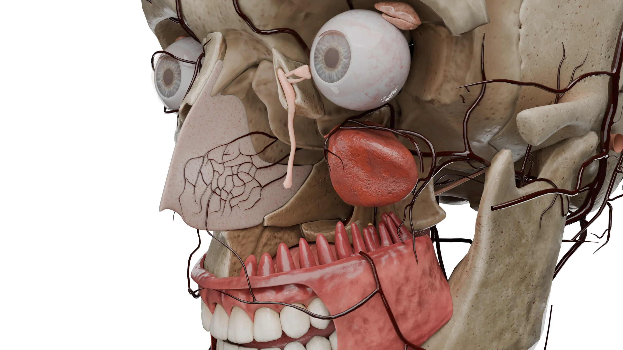
Posterior hemorrhage corresponds to damage directly to the to the anterior or posterior lattice arteries..
In case of traumas, surgeries and local pathological processes in the nasal cavity, traumatization (rupture) of the vascular wall occurs, accompanied by bleeding. Nasal cavity neoplasms and various infectious ulcers can bleed due to their independent disintegration. Diseases of the cardiovascular system are characterized by pathological changes in the vessel wall, there is a dystrophy of the endothelial layer, increased thrombosis, which contributes to additional necrosis of the vascular wall and its sclerosis, all this thins the vessel wall. Against the background of these processes can develop as blood soaking through the vessel wall (diapedesis), and its rupture in the most damaged areas with profuse bleeding, increased blood pressure worsens the course. In diseases of the liver and blood system there is a violation of vascular-thrombocytic hemostasis: the formation of clotting factors is reduced, the process of thrombosis and platelet aggregation, which is important for the construction of the endothelium of the vascular wall, as platelets perform including angiotrophic function, there is a syndrome of localized vascular coagulation. All this also leads to diapedesis or rupture of the vessel, further hemostasis, due to impaired blood coagulation is impaired and bleeding time is prolonged.
Epistaxis with changes in the hormonal background in women during pregnancy occurs due to an increase in progesterone levels, which contributes to vasodilation not only in the placenta, but also in the nasal cavity, as well as increased vascular permeability, the nasal mucosa becomes more friable and easily traumatized. The cause of vicarious bleeding has not yet been determined, it is assumed that endometrial tissue migrates to atypical localizations (nasal cavity, thoracic and abdominal cavities), where it sloughs off and bleeds during menstruation.
Nasal bleeding is manifested by the oozing of blood from the nasal cavity through the vestibule of the nose or through the nasopharynx.
Depending on the amount of blood loss, there is scanty bleeding (a few milliliters), moderate (up to 200-250 ml), profuse (over 250-300 ml). Some patients may experience prodroma before the onset of bleeding: tinnitus, flies in front of the eyes, itching in the nose.
Scanty bleeding stops on its own, no additional medical care is required, more often the source is Kiesselbach area, it develops due to local pathological processes in the nasal cavity, often atrophic, lasts a few minutes, the general condition is not affected. Blood is bright scarlet in color, bleeds through the nasal vestibule in drops or a thin stream.
Moderate and profuse bleeding is characterized by a stream of blood, mostly dark in color, both through the vestibule of the nose and flowing down the back of the pharynx, which may be swallowed, leading to hematemesis (vomiting blood) or melena (black stools). In case of significant blood loss in a short time, more than 1 liter, the general symptoms of hypovolemic shock appear: cold clammy sweat, rapid breathing, drop in blood pressure, increased heart rate, headache, flickers before the eyes, nausea and vomiting, loss of consciousness.
Epistaxis occurring on the background of general diseases is characterized by scanty or moderate periodic bleeding and recurrent course, in which it is impossible to adequately assess the total amount of blood loss, which leads to chronic posthemorrhagic anemia. In nasal injuries with fracture of the lattice labyrinth, the anterior and posterior lattice arteries are damaged, which is accompanied by severe profuse nasal bleeding and the development of hypovolemic shock.
It should be remembered that through the nose can be released blood from the pulmonary or gastrointestinal tract, middle ear cavity through the auditory tube, which can be mistaken as a nosebleed and increase the time to diagnose and prescribe adequate treatment.
To establish the source of bleeding, if possible – after clearing the nose of blood clots, rhinopharyngoscopy is performed, in some cases endoscopy of the nasal cavity is performed.
It is necessary to establish the duration of bleeding, provoking factors, collect a detailed history, including family history (presence of hereditary diseases) and medication (taking anticoagulants, NSAIDs).
It is obligatory to measure blood pressure, heart rate.
Laboratory values are being evaluated:
In case of persistent profuse recurrent bleeding, when the source is not found, angiography of the internal and external carotid arteries is performed, if necessary with further embolization of the branches that are the cause of epistaxis.
First of all, the patient should be given the correct position – verticalization with the head tilted forward and down, a cold compress should be placed on the bridge of the nose and occiput, the wing of the nose should be pressed against the septum, a tampon moistened with 3% hydrogen peroxide or local decongestants can be placed in the nasal passage. In most cases, these actions contribute to stopping bleeding, if this does not happen – resort to the help of medical professionals.
Find more scientifically accurate content on our social media


Anterior nasal tamponade is performed with a hemostatic sponge, balloon catheter, acetate sponge with air ducts, or gauze tubes. Gauze tamponade is the most accessible and popular, but also the most painful.
To perform it, the nasal mucosa is anesthetized with anesthetic solution, a turunda moistened with bactericidal ointment or vaseline oil, 70-100 cm long, is inserted through the nasal vestibule to the choans and placed from bottom to top in layers, densely filling the entire nasal cavity, additionally impregnated with aminocaproic acid or fibrin solution. To achieve the best effect, the tampon remains in the nasal cavity for 3-5 days, until the formation of a dense thrombus, to prevent bacterial complications, an antibiotic is administered inside and regular impregnation of the tampon with antiseptic solutions, after the expiration of the required period the tampon is removed.
In some cases, anterior tamponade is not enough and posterior tamponade is performed, in which a tampon or balloon is inserted through the nasal cavity into the nasopharynx, where it is then inflated by filling it with sterile solution. After the balloon is inserted, an anterior tamponade is performed. This procedure is very painful and is performed under sedation. These patients are usually seen in the intensive care unit or ICU. If this method of stopping bleeding is ineffective, vessel ligation or embolization by angiography is performed.
In addition to local methods of stopping bleeding additionally prescribe styptic drugs (ethamsylate, aminocapron or tranexamic acid, blood components – plasma, platelet mass), replenish the volume of circulating blood, necessarily cancel drugs that may have contributed to the development of bleeding.
In case of recurrent nosebleeds, electrocoagulation of nasal cavity vessels is performedor mucosal detachment, which promotes scarring and sclerosization of the causative vessel.
In cases where the cause of bleeding is a background disease, allied specialists (internists, cardiologists, hepatologists, hepatologists, hematologists, oncohematologists, etc.) join the treatment.
1. Where’s the nosebleed coming from?
2. What are the dangers of a nosebleed?
3. What should not be done during a nosebleed?
4. Causes of frequent nosebleeds in children
List of Sources
1.
VOKA Catalog.
https://catalog.voka.io/
2.
Total Otolaryngology—Head and Neck Surgery, Anthony P. Sclafani, Robin A. Dyleski, Michael J. Pitman, Stimson P. Schantz. Thieme Medical Publishers, Inc., 2015. ISBN 978-1-60406-646-3.
3.
Бербом Х. Болезни уха, горла и носа / Ханс Бербом, Оливер Кашке, Тадеус Навка, Эндрю Свифт; пер. с англ. – 2-е изд. – М. : МЕДпреcс-информ, 2016. – 776 с. : ил. ISBN 978-5-00030- 322-1.
4.
Tabassom A, Dahlstrom JJ. Epistaxis. [Updated 2022 Sep 12]. In: StatPearls [Internet]. Treasure Island (FL): StatPearls Publishing; 2025 Jan-. Available from:
https://www.ncbi.nlm.nih.gov/books/NBK435997/
5.
Tunkel DE, Anne S, Payne SC, Ishman SL, Rosenfeld RM, Abramson PJ, Alikhaani JD, Benoit MM, Bercovitz RS, Brown MD, Chernobilsky B, Feldstein DA, Hackell JM, Holbrook EH, Holdsworth SM, Lin KW, Lind MM, Poetker DM, Riley CA, Schneider JS, Seidman MD, Vadlamudi V, Valdez TA, Nnacheta LC, Monjur TM. Clinical Practice Guideline: Nosebleed (Epistaxis). Otolaryngol Head Neck Surg. 2020 Jan;162(1_suppl):S1-S38. doi: 10.1177/0194599819890327. PMID: 31910111.
6.
Mylonas S, Skoulakis C, Nikolaidis V, Hajiioannou J. Epistaxis Treatment Options: Literature Review. Indian J Otolaryngol Head Neck Surg. 2023 Sep;75(3):2235-2244. doi: 10.1007/s12070-023-03824-z. Epub 2023 May 8. PMID: 37636777; PMCID: PMC10447774.


Summarize article with AI
Choose your preferable AI assistant:
Link successfully copied to clipboard
Thank you!
Your message is sent!
Our experts will contact you shortly. If you have any additional questions, please contact us at info@voka.io


