


Acute Sinusitis (Acute Rhinosinusitis): Classification, Clinical Manifestations, Diagnosis, and Treatment
A detailed review of rhinosinusitis, including classification, symptoms, diagnostic approaches, and current treatment strategies.
Anesthesia
Pain management and sedation techniques
Angiology
Arterial and venous pathologies
Cardiology
Acquired and congenital heart diseases
Dentistry
Diseases of teeth, gums, and the oral cavity
Dermatology
Disorders of the skin and subcutaneous tissue
Endocrinology
Disorders of the glands and hormonal imbalance
Gastroenterology
Stomach, intestinal, and digestive diseases
Gynecology
Diseases of female reproductive organs
Hepatology
Liver, gallbladder, and biliary tract diseases
Neurology
Brain, spinal cord, and peripheral nerve disorders
Obstetrics
Pregnancy complications and abnormal fetal positions
Oncology
Cancer types, benign and malignant tumors
Ophthalmology
Conditions affecting the eyes and vision
Otorhinolaryngology
Ear, nose, and throat diseases
Pediatrics
Child health, development, and clinical conditions
Physiology
Biological processes within organs and systems
Pulmonology
Lung and respiratory tract diseases
Traumatology
Acute injuries and musculoskeletal trauma
Urology
Urinary tract and male reproductive disorders

This article is for informational purposes only
The content on this website, including text, graphics, and other materials, is provided for informational purposes only. It is not intended as advice or guidance. Regarding your specific medical condition or treatment, please consult your healthcare provider.
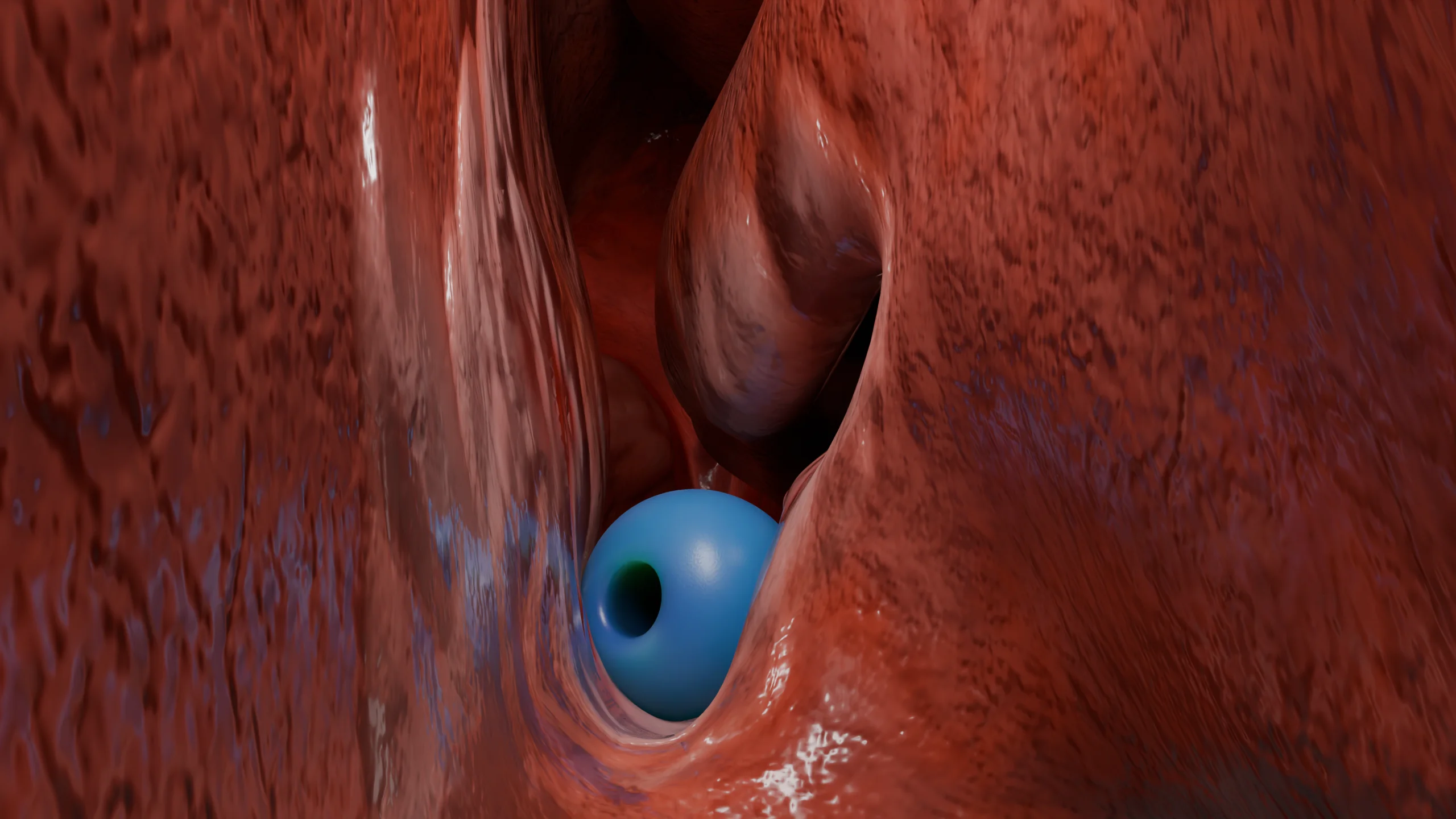
Foreign bodies in the nose, paranasal sinuses, and throat are pathological conditions in which external objects enter the human body. These foreign bodies get stuck in or on the surface of tissues, causing discomfort and unpleasant sensations. If left for a prolonged period, they can contribute to the development of chronic infections.
Living foreign bodies in the nasal cavity are more common among residents of tropical regions, although isolated cases have been described in residents of central regions of Russia. Among living foreign bodies, one can encounter larvae, leeches, helminths, and arthropods. Flies and botflies lay their larvae in the nasal cavity, arthropods crawl in during sleep outdoors, and leeches enter when swimming in water bodies.
Non-living foreign bodies are more common in adults with mental disorders and in children who insert objects during play or exploration of their environment. However, there are cases of foreign bodies entering during vomiting (food remnants), facial injuries, or forgotten during surgical procedures (often dressing materials).
They are categorized as:
Rhinoliths, or nasal stones, form either by the deposition of salts of various microelements on a foreign body or by the accumulation of these salts in poorly ventilated areas of the nasal cavity where mucus accumulates abundantly (floor of the nasal cavity, inferior nasal meatus).
Foreign bodies of the sinuses come from the nasal cavity or from the oral cavity, if there is a pathologic communication(oroantral junction). The maxillary sinuses are most often affected. The presence of filling material in the sinuses indicates earlier dental manipulations, during the performance of which the integrity of the upper jaw was violated with penetration of the wall into the maxillary sinus. Teeth in the maxillary sinuses are extremely rare and may be the result of a violation of embryogenesis (the tooth develops inside the sinus instead of the maxilla), displacement of the tooth or its parts during trauma or surgical intervention.
The cause of foreign bodies in the throat is most often inattention and haste while eating. Small bones, pieces of meat, glass shards, small pills, and plastic wrappers are more likely to get stuck. Young children left unattended may put small household items, toy parts, buttons, or plant parts in their mouths and accidentally swallow them. In hot countries, living foreign bodies (insects, leeches) can enter with food or water.
The presence of foreign bodies in the nasal cavity contributes to the local inflammatory response in the form of infiltration and hyperemia of the mucosa with an increase in mucous-serous discharge.
If the foreign body is not removed soon (up to 2-3 days), the discharge becomes purulent, mucosal sores are formed and the process becomes chronic, spreading to the surrounding structures, chronic rhinitis, rhinosinusitis, nasal septum perforation may develop. A special danger are small lithium batteries in the form of tablets, which for a few hours of being in the nasal cavity leads to severe electrochemical burns, forming ulcers and perforations.
Living foreign bodies, while passing through part of their life cycle in the nasal cavity, can penetrate the soft tissues of the nose, leading to the development of an inflammatory process.
Prolonged presence of inanimate foreign bodies in some patients leads to the formation of rhinoliths, when mineral salts precipitate around the foreign body (nucleus), but the presence of a foreign body is not a prerequisite for the formation of nasal stones. The etiology and triggers are not fully understood. Often, a combination of several factors is necessary: the presence of pathological narrowing in the nasal cavity with impaired ventilation (deviated septum, spurs, hypertrophied turbinates), where a large amount of mucus constantly accumulates, and a focus of chronic infection. All this contributes to the precipitation of mineral salts and the formation of stones, which can be of different sizes, from small and poorly visible to huge ones obstructing several nasal passages, up to the destruction of bone structures, and replicating the nasal cavity in the form of casts.
Foreign bodies of the sinuses are characterized by a local inflammatory response, accompanied by edema and hyperemia of the mucousmembrane, abundantmucous or mucopurulentdischarge, impaired mucociliary transport in the affected sinus. In the presence of objects that have fallen from the oral cavity (teeth or their parts, filling material), there is a violation of the integrity of the lower bony wall of the sinus with the presence of oroantral union, and in some cases – fistula with purulent discharge, the objects themselves are deposited at the bottom of the sinus. Cases have been described in which small elements can evacuate from the sinus independently through natural openings by the movement of cilia and mucus.
Foreign bodies of the pharynx: on examination, the presence of pathologic contents in the soft tissues, usually partially immersed in the mucosa. Depending on the localization, it can be the palatine tonsils (occurs most often due to their loose structure with the presence of lacunae and protruding position in the oropharynx), also foreign bodies can get into the space between the palatine tonsils and the palatine arch, which causes some difficulties in the diagnostic search.
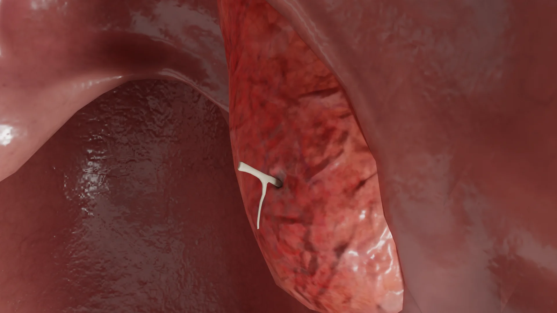
Less common are foreign bodies of the uvula, palatine arches, and lateral pharyngeal folds. If foreign bodies are not removed in time, with a favorable outcome, they can encapsulate and self-reject after a few days. If a foreign body remains in the tissue for a long time, localized inflammation can develop, accompanied by infiltration and hyperemia, in rare cases – the presence of serous-purulent discharge, and parapharyngitis or more serious complications (parapharyngeal abscess and mediastinitis) can also develop. After removing foreign bodies, abrasions and small hemorrhages may remain.
All foreign bodies in the nose are characterized by difficulty in nasal breathing, sneezing, and abundant mucous discharge from one half of the nose. Sometimes pain and discomfort in the nose may appear, as well as bleeding, which is especially characteristic in the presence of living foreign bodies and lithium batteries.
Rhinoliths can remain in the nasal cavity for a long time without clinical manifestations and be discovered during routine otorhinolaryngological examination or during surgical interventions. An ENT doctor should be alerted to nasal stones by complaints of prolonged pain, nasal discharge, and difficulty breathing, which are unilateral in nature, a history of a foreign body entering that half of the nose, as well as frequent recurrent unilateral rhinosinusitis.
Foreign bodies in the paranasal sinuses can proceed for a long time without clinical symptoms and more often manifest after a long period of time under the guise of chronic sinusitis. Patients complain of headache and a feeling of pressure in the affected sinus, congestion, and mucopurulent discharge. When the maxillary sinus is affected, pain may radiate to the upper jaw, and when the sphenoid sinus is affected, patients note pain in the occipital region. The presence of a pathological communication with the oral cavity may manifest as purulent discharge with an unpleasant odor from the mouth.
In the presence of immunodeficiency or chronic endocrine system pathologies (diabetes mellitus), foreign bodies may manifest only with paraorbital or intracranial complications, which is an extremely unfavorable sign.
In some cases, foreign bodies in the paranasal sinuses can be discovered accidentally during CT/MRI of the facial skull or during endoscopic surgeries to improve nasal breathing.
The main complaint in the presence of foreign bodies in the pharynx is painful sensations and discomfort at the site of their localization, intensifying when swallowing, tickling, and coughing. Complaints may persist for some time even after the removal of foreign objects.
Diagnosis of foreign bodies in the nasal cavity is based on medical history data and rhinoscopy. In case of difficulty in diagnosis, nasal cavity fibroscopy, X-ray of the paranasal sinuses or lateral projection of the nasopharynx (for radio-opaque objects), CT scan of the facial bones are performed.
For the diagnosis of foreign bodies in the paranasal sinuses, imaging methods such as CT and MRI are used. Subsequently, endoscopy of the nasal cavity and paranasal sinuses is performed, which in this case is a therapeutic and diagnostic method.
Diagnosis of pharyngeal foreign bodies is based on the data of oropharyngoscopy. In some cases it is quite difficult to detect a foreign body due to its location – in the thickness of the palatine tonsil, when only a small tip is sticking out, which is hidden behind the palatine tonsil and the foreign body itself is translucent, for example, a fish bone.
Hypersalivation can also be troublesome for the patient. In this case, it is necessary to calm the patient, give a drink of clean drinking water, irrigate the mucosa with a local anesthetic (eg, lidocaine 10%), if there is no allergic reaction, and try to rotate the palatine tonsil outward from the palatine glands for a better view. In cases where visual localization of the foreign body can not be established, resort to palpation and, if necessary, radiography (if the foreign body is X-ray contrast). In some cases, there is no foreign body at the appointment, but complaints persist due to residual phenomena such as abrasions and bruises, such patients are recommended dynamic observation and treatment of the mucosa with antiseptic solutions for rapid healing.
Find more scientifically accurate content on our social media


Treatment involves removal of the foreign body from the nose, for which hooks, forceps, clamps, and nasal aspirators are used. The procedure is performed after anemization and, if necessary, local anesthesia of the nasal cavity. In some cases, particularly with especially agitated patients, especially children, intravenous sedation or general anesthesia is used. If foreign bodies are present in the deep parts of the nasal cavity, endoscopy may be required.
Rhinoliths are more often detected and removed during surgical interventions to improve nasal breathing (septoplasty, vasotomy of nasal turbinates), the nasal stone is crushed and evacuated in parts.
Foreign bodies in the paranasal sinuses are removed during surgery under general anesthesia using endoscopic equipment. Sanitation of the paranasal sinuses, restoration of sinus aeration and nasal breathing are performed. If there is an oroantral fistula or sinus, their plastic surgery is performed with the participation of maxillofacial surgeons.
After removal of foreign bodies, it is recommended to rinse the nose with saline solutions, use local decongestants until the edema subsides, and apply local antiseptic or antibacterial medications.
Treatment of a foreign body in the pharynx also involves its removal, for which forceps, tweezers, or mosquito clamps are used. Removal should preferably be performed on an empty stomach after irrigating the site of entry with anesthetic solutions (10% lidocaine solution). If abrasions and hemorrhages are present, local anti-inflammatory therapy and a soft diet are prescribed.
1. What are the symptoms of a foreign body in the nose, pharynx?
2. What are the signs of a foreign body in a child’s nose?
3. What is the first aid when a foreign body enters the nose, paranasal sinuses, pharynx?
a) Foreign body in the nose:
Do not remove the object yourself. If there is no pain and breathing difficulty, blow your nose gently, pinching the healthy nostril. If it is impossible to remove it by yourself, if bleeding starts, consult an ENT doctor.
b) Foreign body in the paranasal sinuses:
Most often does not manifest itself immediately. If you suspect a sinus foreign body (unilateral pain, purulent discharge, connection to teeth or surgery) – see your doctor for diagnosis and removal.
c) Foreign body in the pharynx:
Causes pain, fever, cough, difficulty swallowing. Do not attempt to swallow the item or remove it yourself. See a specialist.
List of Sources
1.
VOKA Catalog.
https://catalog.voka.io/
2.
Total Otolaryngology—Head and Neck Surgery, Anthony P. Sclafani, Robin A. Dyleski, Michael J. Pitman, Stimson P. Schantz. Thieme Medical Publishers, Inc., 2015. ISBN 978-1-60406-646-3.
3.
Бербом Х. Болезни уха, горла и носа / Ханс Бербом, Оливер Кашке, Тадеус Навка, Эндрю Свифт; пер. с англ. – 2-е изд. – М. : МЕДпреcс-информ, 2016. – 776 с. : ил. ISBN 978-5-00030- 322-1.
4.
Baranowski K, Al Aaraj MS, Sinha V. Nasal Foreign Body. [Updated 2023 Jul 3]. In: StatPearls [Internet]. Treasure Island (FL): StatPearls Publishing; 2025 Jan-. Available from:
https://www.ncbi.nlm.nih.gov/books/NBK459279/
5.
Kumar S, Singh AB. An unusual foreign body in the nose of an adult. BMJ Case Rep. 2013 Jun 16;2013:bcr2012007780. doi: 10.1136/bcr-2012-007780. PMID: 23774703; PMCID: PMC3703090.
6.
Endican S, Garap JP, Dubey SP. Ear, nose and throat foreign bodies in Melanesian children: an analysis of 1037 cases. Int J Pediatr Otorhinolaryngol. 2006 Sep;70(9):1539-45. doi: 10.1016/j.ijporl.2006.03.018. Epub 2006 May 16. PMID: 16707167.
7.
Park S, Choi DS, Shin HS, Cho JM, Jeon KN, Bae KS, Koh EH, Park JJ. Fish bone foreign bodies in the pharynx and upper esophagus: evaluation with 64-slice MDCT. Acta Radiol. 2014 Feb;55(1):8-13. doi: 10.1177/0284185113493087. Epub 2013 Jul 24. PMID: 23884842.
Loading test 6 questions
Table of Contents


Summarize article with AI
Choose your preferable AI assistant:
Link successfully copied to clipboard
Thank you!
Your message is sent!
Our experts will contact you shortly. If you have any additional questions, please contact us at info@voka.io


