Structure of a cardiomyocyte: histology, types, and functional morphology

This article is for informational purposes only
The content on this website, including text, graphics, and other materials, is provided for informational purposes only. It is not intended as advice or guidance. Regarding your specific medical condition or treatment, please consult your healthcare provider.
Cardiomyocytes are branched cells integrated into a functional syncytium of cardiac muscle fibers. Like other cells of the body, the cardiomyocyte is covered with a cytoplasmic membrane (sarcolemma) and filled with cytoplasm (sarcoplasm).
The contractile apparatus and microscopic structure
In the sarcoplasm, myofibrils are arranged in parallel; these are threadlike structures composed of sarcomeres (contractile units of striated muscle cells). The sarcomere, in turn, is composed of thin actin protein filaments and thick myosin protein filaments.
By electron microscopy, it can be seen that sarcomeres consist of alternating light (I-bands) and dark (A-bands) zones. Connections of sarcomeres with each other form Z-lines. Alternating light and dark zones in the myofibrils give them and the entire muscle fiber a specific striation.
Intracellular systems and organelles
Myofibrils are surrounded by the sarcoplasmic reticulum and T-tubules. The sarcoplasmic reticulum acts as a reservoir for calcium ions, while the T-tubules provide communication with the sarcolemma and the extracellular environment.
T-tubules encircle the myofibril at the level of Z-lines, and nearby, the sarcoplasmic reticulum forms poorly defined expansions – terminal cisternae. Mitochondria are located between the myofibrils in the sarcoplasm.
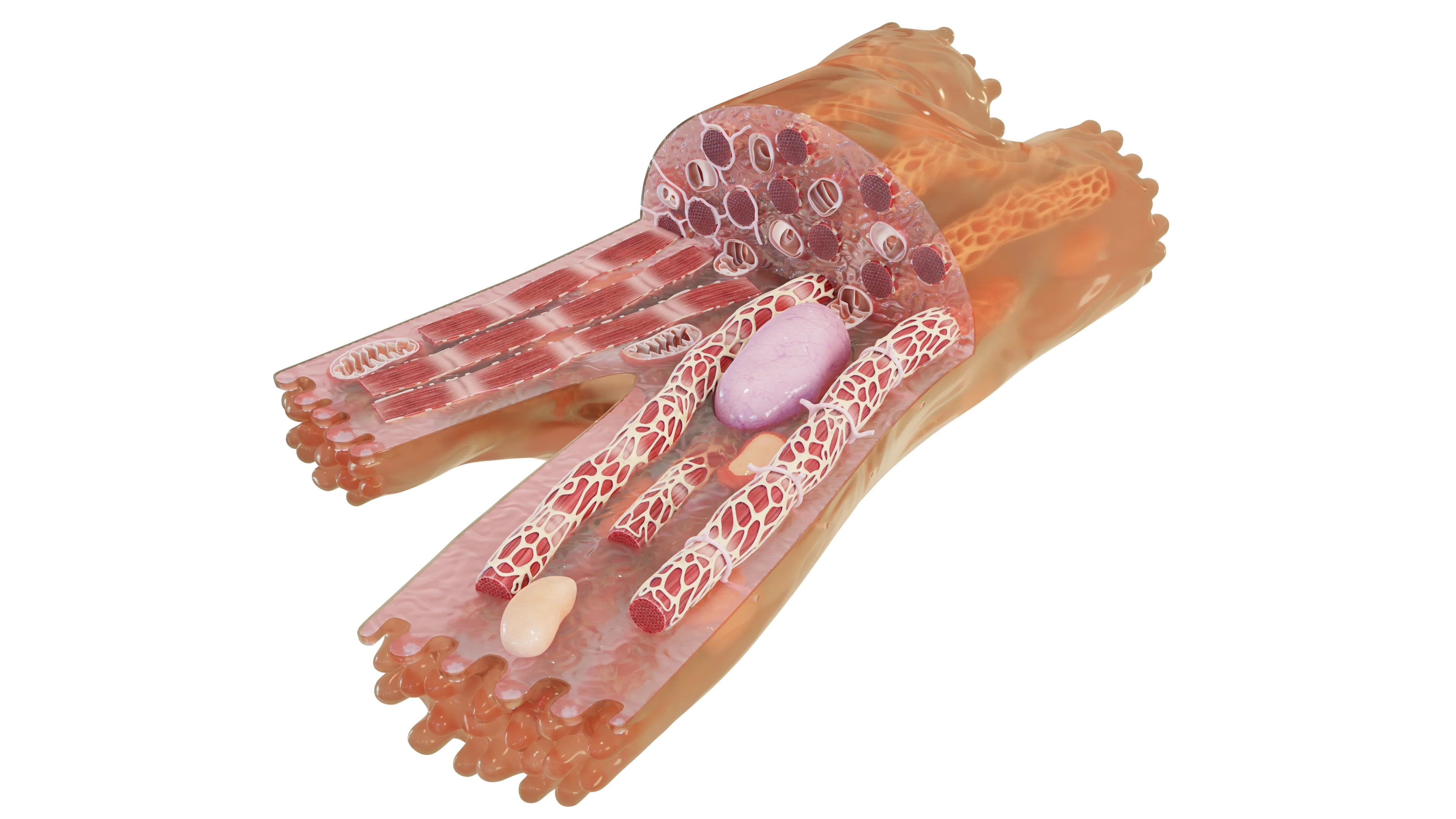
A cardiomyocyte typically has a single oval centrally located nucleus, but binucleated forms can also be found. The nucleus is surrounded by perinuclear sarcoplasm, a zone free of myofibrils. Most of the organelles of a cardiomyocyte are located in the perinuclear sarcoplasm.
Intercellular contacts: intercalated discs
Cardiomyocytes in cardiac muscle are connected to each other via intercalated discs. Intercalated discs are the terminal segments of the sarcolemma, which have a stepped shape with interdigitations and contain the following:
- Desmosomes, linking neighboring cells together;
- Gap junctions (connexon proteins), which facilitate the exchange of ions and molecules between cardiac muscle cells.
Under light microscopy, intercalated discs appear as thick dark bands running across muscle fibers. Thus, intercalated discs are specialized intercellular contacts that ensure the coordinated function of all cardiac muscle cells.
Find more scientifically accurate content on our social media


Differences between cardiomyocytes and skeletal muscle fibers
The above implies that heart muscle cells belong to striated muscle tissue, like skeletal muscle fibers, but have several distinctive differences:
- Single oval centrally located nucleus;
- Projections, with which cardiac muscle fibers connect to each other via intercalated discs, forming a syncytium and functioning as a whole;
- Terminal cisternae of the sarcoplasmic reticulum are weakly defined;
- T-tubules are located at the level of Z-lines;
- The quantity of myofibrils is less than in skeletal muscle fibers.
FAQ
1. What is a cardiomyocyte?
2. What is the structure of the contractile apparatus of such a cell?
3. What is the function of the intercalated discs?
4. Where are the nucleus and main organelles located in the cardiomyocyte?
5. Which systems provide contraction and energy supply to the cell?
References
1.
VOKA 3D Anatomy & Pathology – Complete Anatomy and Pathology 3D Atlas [Internet]. VOKA 3D Anatomy & Pathology.
Available from: https://catalog.voka.io/
2.
Young, B., O’Dowd, G., & Woodford, P. (2013). Wheater’s functional histology: A text and colour atlas (6th ed.). Elsevier Health Sciences.
3.
Eroschenko, V. P. (2017). Atlas of histology with functional correlations (13th ed.). Wolters Kluwer.
4.
Fred E. Hossler (2014). Ultrastructure Atlas of Human Tissues. John Wiley & Sons, Inc. Hoboken.


