Traumatic Epidural Hematomas: Etiology, Pathogenesis, Clinical Presentation, Diagnosis, and Treatment
Artur D.Neurosurgeon, MD
18 min read·January 29, 2026
This article is for informational purposes only
The content on this website, including text, graphics, and other materials, is provided for informational purposes only. It is not intended as advice or guidance. Regarding your specific medical condition or treatment, please consult your healthcare provider.
Traumatic epidural hematomas are a type of focal traumatic brain injury characterized by blood accumulation in the epidural space between the dura mater and the bones of the skull.
The incidence of acute epidural hematomas accounts for about 2 to 11% of all reported traumatic brain injuries (TBIs), with the frequency increasing to 15% among all patients who died from TBIs.
The average age of occurrence of traumatic epidural hematomas is 20–30 years. The age-related characteristics of acute epidural hematoma incidence are due to anatomical features: with age, the dura mater fuses more tightly with the bones of the skull, reducing the likelihood of blood accumulation in the epidural space.
The frequency of surgical treatment for acute epidural hematomas is 56%.
Etiology of traumatic epidural hematomas
The etiological factors in the development of traumatic epidural hematomas include the same causes that underlie the occurrence of traumatic brain injury:
Road traffic accidents;
Falls;
Violent injuries;
Sports injuries.
More than half of all etiological factors in which acute epidural hematomas occur are precisely traffic accidents.
Pathogenesis of acute traumatic epidural hematomas
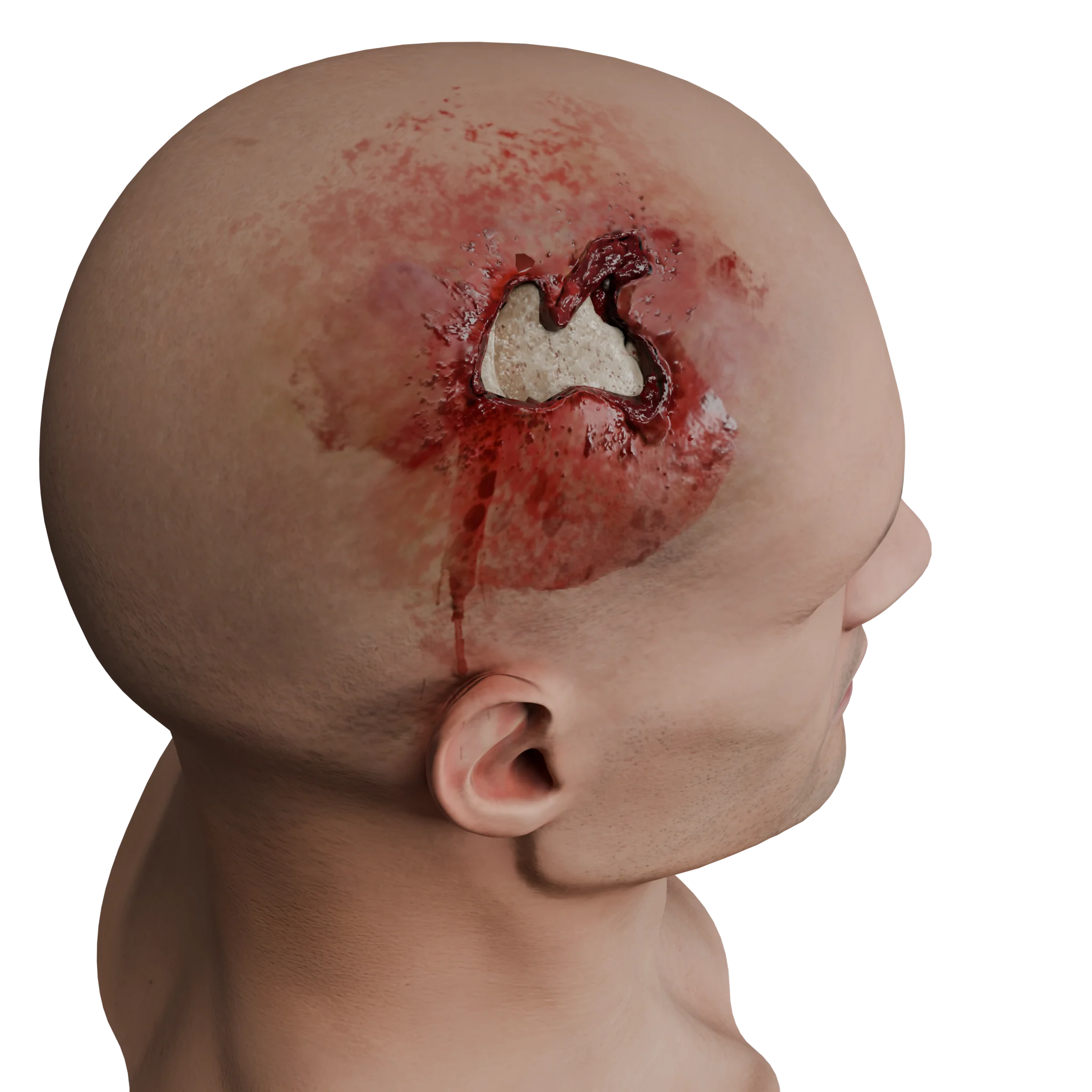
The pathogenesis of acute epidural hematomas is characterized by a coup-contrecoup mechanism, classifying this type of traumatic brain injury as focal. In the course of applying the traumatic agent to the rigid bones of the skullcap, deformation occurs, which, with a high level of kinetic energy, leads to fractures of the cranial bones, tears of blood vessels in the dura mater (commonly the middle meningeal artery), tears in the walls of the dural sinuses, and continued bleeding into the epidural space.
Most often, epidural hematomas form when a traumatic agent is applied to the temporal region (about 75% of all acute epidural hematomas) due to two factors:
The presence of a thin squama of the temporal bone, which is easily prone to fractures due to its low strength;
Passage of the middle meningeal artery in this area, which gets damaged with fractures of the temporal bone squama, leading to active arterial bleeding that then peels off the dura mater from the cranial bones.
Epidural hematoma basotemporal localization: 3D model
Approximately 10% of all acute epidural hematomas form due to ruptures of the dural sinuses and bleeding from these anatomical structures.
In some cases, acute epidural hematomas can form due to damage in the diploic veins within the bones of the skull, leading to bleeding into the epidural space.
Clinical presentation
In the clinical picture of an acute epidural hematoma, global cerebral symptoms are prioritized, the leading one being loss of consciousness.
Consciousness impairment is typically three-phase, featuring initial loss of consciousness, subsequent recovery, and secondary consciousness depression after a period of wakefulness. The so-called “classic lucid interval” is observed in approximately ¼ of patients, whereas the rest either have a blurred lucid interval or none at all, with patients being in a coma from the onset of injury.
If the patient is conscious, they will usually report an intense, paroxysmal headache.
Focal neurological symptoms associated with acute epidural hematomas include:
Hemisyndrome: presence of pyramidal disorders (motor disturbances up to complete movement impairment) on the side opposite to the hematoma location. This syndrome is typically associated either with primary compression of the motor cortex or compression of the brainstem tracts due to a progressive increase in ICP.
Aphasic disorders: with hematoma localization in the dominant hemisphere of the brain, over 50% of cases exhibit various speech disorders (up to complete loss).
Epileptic activity: due to cortical stimulation, patients may experience seizures, reaching generalized episodes.
Mydriasis: a distinctive sign, manifesting as pupil dilation on the side of the epidural hematoma. This symptom appears with progressive ICP increase and development of temporal-tentorial herniation and oculomotor nerve palsy.
When the epidural hematoma is localized in the posterior fossa area, primary symptoms relate to early cerebrospinal fluid outflow obstruction leading to hydrocephalus development.
Coordination disorders take precedence, along with ataxia and bulbar dysfunction, whereas pyramidal insufficiency is less severe.
Diagnosis of acute epidural hematomas
Upon patient admission, the diagnostic algorithm for acute epidural hematomas consists of the following steps:
1. Assessment of consciousness level using the GCS (Glasgow Coma Scale)
This step allows for determining the patient’s level of consciousness and estimating the severity of brain structure damage, suggesting possible treatment options (surgical or non-surgical). It is essential not to forget about the two-phase course, and in the presence of traumatic injuries, particularly in the temporal and occipital regions, an acute epidural hematoma should be suspected within the lucid interval.
2. Medical history
Medical history collection includes the timing and mechanism of the injury and the presence of consciousness loss in the patient after the injury.
This step primarily assesses factors like traumatic brain injury with consciousness loss and duration, examines injury mechanisms, and excludes associated injuries in adjacent regions.
3. Visual examination of the patient
Visual examination involves examining the head area and checking for concomitant soft tissue injuries in areas of the body other than the head.
This phase focuses attention on localizing soft tissue injuries on the head to hypothesize possible epidural hematoma location. Injuries in the temporal and occipital areas should alert doctors to possible bone fractures in these regions, given their low strength, potentially leading to acute epidural hematoma formation. In cases of active bleeding, this stage is crucial for providing initial aid to the patient.
4. Neurological examination of the patient
The neurological examination includes assessment of nonfocal, focal, and meningeal symptoms.
This stage enables preliminary topical diagnosis of brain injury to determine indications for neuroimaging studies.
If the patient presents with non-focal, focal, or meningeal symptoms, neuroimaging is indicated. In cases where neuroimaging is unavailable, a neurological examination is crucial to determine burr hole location and intracranial injury diagnosis.
5. Brain computed tomography
Brain computed tomography is the “gold standard” in diagnosing traumatic brain injury, including acute epidural hematomas.
The advantages of CT in brain evaluation over other methods include:
Examination completion speed;
Ability to clearly evaluate the localization, extent, and nature of brain damage;
Ability to assess associated bone structure damage;
Capability (if necessary) to quickly scan other parts of the body to exclude concurrent injury.
A CT image of acute epidural hematomas is characterized by:
Lentiform clot location above the cerebral hemisphere.
Presence of a cranial fracture zone in the hematoma vicinity (commonly).
During the acute hemorrhage phase, the epidural hematoma displays a hyperintense (50-60 HU) homogeneous signal forming a biconvex lens over the cerebral hemisphere, usually not extending beyond the cerebral sutures (due to the fusion of the dura mater with the bone in the area of the sutures).
6. Laboratory diagnostics
Laboratory diagnostics include a complete blood count, urinalysis, coagulation profile, blood chemistry, and blood group and Rh factor testing.
These tests do not confirm or suspect epidural hemorrhage but allow assessment of the patient’s general condition, associated coagulation disorders, and preparation for potential surgical intervention.
Find more scientifically accurate content on our social media
Subscribe and don’t miss out the latest resources
Treatment of acute traumatic epidural hematomas
Medical therapy
Non-surgical treatment of epidural hematomas is carried out for small hematomas, with no consciousness depression in the patient, and in the absence of a dislocation syndrome. In such cases, mandatory CT monitoring is recommended 24-48 hours following diagnosis.
The primary goal of non-surgical treatment is to relieve symptoms and maintain dynamic observation of the patient to evaluate vital functions and neurological status over time.
Symptomatic therapy includes:
Pain relief (NSAIDs, narcotic analgesics);
Antiemetic drugs;
Anticonvulsants (if seizures manifest).
Osmotic diuretics are prescribed in the presence of cerebral edema.
Surgical therapy
Regardless of the patient’s level of consciousness, surgical treatment of acute epidural hematomas is indicated when the following criteria are present:
Hematoma thickness of 15 mm or more;
Displacement of midline structures of 5 mm or more;
Hematoma volume over 35 ml.
In the case of an epidural hematoma in the posterior cranial fossa, the indication for surgical intervention is a hematoma volume of more than 25 cm³.
Epidural hematoma of the posterior cranial fossa: 3D model
Craniotomy
The standard method of surgical treatment is a craniotomy at the projection of the epidural hematoma with its complete removal, search, and coagulation of the bleeding source (most often the damaged middle meningeal artery or its branches, less commonly venous sinuses).
After meticulous hemostasis, if necessary, a revision of the subdural space is performed, followed by hermetic suturing of the dura mater, the bone flap is positioned back, and the wound is sutured in layers. If necessary, a subgaleal drainage system can be placed.
Decompressive craniectomy
In certain cases (hematoma volume over 150 cm³ and/or severe dislocation syndrome over 12 mm), decompressive craniectomy is indicated.
A wide-cranial trepanation is performed in the frontal-temporal-parietal area with the removal of the temporal bone’s squama from the hematoma side, and the bone flap is sent for conservation. After removal of the hematoma, hemostasis, and plastic repair of the dura mater, the wound is sutured in layers.
FAQ
1. What is an epidural hematoma?
An epidural hematoma is an accumulation of blood between the dura mater and the inner surface of the skull bones resulting from head injury. It belongs to the focal forms of traumatic brain injury and requires urgent diagnosis and treatment.
2. How common are epidural hematomas?
Acute epidural hematomas occur in 2 to 11% of all traumatic brain injury cases.
3. Why do epidural hematomas occur more frequently in the young?
In young individuals, the dura mater is less tightly adhered to the skull bones, facilitating its separation during bleeding and hematoma formation. As age progresses, the membrane becomes denser, reducing the risk of epidural hematomas.
4. What are the primary causes of epidural hematomas?
Acute epidural hematomas arise in the event of traumatic head injuries, most commonly associated with damage to the middle meningeal artery and less frequently to brain venous sinuses.
5. How does an epidural hematoma manifest?
Key symptoms of an epidural hematoma include loss of consciousness with a “lucid interval” and severe headache. Focal disturbances such as hemiparesis, aphasia, and convulsions also develop. A specific sign of brain compression is mydriasis on the side of the injury. Involvement of the posterior cranial fossa leads to coordination disorders, ataxia, and bulbar dysfunctions.
6. How is an epidural hematoma diagnosed?
The gold standard for detecting acute epidural hematomas is a brain CT scan. In rare cases, skull radiography helps to detect skull fractures and raise suspicion for epidural hemorrhage in the fracture area.
7. When is non-surgical treatment of epidural hematomas possible?
Non-surgical strategy is feasible if the hematoma thickness is under 15 mm, the volume doesn’t exceed 35 ml, and there is no displacement of midline structures. The patient should retain consciousness without signs of intracranial hypertension, requiring dynamic observation and CT monitoring within 24–48 hours.
8. When is surgical treatment of epidural hematomas required?
Surgery is indicated if the hematoma thickness reaches 15 mm or more, midline structures are displaced by 5 mm or more, or the volume exceeds 35 ml (in the posterior cranial fossa — more than 25 cm³). Intervention is also required if consciousness deteriorates or focal symptoms appear regardless of the hematoma size.
9. What are the consequences of an epidural hematoma?
The prognosis depends on the volume of bleeding and the speed of surgical intervention. Timely surgical intervention can lead to full recovery without neurological deficit. Delayed diagnosis or severe brain compression can result in persistent complications: motor and speech disorders, cognitive impairments, post-traumatic epilepsy, and, in critical cases, death.
References
1.
VOKA 3D Anatomy & Pathology – Complete Anatomy and Pathology 3D Atlas [Internet]. VOKA 3D Anatomy & Pathology.
Available from: https://catalog.voka.io/
2.
Pisică D., Volovici V., Yue J.K., et al. Clinical and imaging characteristics, care pathways, and outcomes of traumatic epidural hematomas: a collaborative European NeuroTrauma Effectiveness Research in Traumatic Brain Injury study // Neurosurgery. – 2024. – Vol. 95, No. 5. – P. 986–999. – DOI: 10.1227/neu.0000000000002982.
3.
Luo Y., He X., Yang M., Du C., Jin X. A prognostic scoring system for operated acute epidural hematoma based on gray–white matter ratio // Medicine (Baltimore). – 2021. – Vol. 100, No. 33. – e26888. – DOI: 10.1097/MD.0000000000026888.
4.
Yoon S.Y., Kim Y.J., Lee K.H., et al. Surgical management of traumatic supra- and infratentorial extradural hematomas: experience and systematic literature review // Journal of Trauma and Injury. – 2023. – Vol. 36, No. 2. – P. 87–96. – DOI: 10.20408/jti.2022.0051.
5.
Lee K.S., Park Y.S., Kang D.H., et al. Traumatic posterior fossa extradural hematoma in children: case series and meta-analysis // Acta Neurochirurgica. – 2024. – Vol. 166, No. 4. – P. 801–811. – DOI: 10.1007/s00701-024-05987-0.
6.
Hasanpour M., Ghorbani M., Ahmadi S., et al. Predicting epidural hematoma expansion in traumatic brain injury using machine learning models // Frontiers in Neurology. – 2024. – Vol. 15. – Article 1511224. – DOI: 10.3389/fneur.2024.1511224.
7.
Arif S.H., Shah A., Malik A., et al. Benign extradural haemorrhage: scope of conservative trial // Egyptian Journal of Neurosurgery. – 2024. – Vol. 39, No. 1. – P. 1–7. – DOI: 10.1186/s41984-024-00274-5.
8.
Schweitzer A.D., Niogi S.N., Lignelli A., et al. Traumatic brain injury: imaging patterns and complications // Radiographics. – 2019. – Vol. 39, No. 6. – P. 1683–1706. – DOI: 10.1148/rg.2019190030.
9.
Flaherty B.F., Nash T., Taylor G.A., et al. Repeat head CT for expectant management of traumatic pediatric intracranial hemorrhage // Pediatrics. – 2018. – Vol. 142, No. 3. – e20180385. – DOI: 10.1542/peds.2018-0385.
10.
Khairat A., Ziu M. Epidural Hematoma // StatPearls [Internet]. – Treasure Island (FL): StatPearls Publishing; Updated 2023.
Available from: https://www.statpearls.com/point-of-care/21228
11.
Pereira C.U., Vilela L.H., de Souza A.C., et al. Treatment options for intracranial epidural hematoma: a comprehensive review // Neurosurgical Review. – 2024. – DOI: 10.1055/s-0044-1796652.