


Acquired Tricuspid Valve Diseases: Causes, Symptoms, and Treatment Options

Table of Contents
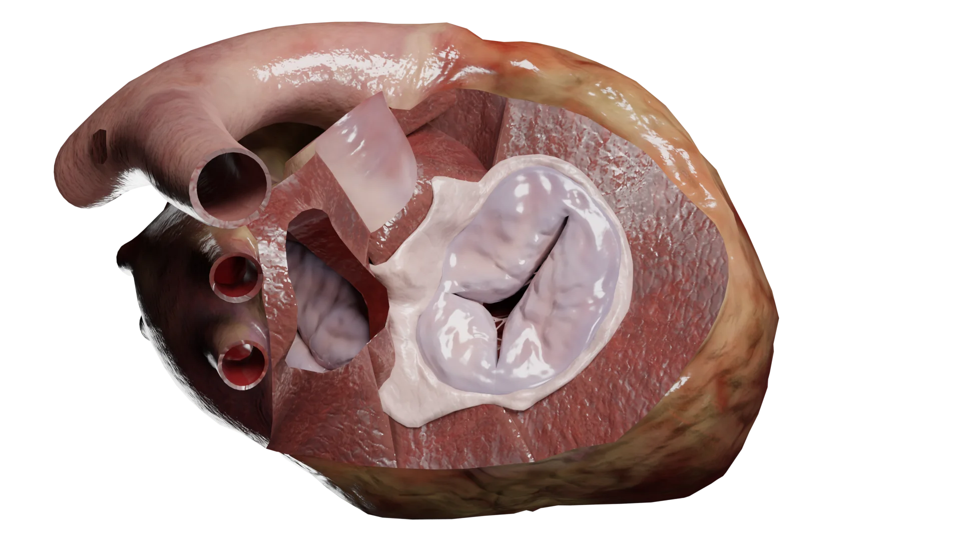
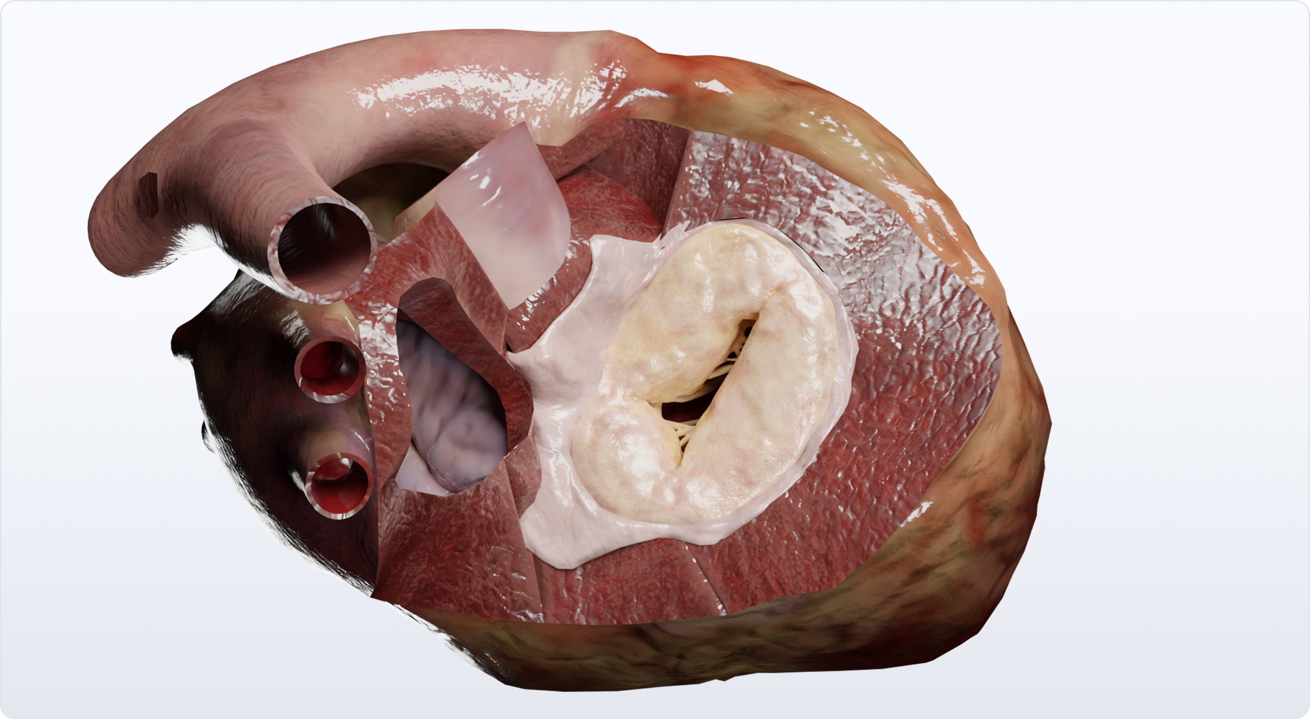
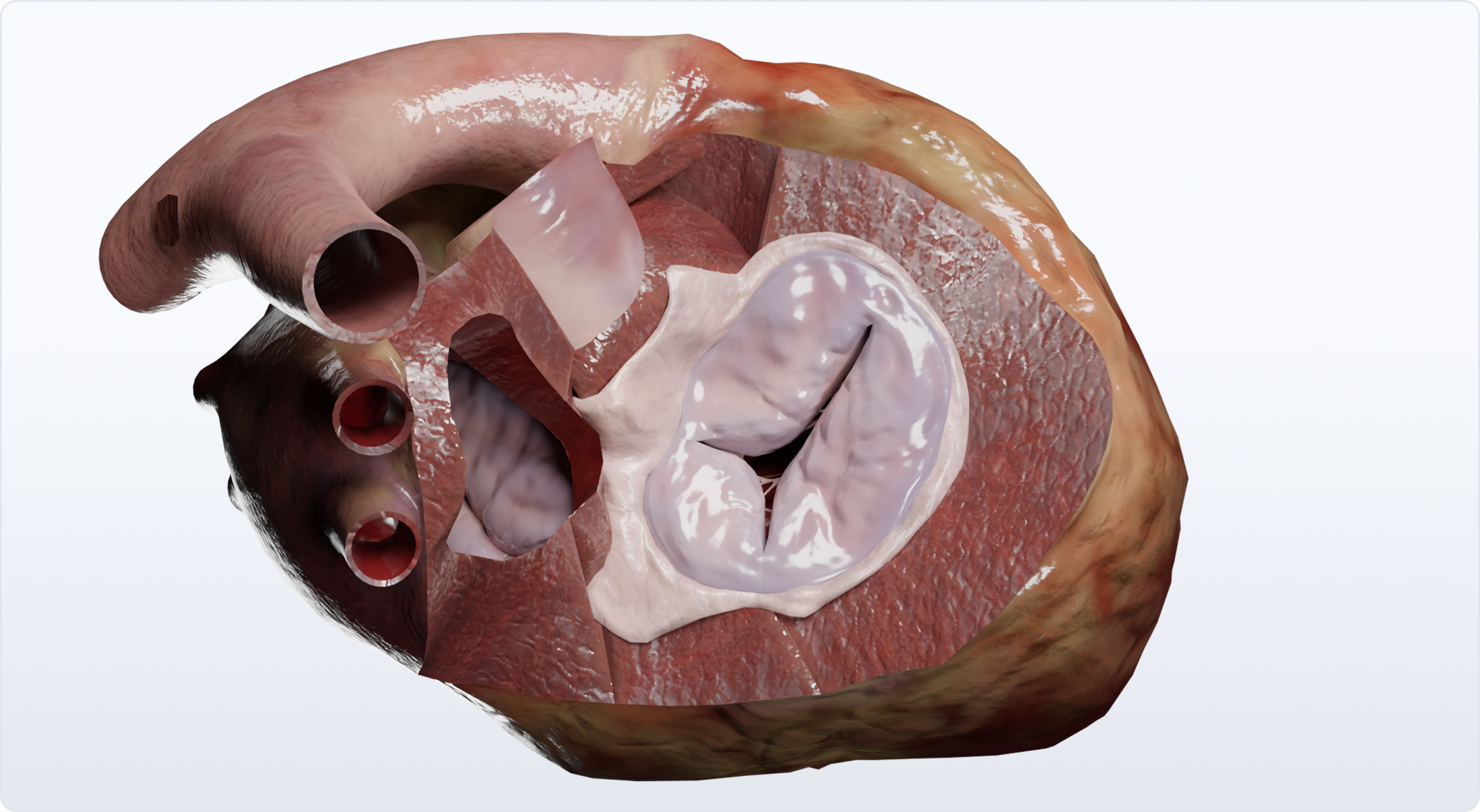
3D models of tricuspid valve insufficiency:


The tricuspid valve regulates blood flow between the right atrium and right ventricle. Acquired tricuspid valve diseases develop after birth and disrupt this flow. The two main types are tricuspid stenosis (TS) and tricuspid regurgitation (TR). While rheumatic fever and infective endocarditis are known causes, other factors like carcinoid syndrome, radiation therapy, connective tissue disorders, pulmonary hypertension, and right heart failure also play a role. Recognizing and treating TR, even when secondary to other cardiac issues, is increasingly important.
What causes tricuspid stenosis (TS)?
TS arises from the narrowing of the tricuspid valve opening, obstructing blood flow from the right atrium (RA) to the right ventricle (RV). Rheumatic fever is the most frequent cause, which leads to thickened and fused valve leaflets. Other less common causes include infective endocarditis, congenital heart defects, tumors, and chest radiation.
Clinical manifestations and diagnosis of tricuspid stenosis
Common TS symptoms include:
- Peripheral edema (swelling in the legs, ankles, and feet): Caused by fluid buildup due to impaired blood flow.
- Hepatomegaly (enlarged liver): Resulting from blood backing up into the liver.
- Cardiac arrhythmias (irregular heartbeat): Due to the strain on the right atrium.
- Neck discomfort/fluttering: Related to the enlarged right atrium and jugular venous distension.
- Cold skin and fatigue: Due to reduced blood flow and oxygen delivery to the body.
Diagnostic procedures for TS:
- ECG (Electrocardiogram): May reveal nonspecific changes and detect cardiac arrhythmias.
- Echocardiography: The primary diagnostic tool, assesses stenosis severity, measures the systolic pressure gradient across the valve, and evaluates valve morphology.
- Chest X-ray: Can show enlargement of the right side of the heart.
- Cardiac catheterization: Sometimes used to accurately measure the pressure gradient and assess the severity of stenosis.
Tricuspid regurgitation (TR): causes, symptoms, and pathophysiology
TR occurs when the tricuspid valve doesn’t close completely, causing blood to leak back into the RA during RV contraction. This can stem from:
- Right ventricular enlargement: Often a consequence of other heart conditions.
- Pulmonary hypertension: High blood pressure in the arteries leading to the lungs.
- Rheumatic fever: Can damage the valve leaflets.
- Infective endocarditis: Infection of the heart valves.
- Other causes: Marfan syndrome, rheumatoid arthritis, injury, carcinoid tumors, and myxomatous degeneration.
Carpentier’s functional classification of TR:
| Type I: | Type II: | Type III: |
|---|---|---|
| Normal leaflet motion (annular dilation, leaflet cleft or perforation). | Excess leaflet motion (leaflet prolapse due to chordal elongation/rupture, papillary muscle elongation/rupture, myxomatous degeneration). | Restricted leaflet motion. IIIa: Restricted opening motion (acute rheumatic fever, degenerative fusion). IIIb: Restricted closing motion (RV dilation, chordal shortening). |
Clinical manifestations of tricuspid regurgitation
TR symptoms can be vague or absent in mild cases, but may include:
- Dyspnea (shortness of breath): Due to fluid buildup in the lungs.
- Fatigue: Resulting from reduced blood flow and oxygen delivery.
- Ascites (fluid buildup in the abdomen): Due to venous congestion.
- Hepatomegaly (enlarged liver): Caused by blood backing up into the liver.
- Active pulsing in neck veins: Due to backflow of blood into the jugular veins.
- Swelling in the legs, ankles, and feet: Caused by fluid buildup.
- Decreased urine output: The body’s attempt to conserve fluid.
Diagnosis of tricuspid regurgitation:
- ECG: May show nonspecific changes or cardiac arrhythmias.
- Echocardiography: Assesses the severity of TR and the size of the RA.
- Chest X-ray: May reveal enlargement of the right side of the heart.
- MRI: Evaluates the RV structure and function.
- Cardiac catheterization: Measures pulmonary artery pressure, especially important in patients with pulmonary hypertension.
Treatment for tricuspid valve diseases
Treatment varies depending on the severity and type of valve disease:
- Medical management: Medications can help manage symptoms like swelling and fluid buildup. Treating underlying conditions like pulmonary hypertension is crucial.
- Surgical intervention: For severe cases, valve repair or replacement may be necessary. Repair techniques include suture techniques, ring annuloplasty, and bicuspidization. If repair isn’t feasible, the valve is replaced with a prosthetic valve.


Comparison table: tricuspid stenosis vs. tricuspid regurgitation
| Feature | Tricuspid stenosis | Tricuspid regurgitation |
|---|---|---|
| Cause | Rheumatic fever, infective endocarditis, congenital heart defects, carcinoid syndrome, radiation therapy | Pulmonary hypertension, right ventricular enlargement, rheumatic fever, infective endocarditis, Marfan syndrome, rheumatoid arthritis, injury, carcinoid tumors, myxomatous degeneration |
| Pathophysiology | Obstruction of blood flow from RA to RV, increased RA pressure | Backward blood flow into RA during RV contraction, increased RA pressure |
| Symptoms | Peripheral edema, hepatomegaly, cardiac arrhythmias, neck discomfort/fluttering, fatigue, cold ski | Dyspnea, fatigue, ascites, hepatomegaly, cardiac arrhythmias, pulsating neck veins, leg swelling, decreased urine output |
| Heart Sound | Diastolic murmur along left sternal border | Holosystolic murmur along left sternal border |
| Diagnosis | Echocardiography, ECG, chest X-ray, cardiac catheterization | Echocardiography, MRI, cardiac catheterization, ECG, chest X-ray |
| Treatment | Drug therapy, valvuloplasty, valve replacement | Drug therapy, valve repair or replacement |
