
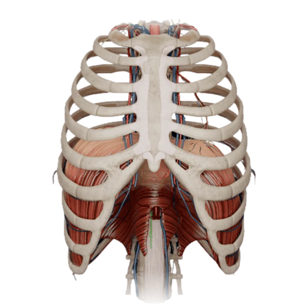
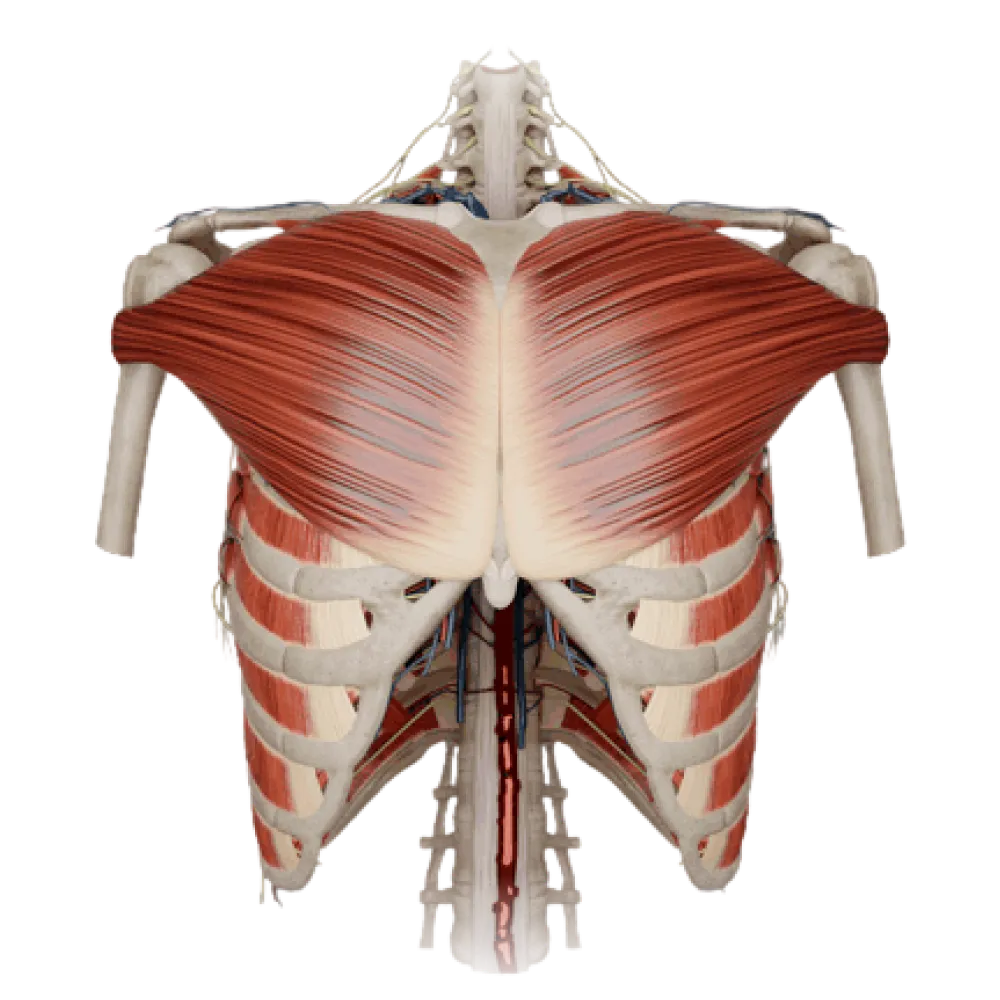
Diaphragm anatomy test
Evaluate your knowledge of diaphragm anatomy. The test evaluates the topography, ligaments, vessels, nerves, and contents of its natural openings.
1/20
bold
text
1. What are the three muscular parts identified in the structure of the diaphragm?
-
Clavicular, sternocostal, and abdominal
In the diaphragm, the muscular bundles are distinguished into sternal, costal, and lumbar parts based on their origin.
-
Costal, vertebral, and lumbar
In the diaphragm, the muscular bundles are distinguished into sternal, costal, and lumbar parts based on their origin.
-
Sternal, costal, and lumbar
In the diaphragm, the muscular bundles are distinguished into sternal, costal, and lumbar parts based on their origin.
-
Sternal, medial, and lateral
In the diaphragm, the muscular bundles are distinguished into sternal, costal, and lumbar parts based on their origin.
-
I find it difficult to answer
In the diaphragm, the muscular bundles are distinguished into sternal, costal, and lumbar parts based on their origin.
2. To which vertebrae does the right crus (crus dextrum) of the lumbar part of the diaphragm attach?
-
To the anterolateral surfaces of the bodies of lumbar vertebrae I-IV
The right crus of the lumbar part of the diaphragm is longer than the left and originates from the anterolateral surfaces of the bodies of lumbar vertebrae I-IV.
-
To the transverse processes of lumbar vertebrae I-III
The right crus of the lumbar part of the diaphragm is longer than the left and originates from the anterolateral surfaces of the bodies of lumbar vertebrae I-IV.
-
To the bodies of thoracic vertebrae XI-XII
The right crus of the lumbar part of the diaphragm is longer than the left and originates from the anterolateral surfaces of the bodies of lumbar vertebrae I-IV.
-
To the anterior surfaces of the bodies of lumbar vertebrae I-II
The right crus of the lumbar part of the diaphragm is longer than the left and originates from the anterolateral surfaces of the bodies of lumbar vertebrae I-IV.
-
I find it difficult to answer
The right crus of the lumbar part of the diaphragm is longer than the left and originates from the anterolateral surfaces of the bodies of lumbar vertebrae I-IV.
3. What anatomical structure passes through the aortic hiatus (hiatus aorticus) of the diaphragm together with the aorta?
-
Azygos vein
Through the aortic hiatus of the diaphragm, formed by its crura and the median arcuate ligament, pass the aorta and thoracic duct.
-
Sympathetic trunk
Through the aortic hiatus of the diaphragm, formed by its crura and the median arcuate ligament, pass the aorta and thoracic duct.
-
Vagus nerve
Through the aortic hiatus of the diaphragm, formed by its crura and the median arcuate ligament, pass the aorta and thoracic duct.
-
Thoracic duct
Through the aortic hiatus of the diaphragm, formed by its crura and the median arcuate ligament, pass the aorta and thoracic duct.
-
I find it difficult to answer
Through the aortic hiatus of the diaphragm, formed by its crura and the median arcuate ligament, pass the aorta and thoracic duct.
4. Which nerves pass through the esophageal hiatus (hiatus oesophageus) of the diaphragm along with the esophagus?
-
Greater splanchnic nerves
The vagus nerves form a plexus on the esophagus, from which the anterior and posterior vagal trunks are formed, passing through the esophageal hiatus into the abdominal cavity.
-
Anterior and posterior vagal trunks
The vagus nerves form a plexus on the esophagus, from which the anterior and posterior vagal trunks are formed, passing through the esophageal hiatus into the abdominal cavity.
-
Right and left phrenic nerves
The vagus nerves form a plexus on the esophagus, from which the anterior and posterior vagal trunks are formed, passing through the esophageal hiatus into the abdominal cavity.
-
Sympathetic trunks
The vagus nerves form a plexus on the esophagus, from which the anterior and posterior vagal trunks are formed, passing through the esophageal hiatus into the abdominal cavity.
-
I find it difficult to answer
The vagus nerves form a plexus on the esophagus, from which the anterior and posterior vagal trunks are formed, passing through the esophageal hiatus into the abdominal cavity.
5. Where is the opening of the inferior vena cava (foramen venae cavae) located in the diaphragm?
-
In the muscular part of the lumbar region
The opening of the inferior vena cava is located in the tendinous center of the diaphragm, slightly to the right of the median plane.
-
Between the sternal and costal parts
The opening of the inferior vena cava is located in the tendinous center of the diaphragm, slightly to the right of the median plane.
-
In the tendinous center, closer to the right leaflet
The opening of the inferior vena cava is located in the tendinous center of the diaphragm, slightly to the right of the median plane.
-
In the region of the costodiaphragmatic sinus
The opening of the inferior vena cava is located in the tendinous center of the diaphragm, slightly to the right of the median plane.
-
I find it difficult to answer
The opening of the inferior vena cava is located in the tendinous center of the diaphragm, slightly to the right of the median plane.
6. What structure forms the median arcuate ligament (ligamentum arcuatum medianum) of the diaphragm?
-
The tendinous edges of the right and left crura of the lumbar part
The median arcuate ligament is formed by the crossing of the medial tendinous edges of the right and left crura of the diaphragm above the aorta.
-
Thickened fascia over the quadratus lumborum muscle
The median arcuate ligament is formed by the crossing of the medial tendinous edges of the right and left crura of the diaphragm above the aorta.
-
Aponeurosis of the transversus abdominis muscle
The median arcuate ligament is formed by the crossing of the medial tendinous edges of the right and left crura of the diaphragm above the aorta.
-
Thickened thoracolumbar fascia over the psoas major muscle
The median arcuate ligament is formed by the crossing of the medial tendinous edges of the right and left crura of the diaphragm above the aorta.
-
I find it difficult to answer
The median arcuate ligament is formed by the crossing of the medial tendinous edges of the right and left crura of the diaphragm above the aorta.
7. Over what muscle does the medial arcuate ligament (ligamentum arcuatum mediale) of the diaphragm arch?
-
Over the quadratus lumborum muscle
The medial arcuate ligament stretches between the body and the transverse process of the first lumbar vertebra, arching over the psoas major muscle (m. psoas major).
-
Over the iliacus muscle
The medial arcuate ligament stretches between the body and the transverse process of the first lumbar vertebra, arching over the psoas major muscle (m. psoas major).
-
Over the transversus abdominis muscle
The medial arcuate ligament stretches between the body and the transverse process of the first lumbar vertebra, arching over the psoas major muscle (m. psoas major).
-
Over the psoas major muscle
The medial arcuate ligament stretches between the body and the transverse process of the first lumbar vertebra, arching over the psoas major muscle (m. psoas major).
-
I find it difficult to answer
The medial arcuate ligament stretches between the body and the transverse process of the first lumbar vertebra, arching over the psoas major muscle (m. psoas major).
8. What passes in the gap between the muscular bundles of the right crus of the lumbar part of the diaphragm?
-
Hemiazygos vein and lesser splanchnic nerve
Through the gap in the right crus of the lumbar part of the diaphragm pass the azygos vein (v. azygos) and the splanchnic nerves (nn. splanchnici major et minor).
-
Azygos vein, greater and lesser splanchnic nerves
Through the gap in the right crus of the lumbar part of the diaphragm pass the azygos vein (v. azygos) and the splanchnic nerves (nn. splanchnici major et minor).
-
Inferior vena cava and right phrenic nerve
Through the gap in the right crus of the lumbar part of the diaphragm pass the azygos vein (v. azygos) and the splanchnic nerves (nn. splanchnici major et minor).
-
Thoracic duct and right vagus nerve
Through the gap in the right crus of the lumbar part of the diaphragm pass the azygos vein (v. azygos) and the splanchnic nerves (nn. splanchnici major et minor).
-
I find it difficult to answer
Through the gap in the right crus of the lumbar part of the diaphragm pass the azygos vein (v. azygos) and the splanchnic nerves (nn. splanchnici major et minor).
9. Which vessels pass through the sternocostal triangle (Larrey-Morgagni gap) of the diaphragm?
-
Inferior epigastric artery and vein
Through the sternocostal triangle pass the superior epigastric vessels — extensions of the internal thoracic arteries and veins.
-
Musculophrenic artery and vein
Through the sternocostal triangle pass the superior epigastric vessels — extensions of the internal thoracic arteries and veins.
-
Superior epigastric artery and vein
Through the sternocostal triangle pass the superior epigastric vessels — extensions of the internal thoracic arteries and veins.
-
Pericardiophrenic vessels
Through the sternocostal triangle pass the superior epigastric vessels — extensions of the internal thoracic arteries and veins.
-
I find it difficult to answer
Through the sternocostal triangle pass the superior epigastric vessels — extensions of the internal thoracic arteries and veins.
10. What is the name of the paired triangular area of the diaphragm, devoid of muscle fibers, located between the costal and lumbar parts?
-
Lumbocostal triangle (Bochdalek's triangle)
The lumbocostal triangle (trigonum lumbocostale), or Bochdalek's triangle, is located between the lumbar and costal parts of the diaphragm.
-
Sternocostal triangle (Larrey's triangle)
The lumbocostal triangle (trigonum lumbocostale), or Bochdalek's triangle, is located between the lumbar and costal parts of the diaphragm.
-
Morgagni's triangle
The lumbocostal triangle (trigonum lumbocostale), or Bochdalek's triangle, is located between the lumbar and costal parts of the diaphragm.
-
Tendinous center
The lumbocostal triangle (trigonum lumbocostale), or Bochdalek's triangle, is located between the lumbar and costal parts of the diaphragm.
-
I find it difficult to answer
The lumbocostal triangle (trigonum lumbocostale), or Bochdalek's triangle, is located between the lumbar and costal parts of the diaphragm.
11. Which vessel do the superior phrenic arteries (aa. phrenicae superiores) arise from?
-
From the internal thoracic artery
The superior phrenic arteries are parietal branches of the thoracic aorta and supply the superior surface of the diaphragm.
-
From the musculophrenic artery
The superior phrenic arteries are parietal branches of the thoracic aorta and supply the superior surface of the diaphragm.
-
From the abdominal part of the aorta
The superior phrenic arteries are parietal branches of the thoracic aorta and supply the superior surface of the diaphragm.
-
From the thoracic aorta.
The superior phrenic arteries are parietal branches of the thoracic aorta and supply the superior surface of the diaphragm.
-
I find it difficult to answer
The superior phrenic arteries are parietal branches of the thoracic aorta and supply the superior surface of the diaphragm.
12. From which source do the inferior phrenic arteries (aa. phrenicae inferiores) predominantly originate?
-
From the thoracic aorta
The inferior phrenic arteries arise from the anterior surface of the abdominal part of the aorta immediately upon its exit from the aortic hiatus.
-
From the abdominal part of the aorta
The inferior phrenic arteries arise from the anterior surface of the abdominal part of the aorta immediately upon its exit from the aortic hiatus.
-
From the celiac trunk
The inferior phrenic arteries arise from the anterior surface of the abdominal part of the aorta immediately upon its exit from the aortic hiatus.
-
From the superior mesenteric artery
The inferior phrenic arteries arise from the anterior surface of the abdominal part of the aorta immediately upon its exit from the aortic hiatus.
-
I find it difficult to answer
The inferior phrenic arteries arise from the anterior surface of the abdominal part of the aorta immediately upon its exit from the aortic hiatus.
13. Which artery, being a branch of the internal thoracic artery, supplies the diaphragm and pericardium?
-
Superior epigastric artery
The pericardiophrenic artery (branch of a. thoracica interna) descends with the phrenic nerve and supplies the pericardium and diaphragm.
-
Anterior intercostal artery
The pericardiophrenic artery (branch of a. thoracica interna) descends with the phrenic nerve and supplies the pericardium and diaphragm.
-
Pericardiacophrenic artery
The pericardiophrenic artery (branch of a. thoracica interna) descends with the phrenic nerve and supplies the pericardium and diaphragm.
-
Posterior intercostal artery
The pericardiophrenic artery (branch of a. thoracica interna) descends with the phrenic nerve and supplies the pericardium and diaphragm.
-
I find it difficult to answer
The pericardiophrenic artery (branch of a. thoracica interna) descends with the phrenic nerve and supplies the pericardium and diaphragm.
14. From which plexus is the phrenic nerve (n. phrenicus), providing motor innervation to the diaphragm, formed?
-
From the cervical plexus
The phrenic nerve is primarily formed from the anterior branches of the spinal nerves C3-C5, belonging to the cervical plexus.
-
From the brachial plexus
The phrenic nerve is primarily formed from the anterior branches of the spinal nerves C3-C5, belonging to the cervical plexus.
-
From the lumbar plexus
The phrenic nerve is primarily formed from the anterior branches of the spinal nerves C3-C5, belonging to the cervical plexus.
-
From the thoracic sympathetic trunk
The phrenic nerve is primarily formed from the anterior branches of the spinal nerves C3-C5, belonging to the cervical plexus.
-
I find it difficult to answer
The phrenic nerve is primarily formed from the anterior branches of the spinal nerves C3-C5, belonging to the cervical plexus.
15. Which nerves provide sensory innervation to the peripheral areas of the diaphragm?
-
Vagus nerves
The peripheral part of the diaphragm receives sensory innervation from the anterior branches of the 6-7 lower thoracic spinal nerves (intercostal nerves)
-
Splanchnic nerves
The peripheral part of the diaphragm receives sensory innervation from the anterior branches of the 6-7 lower thoracic spinal nerves (intercostal nerves)
-
Sympathetic trunks
The peripheral part of the diaphragm receives sensory innervation from the anterior branches of the 6-7 lower thoracic spinal nerves (intercostal nerves)
-
Lower intercostal nerves (VI–XI)
The peripheral part of the diaphragm receives sensory innervation from the anterior branches of the 6-7 lower thoracic spinal nerves (intercostal nerves)
-
I find it difficult to answer
The peripheral part of the diaphragm receives sensory innervation from the anterior branches of the 6-7 lower thoracic spinal nerves (intercostal nerves)
16. At what level is the highest point of the right dome of the diaphragm projected during maximal expiration?
-
At the level of the seventh rib
During maximal expiration, the right dome of the diaphragm rises to the level of the fourth intercostal space (left to the fifth).
-
At the level of the fourth intercostal space
During maximal expiration, the right dome of the diaphragm rises to the level of the fourth intercostal space (left to the fifth).
-
At the level of the sixth intercostal space
During maximal expiration, the right dome of the diaphragm rises to the level of the fourth intercostal space (left to the fifth).
-
At the level of the xiphoid process
During maximal expiration, the right dome of the diaphragm rises to the level of the fourth intercostal space (left to the fifth).
-
I find it difficult to answer
During maximal expiration, the right dome of the diaphragm rises to the level of the fourth intercostal space (left to the fifth).
17. Behind which structure does the thoracic sympathetic trunk transition into the abdominal cavity?
-
Behind the median arcuate ligament
The sympathetic trunk passes from the thoracic cavity into the abdominal cavity behind the medial arcuate ligament (ligamentum arcuatum mediale).
-
Behind the lateral arcuate ligament
The sympathetic trunk passes from the thoracic cavity into the abdominal cavity behind the medial arcuate ligament (ligamentum arcuatum mediale).
-
Behind the medial arcuate ligament
The sympathetic trunk passes from the thoracic cavity into the abdominal cavity behind the medial arcuate ligament (ligamentum arcuatum mediale).
-
Through the aortic hiatus
The sympathetic trunk passes from the thoracic cavity into the abdominal cavity behind the medial arcuate ligament (ligamentum arcuatum mediale).
-
I find it difficult to answer
The sympathetic trunk passes from the thoracic cavity into the abdominal cavity behind the medial arcuate ligament (ligamentum arcuatum mediale).
18. At what level is the aortic hiatus (hiatus aorticus) of the diaphragm typically located?
-
Th12
The aortic hiatus of the diaphragm is projected at the level of the twelfth thoracic vertebra (Th12).
-
Th10
The aortic hiatus of the diaphragm is projected at the level of the twelfth thoracic vertebra (Th12).
-
Th8
The aortic hiatus of the diaphragm is projected at the level of the twelfth thoracic vertebra (Th12).
-
L2
The aortic hiatus of the diaphragm is projected at the level of the twelfth thoracic vertebra (Th12).
-
I find it difficult to answer
The aortic hiatus of the diaphragm is projected at the level of the twelfth thoracic vertebra (Th12).
19. At what level of the spine is the opening of the inferior vena cava (foramen venae cavae) in the diaphragm located?
-
Th12
The opening of the inferior vena cava, located in the tendinous center, is projected at the level of the body of the eighth thoracic vertebra (Th8).
-
Th10
The opening of the inferior vena cava, located in the tendinous center, is projected at the level of the body of the eighth thoracic vertebra (Th8).
-
L1
The opening of the inferior vena cava, located in the tendinous center, is projected at the level of the body of the eighth thoracic vertebra (Th8).
-
Th8
The opening of the inferior vena cava, located in the tendinous center, is projected at the level of the body of the eighth thoracic vertebra (Th8).
-
I find it difficult to answer
The opening of the inferior vena cava, located in the tendinous center, is projected at the level of the body of the eighth thoracic vertebra (Th8).
20. Which part of the diaphragm's muscle bundles form the sphincter around the esophageal opening?
-
Costal part
The esophageal opening is bounded by the intersecting muscular bundles of the right and left crura of the lumbar part of the diaphragm, which function as a sphincter.
-
Lumbar part
The esophageal opening is bounded by the intersecting muscular bundles of the right and left crura of the lumbar part of the diaphragm, which function as a sphincter.
-
Sternal part
The esophageal opening is bounded by the intersecting muscular bundles of the right and left crura of the lumbar part of the diaphragm, which function as a sphincter.
-
Tendinous center
The esophageal opening is bounded by the intersecting muscular bundles of the right and left crura of the lumbar part of the diaphragm, which function as a sphincter.
-
I find it difficult to answer
The esophageal opening is bounded by the intersecting muscular bundles of the right and left crura of the lumbar part of the diaphragm, which function as a sphincter.
Retake this quiz?
Your current progress will be reset.