Hip bone anatomy quiz
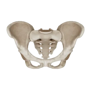
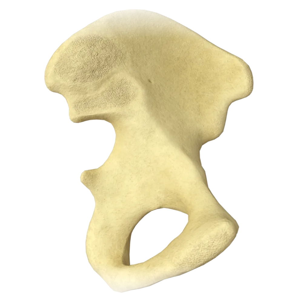
The quiz covers the anatomy of the hip bone: structure, parts, surfaces, foramina, lines, and bony landmarks.
1/20
bold
text
1. The hip bone (os coxae) in an adult is formed by the fusion of how many bones?
-
Two bones
The os coxae is formed through synostosis of three bones: the ilium (os ilium), ischium (os ischii), and pubis (os pubis). Fusion occurs in the region of the acetabulum and is completed at approximately 16–18 years of age.
-
Three bones
The os coxae is formed through synostosis of three bones: the ilium (os ilium), ischium (os ischii), and pubis (os pubis). Fusion occurs in the region of the acetabulum and is completed at approximately 16–18 years of age.
-
Four bones
The os coxae is formed through synostosis of three bones: the ilium (os ilium), ischium (os ischii), and pubis (os pubis). Fusion occurs in the region of the acetabulum and is completed at approximately 16–18 years of age.
-
Five bones
The os coxae is formed through synostosis of three bones: the ilium (os ilium), ischium (os ischii), and pubis (os pubis). Fusion occurs in the region of the acetabulum and is completed at approximately 16–18 years of age.
-
I find it difficult to answer
The os coxae is formed through synostosis of three bones: the ilium (os ilium), ischium (os ischii), and pubis (os pubis). Fusion occurs in the region of the acetabulum and is completed at approximately 16–18 years of age.
2. Which structure of the hip bone is formed by the fusion of all three of its constituent bones?
-
Greater sciatic notch
The acetabulum is the site where the bodies of the ilium, ischium, and pubis converge. The center of synostosis—the Y-shaped cartilage (cartilago ypsiliformis)—is located here and is replaced by bone tissue during skeletal maturation.
-
Obturator foramen
The acetabulum is the site where the bodies of the ilium, ischium, and pubis converge. The center of synostosis—the Y-shaped cartilage (cartilago ypsiliformis)—is located here and is replaced by bone tissue during skeletal maturation.
-
Acetabulum
The acetabulum is the site where the bodies of the ilium, ischium, and pubis converge. The center of synostosis—the Y-shaped cartilage (cartilago ypsiliformis)—is located here and is replaced by bone tissue during skeletal maturation.
-
Arcuate line
The acetabulum is the site where the bodies of the ilium, ischium, and pubis converge. The center of synostosis—the Y-shaped cartilage (cartilago ypsiliformis)—is located here and is replaced by bone tissue during skeletal maturation.
-
I find it difficult to answer
The acetabulum is the site where the bodies of the ilium, ischium, and pubis converge. The center of synostosis—the Y-shaped cartilage (cartilago ypsiliformis)—is located here and is replaced by bone tissue during skeletal maturation.
3. Which part of the acetabulum lacks articular cartilage and is occupied by adipose tissue?
-
Lunate surface (facies lunata)
The acetabular fossa (fossa acetabuli) is the central cartilage-free part of the acetabulum, filled with adipose tissue and containing the ligament of the head of the femur (lig. capitis femoris). Only the lunate surface (facies lunata) is covered with articular cartilage.
-
Acetabular fossa (fossa acetabuli)
The acetabular fossa (fossa acetabuli) is the central cartilage-free part of the acetabulum, filled with adipose tissue and containing the ligament of the head of the femur (lig. capitis femoris). Only the lunate surface (facies lunata) is covered with articular cartilage.
-
Acetabular labrum (labrum acetabulare)
The acetabular fossa (fossa acetabuli) is the central cartilage-free part of the acetabulum, filled with adipose tissue and containing the ligament of the head of the femur (lig. capitis femoris). Only the lunate surface (facies lunata) is covered with articular cartilage.
-
Acetabular margin (limbus acetabuli)
The acetabular fossa (fossa acetabuli) is the central cartilage-free part of the acetabulum, filled with adipose tissue and containing the ligament of the head of the femur (lig. capitis femoris). Only the lunate surface (facies lunata) is covered with articular cartilage.
-
I find it difficult to answer
The acetabular fossa (fossa acetabuli) is the central cartilage-free part of the acetabulum, filled with adipose tissue and containing the ligament of the head of the femur (lig. capitis femoris). Only the lunate surface (facies lunata) is covered with articular cartilage.
4. What is the inferior part of the acetabulum where the lunate surface is interrupted called?
-
Acetabular notch (incisura acetabuli)
The incisura acetabuli is a notch in the inferior part of the acetabular margin through which vessels and nerves pass to the ligament of the head of the femur. The transverse acetabular ligament (lig. transversum acetabuli) spans it.
-
Greater sciatic notch (incisura ischiadica major)
The incisura acetabuli is a notch in the inferior part of the acetabular margin through which vessels and nerves pass to the ligament of the head of the femur. The transverse acetabular ligament (lig. transversum acetabuli) spans it.
-
Lesser sciatic notch (incisura ischiadica minor)
The incisura acetabuli is a notch in the inferior part of the acetabular margin through which vessels and nerves pass to the ligament of the head of the femur. The transverse acetabular ligament (lig. transversum acetabuli) spans it.
-
Obturator groove (sulcus obturatorius)
The incisura acetabuli is a notch in the inferior part of the acetabular margin through which vessels and nerves pass to the ligament of the head of the femur. The transverse acetabular ligament (lig. transversum acetabuli) spans it.
-
I find it difficult to answer
The incisura acetabuli is a notch in the inferior part of the acetabular margin through which vessels and nerves pass to the ligament of the head of the femur. The transverse acetabular ligament (lig. transversum acetabuli) spans it.
5. Which line of the ilium separates the greater and lesser pelvis?
-
Intertrochanteric line (linea intertrochanterica)
The linea arcuata is the arcuate line on the medial surface of the ala of the ilium. Together with the pecten pubis and the sacral promontory, it forms the pelvic brim (linea terminalis), which separates the greater and lesser pelvis.
-
Arcuate line (linea arcuata)
The linea arcuata is the arcuate line on the medial surface of the ala of the ilium. Together with the pecten pubis and the sacral promontory, it forms the pelvic brim (linea terminalis), which separates the greater and lesser pelvis.
-
Iliac crest (crista iliaca)
The linea arcuata is the arcuate line on the medial surface of the ala of the ilium. Together with the pecten pubis and the sacral promontory, it forms the pelvic brim (linea terminalis), which separates the greater and lesser pelvis.
-
Semilunar line (linea semilunaris)
The linea arcuata is the arcuate line on the medial surface of the ala of the ilium. Together with the pecten pubis and the sacral promontory, it forms the pelvic brim (linea terminalis), which separates the greater and lesser pelvis.
-
I find it difficult to answer
The linea arcuata is the arcuate line on the medial surface of the ala of the ilium. Together with the pecten pubis and the sacral promontory, it forms the pelvic brim (linea terminalis), which separates the greater and lesser pelvis.
6. Where on the ilium is the iliac fossa (fossa iliaca) located?
-
On the dorsal (gluteal) surface of the ala
The fossa iliaca is the concave anterior (ventral, medial) surface of the ala of the ilium. The iliacus muscle (m. iliacus) originates here. The dorsal surface of the ala is called the gluteal surface (facies glutealis).
-
On the ventral (medial), concave surface of the ala
The fossa iliaca is the concave anterior (ventral, medial) surface of the ala of the ilium. The iliacus muscle (m. iliacus) originates here. The dorsal surface of the ala is called the gluteal surface (facies glutealis).
-
On the body of the ilium, superior to the acetabulum
The fossa iliaca is the concave anterior (ventral, medial) surface of the ala of the ilium. The iliacus muscle (m. iliacus) originates here. The dorsal surface of the ala is called the gluteal surface (facies glutealis).
-
On the anterior inferior iliac spine
The fossa iliaca is the concave anterior (ventral, medial) surface of the ala of the ilium. The iliacus muscle (m. iliacus) originates here. The dorsal surface of the ala is called the gluteal surface (facies glutealis).
-
I find it difficult to answer
The fossa iliaca is the concave anterior (ventral, medial) surface of the ala of the ilium. The iliacus muscle (m. iliacus) originates here. The dorsal surface of the ala is called the gluteal surface (facies glutealis).
7. How many gluteal lines are located on the external (gluteal) surface of the ala of the ilium?
-
One
Three gluteal lines are distinguished on the facies glutealis of the ilium: the anterior (linea glutealis anterior), posterior (linea glutealis posterior), and inferior (linea glutealis inferior) gluteal lines. They demarcate the attachment areas of the three gluteal muscles.
-
Two
Three gluteal lines are distinguished on the facies glutealis of the ilium: the anterior (linea glutealis anterior), posterior (linea glutealis posterior), and inferior (linea glutealis inferior) gluteal lines. They demarcate the attachment areas of the three gluteal muscles.
-
Three
Three gluteal lines are distinguished on the facies glutealis of the ilium: the anterior (linea glutealis anterior), posterior (linea glutealis posterior), and inferior (linea glutealis inferior) gluteal lines. They demarcate the attachment areas of the three gluteal muscles.
-
Four
Three gluteal lines are distinguished on the facies glutealis of the ilium: the anterior (linea glutealis anterior), posterior (linea glutealis posterior), and inferior (linea glutealis inferior) gluteal lines. They demarcate the attachment areas of the three gluteal muscles.
-
I find it difficult to answer
Three gluteal lines are distinguished on the facies glutealis of the ilium: the anterior (linea glutealis anterior), posterior (linea glutealis posterior), and inferior (linea glutealis inferior) gluteal lines. They demarcate the attachment areas of the three gluteal muscles.
8. What is the posterior inferior projection of the ala of the ilium that bounds the greater sciatic notch superiorly called?
-
Anterior superior iliac spine (spina iliaca anterior superior)
The greater sciatic notch (incisura ischiadica major) is bounded superiorly by the posterior inferior iliac spine (spina iliaca posterior inferior) and inferiorly by the ischial spine (spina ischiadica). The posterior superior iliac spine is located superiorly and forms the superior boundary of the auricular surface.
-
Posterior inferior iliac spine (spina iliaca posterior inferior)
The greater sciatic notch (incisura ischiadica major) is bounded superiorly by the posterior inferior iliac spine (spina iliaca posterior inferior) and inferiorly by the ischial spine (spina ischiadica). The posterior superior iliac spine is located superiorly and forms the superior boundary of the auricular surface.
-
Posterior superior iliac spine (spina iliaca posterior superior)
The greater sciatic notch (incisura ischiadica major) is bounded superiorly by the posterior inferior iliac spine (spina iliaca posterior inferior) and inferiorly by the ischial spine (spina ischiadica). The posterior superior iliac spine is located superiorly and forms the superior boundary of the auricular surface.
-
Anterior inferior iliac spine (spina iliaca anterior inferior)
The greater sciatic notch (incisura ischiadica major) is bounded superiorly by the posterior inferior iliac spine (spina iliaca posterior inferior) and inferiorly by the ischial spine (spina ischiadica). The posterior superior iliac spine is located superiorly and forms the superior boundary of the auricular surface.
-
I find it difficult to answer
The greater sciatic notch (incisura ischiadica major) is bounded superiorly by the posterior inferior iliac spine (spina iliaca posterior inferior) and inferiorly by the ischial spine (spina ischiadica). The posterior superior iliac spine is located superiorly and forms the superior boundary of the auricular surface.
9. Which surface of the ilium articulates with the sacrum?
-
Auricular surface (facies auricularis)
The facies auricularis is the auricular articular surface on the medial aspect of the ala of the ilium. It articulates with the corresponding surface of the sacrum, forming the sacroiliac joint (articulatio sacroiliaca). The iliac tuberosity (tuberositas iliaca) is located posterior to it.
-
Gluteal surface (facies glutealis)
The facies auricularis is the auricular articular surface on the medial aspect of the ala of the ilium. It articulates with the corresponding surface of the sacrum, forming the sacroiliac joint (articulatio sacroiliaca). The iliac tuberosity (tuberositas iliaca) is located posterior to it.
-
Iliac fossa (fossa iliaca)
The facies auricularis is the auricular articular surface on the medial aspect of the ala of the ilium. It articulates with the corresponding surface of the sacrum, forming the sacroiliac joint (articulatio sacroiliaca). The iliac tuberosity (tuberositas iliaca) is located posterior to it.
-
Pelvic surface (facies pelvica)
The facies auricularis is the auricular articular surface on the medial aspect of the ala of the ilium. It articulates with the corresponding surface of the sacrum, forming the sacroiliac joint (articulatio sacroiliaca). The iliac tuberosity (tuberositas iliaca) is located posterior to it.
-
I find it difficult to answer
The facies auricularis is the auricular articular surface on the medial aspect of the ala of the ilium. It articulates with the corresponding surface of the sacrum, forming the sacroiliac joint (articulatio sacroiliaca). The iliac tuberosity (tuberositas iliaca) is located posterior to it.
10. Which part of the hip bone forms the posteroinferior wall of the obturator foramen?
-
Body of pubis
The obturator foramen (foramen obturatum) is bounded superiorly and anteriorly by the superior and inferior rami of the pubis and posteriorly and inferiorly by the ramus of the ischium (ramus ossis ischii). Thus, the posteroinferior boundary is formed specifically by the ramus of the ischium.
-
Ramus of the ilium
The obturator foramen (foramen obturatum) is bounded superiorly and anteriorly by the superior and inferior rami of the pubis and posteriorly and inferiorly by the ramus of the ischium (ramus ossis ischii). Thus, the posteroinferior boundary is formed specifically by the ramus of the ischium.
-
Ramus of the ischium (ramus ossis ischii)
The obturator foramen (foramen obturatum) is bounded superiorly and anteriorly by the superior and inferior rami of the pubis and posteriorly and inferiorly by the ramus of the ischium (ramus ossis ischii). Thus, the posteroinferior boundary is formed specifically by the ramus of the ischium.
-
Inferior ramus of the pubis (ramus inferior ossis pubis)
The obturator foramen (foramen obturatum) is bounded superiorly and anteriorly by the superior and inferior rami of the pubis and posteriorly and inferiorly by the ramus of the ischium (ramus ossis ischii). Thus, the posteroinferior boundary is formed specifically by the ramus of the ischium.
-
I find it difficult to answer
The obturator foramen (foramen obturatum) is bounded superiorly and anteriorly by the superior and inferior rami of the pubis and posteriorly and inferiorly by the ramus of the ischium (ramus ossis ischii). Thus, the posteroinferior boundary is formed specifically by the ramus of the ischium.
11. Where on the hip bone is the obturator crest (crista obturatoria) located?
-
On the superior ramus of the pubis
The crista obturatoria is a crest on the superior surface of the superior ramus of the pubis (ramus superior ossis pubis).
-
On the internal surface of the body of the ischium
The crista obturatoria is a crest on the superior surface of the superior ramus of the pubis (ramus superior ossis pubis).
-
On the inferior ramus of the pubis
The crista obturatoria is a crest on the superior surface of the superior ramus of the pubis (ramus superior ossis pubis).
-
On the medial surface of the iliac fossa
The crista obturatoria is a crest on the superior surface of the superior ramus of the pubis (ramus superior ossis pubis).
-
I find it difficult to answer
The crista obturatoria is a crest on the superior surface of the superior ramus of the pubis (ramus superior ossis pubis).
12. What is the thickened superior border of the superior ramus of the pubis that serves as a landmark of the pelvic brim called?
-
Pubic tubercle (tuberculum pubicum)
The pecten ossis pubis (pecten pubis) is the sharp border of the superior ramus of the pubis and forms part of the pelvic brim (linea terminalis). Medially, it continues into the pubic crest (crista pubica), and laterally, into the iliopubic eminence (eminentia iliopubica).
-
Pecten pubis (pecten ossis pubis)
The pecten ossis pubis (pecten pubis) is the sharp border of the superior ramus of the pubis and forms part of the pelvic brim (linea terminalis). Medially, it continues into the pubic crest (crista pubica), and laterally, into the iliopubic eminence (eminentia iliopubica).
-
Pubic symphysis (symphysis pubica)
The pecten ossis pubis (pecten pubis) is the sharp border of the superior ramus of the pubis and forms part of the pelvic brim (linea terminalis). Medially, it continues into the pubic crest (crista pubica), and laterally, into the iliopubic eminence (eminentia iliopubica).
-
Iliopubic eminence (eminentia iliopubica)
The pecten ossis pubis (pecten pubis) is the sharp border of the superior ramus of the pubis and forms part of the pelvic brim (linea terminalis). Medially, it continues into the pubic crest (crista pubica), and laterally, into the iliopubic eminence (eminentia iliopubica).
-
I find it difficult to answer
The pecten ossis pubis (pecten pubis) is the sharp border of the superior ramus of the pubis and forms part of the pelvic brim (linea terminalis). Medially, it continues into the pubic crest (crista pubica), and laterally, into the iliopubic eminence (eminentia iliopubica).
13. Which bony projection is located on the superior ramus of the pubis at the medial border of the obturator foramen?
-
Ischial spine (spina ischiadica)
The tuberculum pubicum is a small projection on the superior ramus of the pubis, located at the medial boundary of the obturator foramen. The medial part of the inguinal ligament (lig. inguinale) attaches to it, and it is also an important landmark when describing the walls of the inguinal canal.
-
Pubic tubercle (tuberculum pubicum)
The tuberculum pubicum is a small projection on the superior ramus of the pubis, located at the medial boundary of the obturator foramen. The medial part of the inguinal ligament (lig. inguinale) attaches to it, and it is also an important landmark when describing the walls of the inguinal canal.
-
Anterior inferior iliac spine (spina iliaca anterior inferior)
The tuberculum pubicum is a small projection on the superior ramus of the pubis, located at the medial boundary of the obturator foramen. The medial part of the inguinal ligament (lig. inguinale) attaches to it, and it is also an important landmark when describing the walls of the inguinal canal.
-
Iliopubic eminence (eminentia iliopubica)
The tuberculum pubicum is a small projection on the superior ramus of the pubis, located at the medial boundary of the obturator foramen. The medial part of the inguinal ligament (lig. inguinale) attaches to it, and it is also an important landmark when describing the walls of the inguinal canal.
-
I find it difficult to answer
The tuberculum pubicum is a small projection on the superior ramus of the pubis, located at the medial boundary of the obturator foramen. The medial part of the inguinal ligament (lig. inguinale) attaches to it, and it is also an important landmark when describing the walls of the inguinal canal.
14. What is the ischial tuberosity (tuber ischiadicum)?
-
A sharp projection on the posterior surface of the body of the ischium that bounds the greater sciatic notch inferiorly
The tuber ischiadicum is a large, massive projection of the posteroinferior part of the body of the ischium. It bears body weight in the seated position. The posterior thigh muscles (m. biceps femoris, m. semitendinosus, m. semimembranosus) and the sacrotuberous ligament (lig. sacrotuberale) attach to it.
-
A massive thickening of the posteroinferior part of the body of the ischium that bears body weight in the seated position
The tuber ischiadicum is a large, massive projection of the posteroinferior part of the body of the ischium. It bears body weight in the seated position. The posterior thigh muscles (m. biceps femoris, m. semitendinosus, m. semimembranosus) and the sacrotuberous ligament (lig. sacrotuberale) attach to it.
-
An articular surface of the ischium that forms part of the acetabulum
The tuber ischiadicum is a large, massive projection of the posteroinferior part of the body of the ischium. It bears body weight in the seated position. The posterior thigh muscles (m. biceps femoris, m. semitendinosus, m. semimembranosus) and the sacrotuberous ligament (lig. sacrotuberale) attach to it.
-
A projection on the ramus of the ischium at its junction with the inferior ramus of the pubis
The tuber ischiadicum is a large, massive projection of the posteroinferior part of the body of the ischium. It bears body weight in the seated position. The posterior thigh muscles (m. biceps femoris, m. semitendinosus, m. semimembranosus) and the sacrotuberous ligament (lig. sacrotuberale) attach to it.
-
I find it difficult to answer
The tuber ischiadicum is a large, massive projection of the posteroinferior part of the body of the ischium. It bears body weight in the seated position. The posterior thigh muscles (m. biceps femoris, m. semitendinosus, m. semimembranosus) and the sacrotuberous ligament (lig. sacrotuberale) attach to it.
15. Which structure transforms the lesser sciatic notch into a foramen?
-
Sacrotuberous ligament (lig. sacrotuberale)
The sacrospinous ligament (lig. sacrospinale), which extends between the sacrum/coccyx and the ischial spine, transforms the lesser sciatic notch (incisura ischiadica minor) into the lesser sciatic foramen (foramen ischiadicum minus). The sacrotuberous ligament forms the greater sciatic foramen.
-
Sacrospinous ligament (lig. sacrospinale)
The sacrospinous ligament (lig. sacrospinale), which extends between the sacrum/coccyx and the ischial spine, transforms the lesser sciatic notch (incisura ischiadica minor) into the lesser sciatic foramen (foramen ischiadicum minus). The sacrotuberous ligament forms the greater sciatic foramen.
-
Obturator membrane (membrana obturatoria)
The sacrospinous ligament (lig. sacrospinale), which extends between the sacrum/coccyx and the ischial spine, transforms the lesser sciatic notch (incisura ischiadica minor) into the lesser sciatic foramen (foramen ischiadicum minus). The sacrotuberous ligament forms the greater sciatic foramen.
-
Iliolumbar ligament (lig. iliolumbale)
The sacrospinous ligament (lig. sacrospinale), which extends between the sacrum/coccyx and the ischial spine, transforms the lesser sciatic notch (incisura ischiadica minor) into the lesser sciatic foramen (foramen ischiadicum minus). The sacrotuberous ligament forms the greater sciatic foramen.
-
I find it difficult to answer
The sacrospinous ligament (lig. sacrospinale), which extends between the sacrum/coccyx and the ischial spine, transforms the lesser sciatic notch (incisura ischiadica minor) into the lesser sciatic foramen (foramen ischiadicum minus). The sacrotuberous ligament forms the greater sciatic foramen.
16. Which anatomical structure of the hip bone serves as the attachment site for the inguinal ligament (ligamentum inguinale) and is an anterosuperior landmark on palpation?
-
Anterior inferior iliac spine (Spina iliaca anterior inferior)
The spina iliaca anterior superior (SIAS) is the most anteriorly projecting landmark of the ala of the ilium. The inguinal ligament attaches to it, and it is readily palpable in lean individuals.
-
Pubic tubercle (Tuberculum pubicum)
The spina iliaca anterior superior (SIAS) is the most anteriorly projecting landmark of the ala of the ilium. The inguinal ligament attaches to it, and it is readily palpable in lean individuals.
-
Anterior superior iliac spine (Spina iliaca anterior superior)
The spina iliaca anterior superior (SIAS) is the most anteriorly projecting landmark of the ala of the ilium. The inguinal ligament attaches to it, and it is readily palpable in lean individuals.
-
Iliac crest (Crista iliaca)
The spina iliaca anterior superior (SIAS) is the most anteriorly projecting landmark of the ala of the ilium. The inguinal ligament attaches to it, and it is readily palpable in lean individuals.
-
I find it difficult to answer
The spina iliaca anterior superior (SIAS) is the most anteriorly projecting landmark of the ala of the ilium. The inguinal ligament attaches to it, and it is readily palpable in lean individuals.
17. What is the cartilaginous surface covering the pubic articulation on each side called?
-
Facies symphysialis
The facies symphysialis is the symphyseal surface of the body of the pubis, covered with hyaline cartilage. The two symphyseal surfaces are joined by the fibrocartilaginous interpubic disc (discus interpubicus), forming the pubic symphysis (symphysis pubica).
-
Facies auricularis
The facies symphysialis is the symphyseal surface of the body of the pubis, covered with hyaline cartilage. The two symphyseal surfaces are joined by the fibrocartilaginous interpubic disc (discus interpubicus), forming the pubic symphysis (symphysis pubica).
-
Facies lunata
The facies symphysialis is the symphyseal surface of the body of the pubis, covered with hyaline cartilage. The two symphyseal surfaces are joined by the fibrocartilaginous interpubic disc (discus interpubicus), forming the pubic symphysis (symphysis pubica).
-
Facies pelvica
The facies symphysialis is the symphyseal surface of the body of the pubis, covered with hyaline cartilage. The two symphyseal surfaces are joined by the fibrocartilaginous interpubic disc (discus interpubicus), forming the pubic symphysis (symphysis pubica).
-
I find it difficult to answer
The facies symphysialis is the symphyseal surface of the body of the pubis, covered with hyaline cartilage. The two symphyseal surfaces are joined by the fibrocartilaginous interpubic disc (discus interpubicus), forming the pubic symphysis (symphysis pubica).
18. Which landmark of the hip bone corresponds to the anterior end of the iliac crest?
-
Posterior superior iliac spine (spina iliaca posterior superior)
The spina iliaca anterior superior is the anterior end of the iliac crest (crista iliaca). It is an important palpable landmark: the inguinal ligament and m. sartorius attach to it. The anterior inferior iliac spine is located inferiorly and serves as the attachment site for m. rectus femoris and lig. iliofemorale.
-
Anterior superior iliac spine (spina iliaca anterior superior)
The spina iliaca anterior superior is the anterior end of the iliac crest (crista iliaca). It is an important palpable landmark: the inguinal ligament and m. sartorius attach to it. The anterior inferior iliac spine is located inferiorly and serves as the attachment site for m. rectus femoris and lig. iliofemorale.
-
Anterior inferior iliac spine (spina iliaca anterior inferior)
The spina iliaca anterior superior is the anterior end of the iliac crest (crista iliaca). It is an important palpable landmark: the inguinal ligament and m. sartorius attach to it. The anterior inferior iliac spine is located inferiorly and serves as the attachment site for m. rectus femoris and lig. iliofemorale.
-
Iliopubic eminence (eminentia iliopubica)
The spina iliaca anterior superior is the anterior end of the iliac crest (crista iliaca). It is an important palpable landmark: the inguinal ligament and m. sartorius attach to it. The anterior inferior iliac spine is located inferiorly and serves as the attachment site for m. rectus femoris and lig. iliofemorale.
-
I find it difficult to answer
The spina iliaca anterior superior is the anterior end of the iliac crest (crista iliaca). It is an important palpable landmark: the inguinal ligament and m. sartorius attach to it. The anterior inferior iliac spine is located inferiorly and serves as the attachment site for m. rectus femoris and lig. iliofemorale.
19. Which part of the hip bone contributes to the formation of the iliopubic eminence (eminentia iliopubica)?
-
Body of the ischium and superior ramus of the pubis
The eminentia iliopubica is a bony eminence at the junction of the body of the ilium (os ilium) and the superior ramus of the pubis (ramus superior ossis pubis). It is located on the pelvic brim, lateral to the pecten pubis, and serves as a landmark for the course of the iliopsoas muscle.
-
Body of the ilium and superior ramus of the pubis
The eminentia iliopubica is a bony eminence at the junction of the body of the ilium (os ilium) and the superior ramus of the pubis (ramus superior ossis pubis). It is located on the pelvic brim, lateral to the pecten pubis, and serves as a landmark for the course of the iliopsoas muscle.
-
Ala of the ilium and body of the pubis
The eminentia iliopubica is a bony eminence at the junction of the body of the ilium (os ilium) and the superior ramus of the pubis (ramus superior ossis pubis). It is located on the pelvic brim, lateral to the pecten pubis, and serves as a landmark for the course of the iliopsoas muscle.
-
Inferior ramus of the pubis and ramus of the ischium
The eminentia iliopubica is a bony eminence at the junction of the body of the ilium (os ilium) and the superior ramus of the pubis (ramus superior ossis pubis). It is located on the pelvic brim, lateral to the pecten pubis, and serves as a landmark for the course of the iliopsoas muscle.
-
I find it difficult to answer
The eminentia iliopubica is a bony eminence at the junction of the body of the ilium (os ilium) and the superior ramus of the pubis (ramus superior ossis pubis). It is located on the pelvic brim, lateral to the pecten pubis, and serves as a landmark for the course of the iliopsoas muscle.
20. What is the shape of the obturator foramen (foramen obturatum) in males compared with females?
-
Triangular in males and oval in females
Sex differences in the foramen obturatum are well marked: in males, the foramen is larger and rounded-oval, whereas in females, it is smaller and approaches a triangular-oval shape (due to differences in the angle of the pubic arch and the proportions of the pubic and ischial rami), reflecting the general sex differences in pelvic structure.
-
Slit-shaped in males and round in females
Sex differences in the foramen obturatum are well marked: in males, the foramen is larger and rounded-oval, whereas in females, it is smaller and approaches a triangular-oval shape (due to differences in the angle of the pubic arch and the proportions of the pubic and ischial rami), reflecting the general sex differences in pelvic structure.
-
Rounded-oval in males and triangular-oval in females
Sex differences in the foramen obturatum are well marked: in males, the foramen is larger and rounded-oval, whereas in females, it is smaller and approaches a triangular-oval shape (due to differences in the angle of the pubic arch and the proportions of the pubic and ischial rami), reflecting the general sex differences in pelvic structure.
-
The shape is identical in males and females—perfectly round
Sex differences in the foramen obturatum are well marked: in males, the foramen is larger and rounded-oval, whereas in females, it is smaller and approaches a triangular-oval shape (due to differences in the angle of the pubic arch and the proportions of the pubic and ischial rami), reflecting the general sex differences in pelvic structure.
-
I find it difficult to answer
Sex differences in the foramen obturatum are well marked: in males, the foramen is larger and rounded-oval, whereas in females, it is smaller and approaches a triangular-oval shape (due to differences in the angle of the pubic arch and the proportions of the pubic and ischial rami), reflecting the general sex differences in pelvic structure.
Retake this quiz?
Your current progress will be reset.
