1/20
Quiz on the Anatomy of the Pelvic Girdle Bones
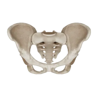
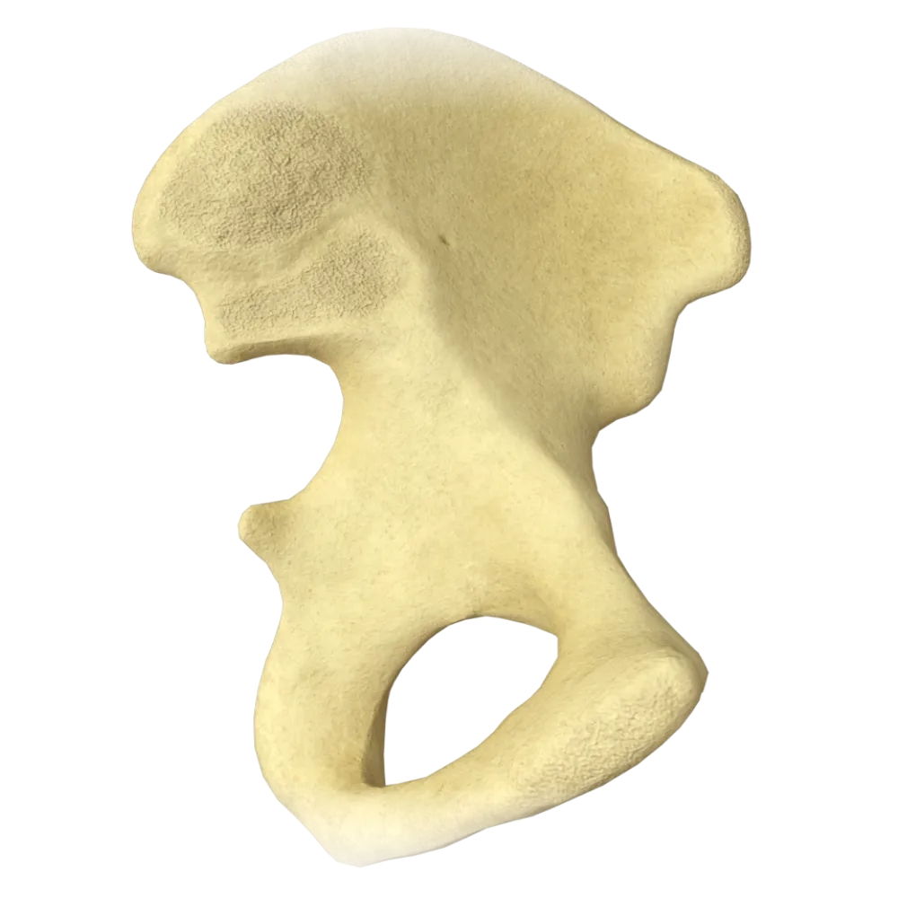
The quiz covers the structure of the pelvic girdle bones: ilium, ischium, pubis, and sacrum.
1. Which of the listed bones is NOT part of the hip bone?
-
Os ilium
The hip bone is formed by three bones: the ilium, the ischium, and the pubis. The sacrum is a separate bone of the axial skeleton and is not part of the hip bone.
-
Os ischium
The hip bone is formed by three bones: the ilium, the ischium, and the pubis. The sacrum is a separate bone of the axial skeleton and is not part of the hip bone.
-
Os pubis
The hip bone is formed by three bones: the ilium, the ischium, and the pubis. The sacrum is a separate bone of the axial skeleton and is not part of the hip bone.
-
Os sacrum
The hip bone is formed by three bones: the ilium, the ischium, and the pubis. The sacrum is a separate bone of the axial skeleton and is not part of the hip bone.
-
I find it difficult to answer
The hip bone is formed by three bones: the ilium, the ischium, and the pubis. The sacrum is a separate bone of the axial skeleton and is not part of the hip bone.
2. At what point do the three parts of the hip bone fuse, forming the acetabulum?
-
In the region of the ischial tuberosity
All three bones – ilium, ischium, and pubis – fuse in the region of the acetabulum. Their line of fusion passes through the acetabulum. The synchondrosis between them obliterates by the age of 16-18.
-
In the region of the pubic symphysis
All three bones – ilium, ischium, and pubis – fuse in the region of the acetabulum. Their line of fusion passes through the acetabulum. The synchondrosis between them obliterates by the age of 16-18.
-
In the region of the pubic rami
All three bones – ilium, ischium, and pubis – fuse in the region of the acetabulum. Their line of fusion passes through the acetabulum. The synchondrosis between them obliterates by the age of 16-18.
-
At the confluence of all three bones within the acetabulum
All three bones – ilium, ischium, and pubis – fuse in the region of the acetabulum. Their line of fusion passes through the acetabulum. The synchondrosis between them obliterates by the age of 16-18.
-
I find it difficult to answer
All three bones – ilium, ischium, and pubis – fuse in the region of the acetabulum. Their line of fusion passes through the acetabulum. The synchondrosis between them obliterates by the age of 16-18.
3. Which structure bounds the obturator foramen inferiorly and posteriorly?
-
Pubis
The obturator foramen is bounded by the pubic bone (superiorly and anteriorly) and the ischial bone (inferiorly and posteriorly). The ilium does not contribute directly to the formation of the obturator foramen.
-
Ilium
The obturator foramen is bounded by the pubic bone (superiorly and anteriorly) and the ischial bone (inferiorly and posteriorly). The ilium does not contribute directly to the formation of the obturator foramen.
-
Ischium
The obturator foramen is bounded by the pubic bone (superiorly and anteriorly) and the ischial bone (inferiorly and posteriorly). The ilium does not contribute directly to the formation of the obturator foramen.
-
Sacrum
The obturator foramen is bounded by the pubic bone (superiorly and anteriorly) and the ischial bone (inferiorly and posteriorly). The ilium does not contribute directly to the formation of the obturator foramen.
-
I find it difficult to answer
The obturator foramen is bounded by the pubic bone (superiorly and anteriorly) and the ischial bone (inferiorly and posteriorly). The ilium does not contribute directly to the formation of the obturator foramen.
4. What is the name of the articular surface on the ilium that contributes to the formation of the sacroiliac joint?
-
Facies symphysialis
The auricular surface of the ilium articulates with the corresponding surface of the sacrum, forming the sacroiliac joint. Facies symphysialis refers to the surface of the pubic symphysis, and facies lunata refers to the crescent-shaped surface of the acetabulum.
-
Facies auricularis
The auricular surface of the ilium articulates with the corresponding surface of the sacrum, forming the sacroiliac joint. Facies symphysialis refers to the surface of the pubic symphysis, and facies lunata refers to the crescent-shaped surface of the acetabulum.
-
Facies lunata
The auricular surface of the ilium articulates with the corresponding surface of the sacrum, forming the sacroiliac joint. Facies symphysialis refers to the surface of the pubic symphysis, and facies lunata refers to the crescent-shaped surface of the acetabulum.
-
Facies poplitea
The auricular surface of the ilium articulates with the corresponding surface of the sacrum, forming the sacroiliac joint. Facies symphysialis refers to the surface of the pubic symphysis, and facies lunata refers to the crescent-shaped surface of the acetabulum.
-
I find it difficult to answer
The auricular surface of the ilium articulates with the corresponding surface of the sacrum, forming the sacroiliac joint. Facies symphysialis refers to the surface of the pubic symphysis, and facies lunata refers to the crescent-shaped surface of the acetabulum.
5. What is the linea terminalis of the pelvis?
-
Crest of the ilium
The linea terminalis separates the greater pelvis from the lesser pelvis. It extends from the promontory of the sacrum, along the arcuate line of the ilium, and then along the pecten ossis pubis and tuberculum pubicum to the pubic symphysis
-
The boundary between the greater and lesser pelvis, formed by the promontory of the sacrum, arcuate lines, and crest of the pubic bones
The linea terminalis separates the greater pelvis from the lesser pelvis. It extends from the promontory of the sacrum, along the arcuate line of the ilium, and then along the pecten ossis pubis and tuberculum pubicum to the pubic symphysis
-
The lower edge of the symphysis
The linea terminalis separates the greater pelvis from the lesser pelvis. It extends from the promontory of the sacrum, along the arcuate line of the ilium, and then along the pecten ossis pubis and tuberculum pubicum to the pubic symphysis
-
The inner edge of the obturator foramen
The linea terminalis separates the greater pelvis from the lesser pelvis. It extends from the promontory of the sacrum, along the arcuate line of the ilium, and then along the pecten ossis pubis and tuberculum pubicum to the pubic symphysis
-
I find it difficult to answer
The linea terminalis separates the greater pelvis from the lesser pelvis. It extends from the promontory of the sacrum, along the arcuate line of the ilium, and then along the pecten ossis pubis and tuberculum pubicum to the pubic symphysis
6. Which surface of the acetabulum is covered with articular cartilage?
-
Fossa acetabuli
The lunate surface of the acetabulum is lined with hyaline articular cartilage and articulates with the head of the femur. The acetabular fossa is a depression in the center covered with adipose tissue and synovial membrane, but not cartilage.
-
Facies lunata
The lunate surface of the acetabulum is lined with hyaline articular cartilage and articulates with the head of the femur. The acetabular fossa is a depression in the center covered with adipose tissue and synovial membrane, but not cartilage.
-
Incisura acetabuli
The lunate surface of the acetabulum is lined with hyaline articular cartilage and articulates with the head of the femur. The acetabular fossa is a depression in the center covered with adipose tissue and synovial membrane, but not cartilage.
-
Foramen obturatum
The lunate surface of the acetabulum is lined with hyaline articular cartilage and articulates with the head of the femur. The acetabular fossa is a depression in the center covered with adipose tissue and synovial membrane, but not cartilage.
-
I find it difficult to answer
The lunate surface of the acetabulum is lined with hyaline articular cartilage and articulates with the head of the femur. The acetabular fossa is a depression in the center covered with adipose tissue and synovial membrane, but not cartilage.
7. Where is the ischial tuberosity located?
-
On the body of the ilium
The ischial tuberosity is a robust protrusion of the inferoposterior part of the body of the ischium. It serves as a support point when sitting and as the site of origin for the posterior thigh muscles (biceps femoris, semimembranosus, semitendinosus).
-
On the inferoposterior part of the body of the ischium
The ischial tuberosity is a robust protrusion of the inferoposterior part of the body of the ischium. It serves as a support point when sitting and as the site of origin for the posterior thigh muscles (biceps femoris, semimembranosus, semitendinosus).
-
On the inferior pubic ramus
The ischial tuberosity is a robust protrusion of the inferoposterior part of the body of the ischium. It serves as a support point when sitting and as the site of origin for the posterior thigh muscles (biceps femoris, semimembranosus, semitendinosus).
-
On the posterior surface of the sacrum
The ischial tuberosity is a robust protrusion of the inferoposterior part of the body of the ischium. It serves as a support point when sitting and as the site of origin for the posterior thigh muscles (biceps femoris, semimembranosus, semitendinosus).
-
I find it difficult to answer
The ischial tuberosity is a robust protrusion of the inferoposterior part of the body of the ischium. It serves as a support point when sitting and as the site of origin for the posterior thigh muscles (biceps femoris, semimembranosus, semitendinosus).
8. Which element of the ilium forms the upper boundary of the greater pelvis?
-
Spina iliaca anterior superior
The iliac crest — the curved superior edge of the ilium — is the upper boundary of the great wing of the pelvis.
-
Crista iliaca
The iliac crest — the curved superior edge of the ilium — is the upper boundary of the great wing of the pelvis.
-
Linea arcuata
The iliac crest — the curved superior edge of the ilium — is the upper boundary of the great wing of the pelvis.
-
Facies auricularis
The iliac crest — the curved superior edge of the ilium — is the upper boundary of the great wing of the pelvis.
-
I find it difficult to answer
The iliac crest — the curved superior edge of the ilium — is the upper boundary of the great wing of the pelvis.
9. What is the name of the foramen formed by the greater sciatic notch and the sacrospinous ligament?
-
Foramen obturatum
The greater sciatic foramen is formed by the greater sciatic notch of the ilium and the sacrospinous ligament. The lesser sciatic foramen is bounded by the lesser sciatic notch and the sacrotuberous ligament.
-
Foramen ischiadicum minus
The greater sciatic foramen is formed by the greater sciatic notch of the ilium and the sacrospinous ligament. The lesser sciatic foramen is bounded by the lesser sciatic notch and the sacrotuberous ligament.
-
Foramen ischiadicum majus
The greater sciatic foramen is formed by the greater sciatic notch of the ilium and the sacrospinous ligament. The lesser sciatic foramen is bounded by the lesser sciatic notch and the sacrotuberous ligament.
-
Canalis pudendalis
The greater sciatic foramen is formed by the greater sciatic notch of the ilium and the sacrospinous ligament. The lesser sciatic foramen is bounded by the lesser sciatic notch and the sacrotuberous ligament.
-
I find it difficult to answer
The greater sciatic foramen is formed by the greater sciatic notch of the ilium and the sacrospinous ligament. The lesser sciatic foramen is bounded by the lesser sciatic notch and the sacrotuberous ligament.
10. Where is the pubic tubercle located?
-
On the superior pubic ramus, at the medial end of the pubic crest
The pubic tubercle is located on the superior pubic ramus at the medial end of the pecten ossis pubis. It is an important anatomical landmark where the medial part of the inguinal ligament attaches.
-
On the inferior pubic ramus
The pubic tubercle is located on the superior pubic ramus at the medial end of the pecten ossis pubis. It is an important anatomical landmark where the medial part of the inguinal ligament attaches.
-
On the body of the ilium
The pubic tubercle is located on the superior pubic ramus at the medial end of the pecten ossis pubis. It is an important anatomical landmark where the medial part of the inguinal ligament attaches.
-
On the crista iliaca
The pubic tubercle is located on the superior pubic ramus at the medial end of the pecten ossis pubis. It is an important anatomical landmark where the medial part of the inguinal ligament attaches.
-
I find it difficult to answer
The pubic tubercle is located on the superior pubic ramus at the medial end of the pecten ossis pubis. It is an important anatomical landmark where the medial part of the inguinal ligament attaches.
11. What is the incisura acetabuli?
-
The fossa in the center of the acetabulum
The incisura acetabuli is a notch on the inferior part of the acetabular margin. It allows for passage of blood vessels supplying the head of the femur and the attachment of the ligamentum capitis femoris. It is covered by the transverse acetabular ligament.
-
A notch in the lower part of the acetabular margin through which the ligamentum capitis femoris passes
The incisura acetabuli is a notch on the inferior part of the acetabular margin. It allows for passage of blood vessels supplying the head of the femur and the attachment of the ligamentum capitis femoris. It is covered by the transverse acetabular ligament.
-
The anterior edge of the acetabulum
The incisura acetabuli is a notch on the inferior part of the acetabular margin. It allows for passage of blood vessels supplying the head of the femur and the attachment of the ligamentum capitis femoris. It is covered by the transverse acetabular ligament.
-
The articular surface of the acetabulum
The incisura acetabuli is a notch on the inferior part of the acetabular margin. It allows for passage of blood vessels supplying the head of the femur and the attachment of the ligamentum capitis femoris. It is covered by the transverse acetabular ligament.
-
I find it difficult to answer
The incisura acetabuli is a notch on the inferior part of the acetabular margin. It allows for passage of blood vessels supplying the head of the femur and the attachment of the ligamentum capitis femoris. It is covered by the transverse acetabular ligament.
12. Which element of the sacrum contributes to the formation of the linea terminalis?
-
Crista sacralis mediana
The promontory of the sacrum — the forward-projecting upper edge of the body of the first sacral vertebra — serves as the starting point of the linea terminalis. It serves as an important obstetric landmark for measuring the diagonal conjugate.
-
Promontory
The promontory of the sacrum — the forward-projecting upper edge of the body of the first sacral vertebra — serves as the starting point of the linea terminalis. It serves as an important obstetric landmark for measuring the diagonal conjugate.
-
Hiatus sacralis
The promontory of the sacrum — the forward-projecting upper edge of the body of the first sacral vertebra — serves as the starting point of the linea terminalis. It serves as an important obstetric landmark for measuring the diagonal conjugate.
-
Cornua sacralia
The promontory of the sacrum — the forward-projecting upper edge of the body of the first sacral vertebra — serves as the starting point of the linea terminalis. It serves as an important obstetric landmark for measuring the diagonal conjugate.
-
I find it difficult to answer
The promontory of the sacrum — the forward-projecting upper edge of the body of the first sacral vertebra — serves as the starting point of the linea terminalis. It serves as an important obstetric landmark for measuring the diagonal conjugate.
13. Which of the sacral foramina opens on its pelvic (anterior) surface?
-
Foramina sacralia posteriora
The pelvic (anterior) sacral foramina — foramina sacralia anteriora (pelvica) — are located on the pelvic surface of the sacrum. The anterior branches of the sacral nerves exit through them. The posterior sacral foramina (foramina sacralia posteriora) open on the dorsal surface.
-
Canalis sacralis
The pelvic (anterior) sacral foramina — foramina sacralia anteriora (pelvica) — are located on the pelvic surface of the sacrum. The anterior branches of the sacral nerves exit through them. The posterior sacral foramina (foramina sacralia posteriora) open on the dorsal surface.
-
Foramina sacralia anteriora (pelvica)
The pelvic (anterior) sacral foramina — foramina sacralia anteriora (pelvica) — are located on the pelvic surface of the sacrum. The anterior branches of the sacral nerves exit through them. The posterior sacral foramina (foramina sacralia posteriora) open on the dorsal surface.
-
Hiatus sacralis
The pelvic (anterior) sacral foramina — foramina sacralia anteriora (pelvica) — are located on the pelvic surface of the sacrum. The anterior branches of the sacral nerves exit through them. The posterior sacral foramina (foramina sacralia posteriora) open on the dorsal surface.
-
I find it difficult to answer
The pelvic (anterior) sacral foramina — foramina sacralia anteriora (pelvica) — are located on the pelvic surface of the sacrum. The anterior branches of the sacral nerves exit through them. The posterior sacral foramina (foramina sacralia posteriora) open on the dorsal surface.
14. What forms the pelvic outlet?
-
Linea terminalis.
The pelvic outlet is bounded by the following: anteriorly – by the inferior edge of the symphysis, laterally – by the inferior pubic and ischial rami and ischial tuberosities, and posteriorly – by the sacrotuberous ligaments and coccyx.
-
Pubic symphysis, inferior pubic and ischial rami, ischial tuberosities, sacrotuberous ligaments, and coccyx
The pelvic outlet is bounded by the following: anteriorly – by the inferior edge of the symphysis, laterally – by the inferior pubic and ischial rami and ischial tuberosities, and posteriorly – by the sacrotuberous ligaments and coccyx.
-
Sacrum and ilium
The pelvic outlet is bounded by the following: anteriorly – by the inferior edge of the symphysis, laterally – by the inferior pubic and ischial rami and ischial tuberosities, and posteriorly – by the sacrotuberous ligaments and coccyx.
-
Pubic rami and obturator foramina
The pelvic outlet is bounded by the following: anteriorly – by the inferior edge of the symphysis, laterally – by the inferior pubic and ischial rami and ischial tuberosities, and posteriorly – by the sacrotuberous ligaments and coccyx.
-
I find it difficult to answer
The pelvic outlet is bounded by the following: anteriorly – by the inferior edge of the symphysis, laterally – by the inferior pubic and ischial rami and ischial tuberosities, and posteriorly – by the sacrotuberous ligaments and coccyx.
15. What is the name of the angle formed by the inferior branches of the pubic bones in men?
-
Arcus pubicus
In men, the angle between the inferior branches of the pubic bones is called the angulus subpubicus and is approximately 70–75°. In females, it is more obtuse (90–100°) and is known as the arcus pubicus. It is one of the sexual dimorphism indicators of the pelvis.
-
Angulus subpubicus
In men, the angle between the inferior branches of the pubic bones is called the angulus subpubicus and is approximately 70–75°. In females, it is more obtuse (90–100°) and is known as the arcus pubicus. It is one of the sexual dimorphism indicators of the pelvis.
-
Arcus ischiadicus
In men, the angle between the inferior branches of the pubic bones is called the angulus subpubicus and is approximately 70–75°. In females, it is more obtuse (90–100°) and is known as the arcus pubicus. It is one of the sexual dimorphism indicators of the pelvis.
-
Angulus pubicus
In men, the angle between the inferior branches of the pubic bones is called the angulus subpubicus and is approximately 70–75°. In females, it is more obtuse (90–100°) and is known as the arcus pubicus. It is one of the sexual dimorphism indicators of the pelvis.
-
I find it difficult to answer
In men, the angle between the inferior branches of the pubic bones is called the angulus subpubicus and is approximately 70–75°. In females, it is more obtuse (90–100°) and is known as the arcus pubicus. It is one of the sexual dimorphism indicators of the pelvis.
16. Where is the ischial spine located?
-
On the body of the ilium above the acetabulum
The ischial spine is a pointed projection on the posterior edge of the ischial body, located between the greater sciatic notch (above) and the lesser sciatic notch (below). The sacrospinous ligament is attached to it.
-
On the posterior edge of the body of the ischium between the greater and lesser sciatic notches
The ischial spine is a pointed projection on the posterior edge of the ischial body, located between the greater sciatic notch (above) and the lesser sciatic notch (below). The sacrospinous ligament is attached to it.
-
On the inferior ramus of the ischium
The ischial spine is a pointed projection on the posterior edge of the ischial body, located between the greater sciatic notch (above) and the lesser sciatic notch (below). The sacrospinous ligament is attached to it.
-
On the sacrum
The ischial spine is a pointed projection on the posterior edge of the ischial body, located between the greater sciatic notch (above) and the lesser sciatic notch (below). The sacrospinous ligament is attached to it.
-
I find it difficult to answer
The ischial spine is a pointed projection on the posterior edge of the ischial body, located between the greater sciatic notch (above) and the lesser sciatic notch (below). The sacrospinous ligament is attached to it.
17. Which line delineates the gluteal surface and the iliac surface of the ilium's wing from the outer side?
-
Linea arcuata
On the gluteal surface of the ilium, there are three gluteal lines: anterior gluteal line, posterior and inferior. They delimit the zones of origin for the three gluteal muscles. Linea arcuata is located on the inner surface of the bone.
-
Linea glutealis anterior
On the gluteal surface of the ilium, there are three gluteal lines: anterior gluteal line, posterior and inferior. They delimit the zones of origin for the three gluteal muscles. Linea arcuata is located on the inner surface of the bone.
-
Linea terminalis.
On the gluteal surface of the ilium, there are three gluteal lines: anterior gluteal line, posterior and inferior. They delimit the zones of origin for the three gluteal muscles. Linea arcuata is located on the inner surface of the bone.
-
Linea aspera.
On the gluteal surface of the ilium, there are three gluteal lines: anterior gluteal line, posterior and inferior. They delimit the zones of origin for the three gluteal muscles. Linea arcuata is located on the inner surface of the bone.
-
I find it difficult to answer
On the gluteal surface of the ilium, there are three gluteal lines: anterior gluteal line, posterior and inferior. They delimit the zones of origin for the three gluteal muscles. Linea arcuata is located on the inner surface of the bone.
18. What is the pecten ossis pubis?
-
Lower edge of the symphysis
The pecten ossis pubis (pubic crest) is a sharp crest on the superior pubic ramus, extending medially into the pubic tubercle and laterally into the arcuate line of the ilium. It is part of the linea terminalis.
-
The crest of the superior pubic ramus, which is part of the linea terminalis
The pecten ossis pubis (pubic crest) is a sharp crest on the superior pubic ramus, extending medially into the pubic tubercle and laterally into the arcuate line of the ilium. It is part of the linea terminalis.
-
Articular surface of the pubic bone
The pecten ossis pubis (pubic crest) is a sharp crest on the superior pubic ramus, extending medially into the pubic tubercle and laterally into the arcuate line of the ilium. It is part of the linea terminalis.
-
Outer crest of the ilium
The pecten ossis pubis (pubic crest) is a sharp crest on the superior pubic ramus, extending medially into the pubic tubercle and laterally into the arcuate line of the ilium. It is part of the linea terminalis.
-
I find it difficult to answer
The pecten ossis pubis (pubic crest) is a sharp crest on the superior pubic ramus, extending medially into the pubic tubercle and laterally into the arcuate line of the ilium. It is part of the linea terminalis.
19. Which structure of the pelvis is the origin of the rectus abdominis muscle?
-
Crista iliaca
The rectus abdominis muscle originates from the pubic crest and the pubic tubercle, i.e. from the anterior surface of the pubic symphysis. This is a direct relationship between the muscle and the bones of the pelvic girdle.
-
Spina iliaca anterior superior
The rectus abdominis muscle originates from the pubic crest and the pubic tubercle, i.e. from the anterior surface of the pubic symphysis. This is a direct relationship between the muscle and the bones of the pelvic girdle.
-
Symphysis pubica (crista and tuberculum ossis pubis)
The rectus abdominis muscle originates from the pubic crest and the pubic tubercle, i.e. from the anterior surface of the pubic symphysis. This is a direct relationship between the muscle and the bones of the pelvic girdle.
-
Linea arcuata
The rectus abdominis muscle originates from the pubic crest and the pubic tubercle, i.e. from the anterior surface of the pubic symphysis. This is a direct relationship between the muscle and the bones of the pelvic girdle.
-
I find it difficult to answer
The rectus abdominis muscle originates from the pubic crest and the pubic tubercle, i.e. from the anterior surface of the pubic symphysis. This is a direct relationship between the muscle and the bones of the pelvic girdle.
20. Which of the listed dimensions is the true (obstetric) conjugate of the pelvis?
-
Distance between the anterior superior iliac spines
The true conjugate is the shortest distance from the promontory of the sacrum to the most prominent point of the posterior surface of the pubic symphysis, and is typically around 11 cm. This dimension determines the minimum anteroposterior diameter of the pelvic inlet.
-
Distance from the promontory to the lower edge of the pubic symphysis
The true conjugate is the shortest distance from the promontory of the sacrum to the most prominent point of the posterior surface of the pubic symphysis, and is typically around 11 cm. This dimension determines the minimum anteroposterior diameter of the pelvic inlet.
-
Distance between the ischial tuberosities
The true conjugate is the shortest distance from the promontory of the sacrum to the most prominent point of the posterior surface of the pubic symphysis, and is typically around 11 cm. This dimension determines the minimum anteroposterior diameter of the pelvic inlet.
-
Distance between the iliac crests
The true conjugate is the shortest distance from the promontory of the sacrum to the most prominent point of the posterior surface of the pubic symphysis, and is typically around 11 cm. This dimension determines the minimum anteroposterior diameter of the pelvic inlet.
-
I find it difficult to answer
The true conjugate is the shortest distance from the promontory of the sacrum to the most prominent point of the posterior surface of the pubic symphysis, and is typically around 11 cm. This dimension determines the minimum anteroposterior diameter of the pelvic inlet.
Retake this quiz?
Your current progress will be reset.

Cookie Consent
We use cookies to enhance your browsing experience, analyze site traffic, and deliver content. Please choose whether you accept all cookies or wish to reject non-essential tracking.
Cookie Preferences
Manage your cookie preferences below:
Essential cookies enable basic functions and are necessary for the proper function of the website.
Name
Description
Duration
Geolocation Config
This cookie is used to store the consent settings based on the visitor's location.
30 days
Cookie Preferences
This cookie is used to store the user's cookie consent preferences.
30 days
Google reCAPTCHA helps protect websites from spam and abuse by verifying user interactions through challenges.
Name
Description
Duration
_GRECAPTCHA
Google reCAPTCHA sets a necessary cookie (_GRECAPTCHA) when executed for the purpose of providing its risk analysis.
179 days
Statistics cookies collect information anonymously. This information helps us understand how visitors use our website.
Google Analytics is a powerful tool that tracks and analyzes website traffic for informed marketing decisions.
Service URL: policies.google.com (opens in a new window)
Name
Description
Duration
__utma
ID used to identify users and sessions
2 years after last activity
__utmt
Used to monitor number of Google Analytics server requests
10 minutes
__utmb
Used to distinguish new sessions and visits. This cookie is set when the GA.js javascript library is loaded and there is no existing __utmb cookie. The cookie is updated every time data is sent to the Google Analytics server.
30 minutes after last activity
__utmc
Used only with old Urchin versions of Google Analytics and not with GA.js. Was used to distinguish between new sessions and visits at the end of a session.
End of session (browser)
__utmz
Contains information about the traffic source or campaign that directed user to the website. The cookie is set when the GA.js javascript is loaded and updated when data is sent to the Google Anaytics server
6 months after last activity
__utmv
Contains custom information set by the web developer via the _setCustomVar method in Google Analytics. This cookie is updated every time new data is sent to the Google Analytics server.
2 years after last activity
__utmx
Used to determine whether a user is included in an A / B or Multivariate test.
18 months
_ga
ID used to identify users
2 years
_gali
Used by Google Analytics to determine which links on a page are being clicked
30 seconds
_ga_
ID used to identify users
2 years
_gid
ID used to identify users for 24 hours after last activity
24 hours
_gat
Used to monitor number of Google Analytics server requests when using Google Tag Manager
1 minute
_gac_
Contains information related to marketing campaigns of the user. These are shared with Google AdWords / Google Ads when the Google Ads and Google Analytics accounts are linked together.
90 days
Clarity is a web analytics service that tracks and reports website traffic.
Service URL: clarity.microsoft.com (opens in a new window)
Name
Description
Duration
_clsk
Connects multiple page views by a user into a single Clarity session recording.
1 day
_clck
Stores a unique user identifier to analyze visitor behavior through heatmaps and session recordings.
1 year
ANONCHK
Indicates whether MUID is transferred to ANID, a cookie used for advertising. Clarity doesn't use ANID and so this is always set to 0.
Session
CLID
Identifies the first-time Clarity saw this user on any site using Clarity.
12 months
_clsk
Connects multiple page views by a user into a single Clarity session recording.
12 months
_clck
Persists the Clarity User ID and preferences, unique to that site is attributed to the same user ID.
12 months