Mandible anatomy quiz
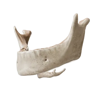
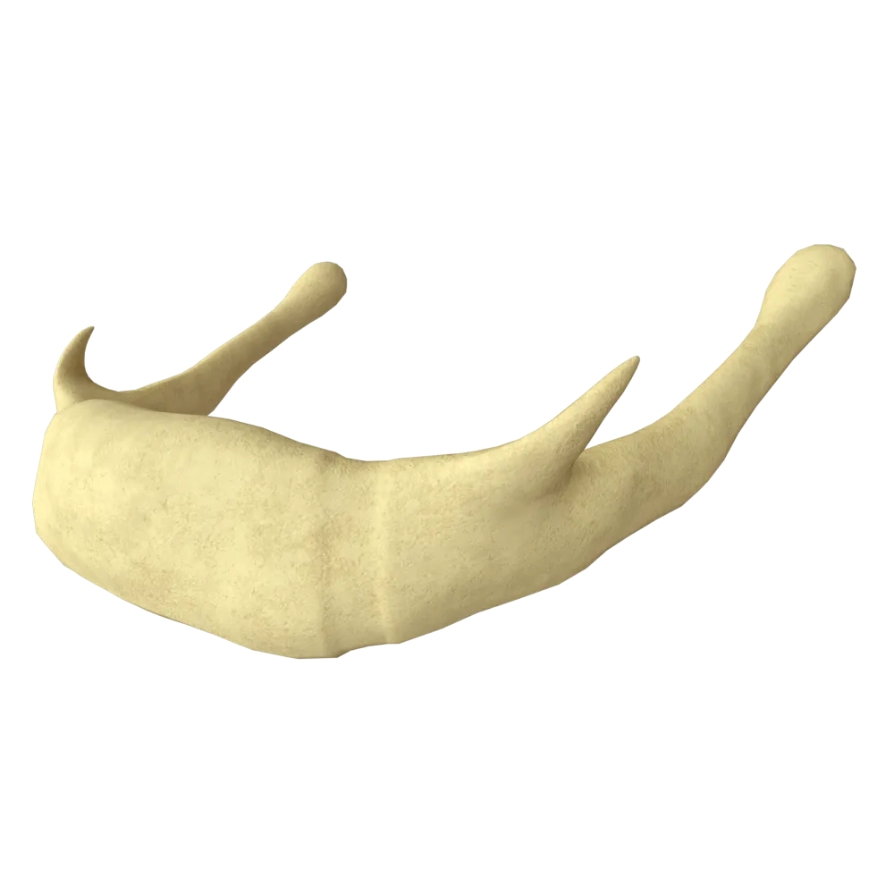
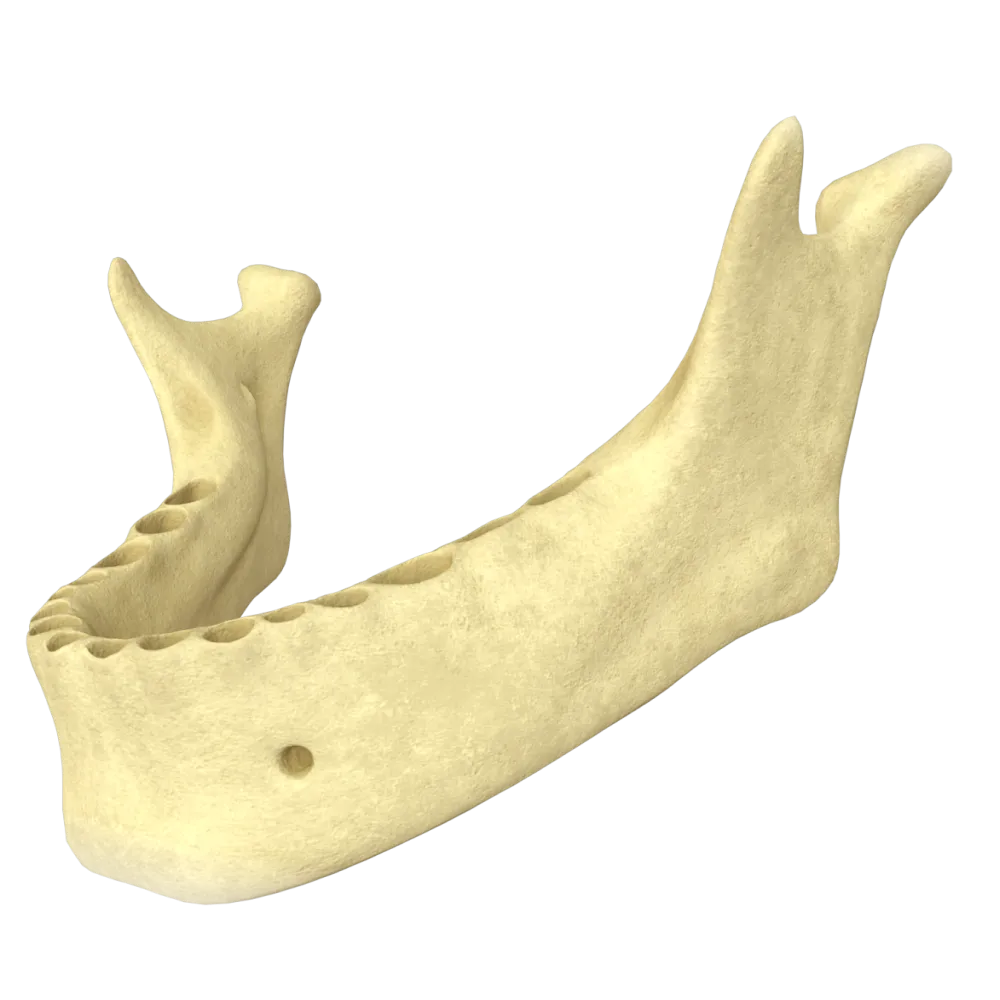
Systemic anatomy quiz on the mandible: structure, processes, canals, articular surfaces, muscle attachments, and neurovascular supply.
1/20
bold
text
1. Which part of the mandible is the attachment site of the masseter muscle (m. masseter)?
-
Coronoid process (processus coronoideus)
M. masseter attaches to the masseteric tuberosity (tuberositas masseterica), located on the external surface of the angle of the mandible. The coronoid process is the attachment site of m. temporalis, the condylar process participates in the formation of the TMJ, and the digastric fossa is the attachment site of the anterior belly of m. digastric muscle.
-
Condylar process (processus condylaris)
M. masseter attaches to the masseteric tuberosity (tuberositas masseterica), located on the external surface of the angle of the mandible. The coronoid process is the attachment site of m. temporalis, the condylar process participates in the formation of the TMJ, and the digastric fossa is the attachment site of the anterior belly of m. digastric muscle.
-
Masseteric tuberosity (tuberositas masseterica)
M. masseter attaches to the masseteric tuberosity (tuberositas masseterica), located on the external surface of the angle of the mandible. The coronoid process is the attachment site of m. temporalis, the condylar process participates in the formation of the TMJ, and the digastric fossa is the attachment site of the anterior belly of m. digastric muscle.
-
Digastric fossa (fossa digastrica)
M. masseter attaches to the masseteric tuberosity (tuberositas masseterica), located on the external surface of the angle of the mandible. The coronoid process is the attachment site of m. temporalis, the condylar process participates in the formation of the TMJ, and the digastric fossa is the attachment site of the anterior belly of m. digastric muscle.
-
I find it difficult to answer
M. masseter attaches to the masseteric tuberosity (tuberositas masseterica), located on the external surface of the angle of the mandible. The coronoid process is the attachment site of m. temporalis, the condylar process participates in the formation of the TMJ, and the digastric fossa is the attachment site of the anterior belly of m. digastric muscle.
2. The mandibular canal (canalis mandibulae) begins with an opening on the:
-
External surface of the ramus of the mandible
The mandibular foramen (foramen mandibulae) is located on the medial (internal) surface of the ramus of the mandible. The inferior alveolar nerve and vessels enter the canalis mandibulae through it.
-
Internal surface of the ramus of the mandible
The mandibular foramen (foramen mandibulae) is located on the medial (internal) surface of the ramus of the mandible. The inferior alveolar nerve and vessels enter the canalis mandibulae through it.
-
Base of the body of the mandible
The mandibular foramen (foramen mandibulae) is located on the medial (internal) surface of the ramus of the mandible. The inferior alveolar nerve and vessels enter the canalis mandibulae through it.
-
Anterior border of the ramus of the mandible
The mandibular foramen (foramen mandibulae) is located on the medial (internal) surface of the ramus of the mandible. The inferior alveolar nerve and vessels enter the canalis mandibulae through it.
-
I find it difficult to answer
The mandibular foramen (foramen mandibulae) is located on the medial (internal) surface of the ramus of the mandible. The inferior alveolar nerve and vessels enter the canalis mandibulae through it.
3. Which nerve exits through the mental foramen (foramen mentale)?
-
inferior alveolar nerve
N. mentalis is a branch of n. alveolaris inferior; it exits through the foramen mentale, located on the external surface of the body of the mandible between the first and second premolars, providing sensory innervation to the chin and lower lip.
-
N. mylohyoideus
N. mentalis is a branch of n. alveolaris inferior; it exits through the foramen mentale, located on the external surface of the body of the mandible between the first and second premolars, providing sensory innervation to the chin and lower lip.
-
mental nerve
N. mentalis is a branch of n. alveolaris inferior; it exits through the foramen mentale, located on the external surface of the body of the mandible between the first and second premolars, providing sensory innervation to the chin and lower lip.
-
N. buccalis
N. mentalis is a branch of n. alveolaris inferior; it exits through the foramen mentale, located on the external surface of the body of the mandible between the first and second premolars, providing sensory innervation to the chin and lower lip.
-
I find it difficult to answer
N. mentalis is a branch of n. alveolaris inferior; it exits through the foramen mentale, located on the external surface of the body of the mandible between the first and second premolars, providing sensory innervation to the chin and lower lip.
4. The attachment line of m. mylohyoideus on the internal surface of the body of the mandible is called:
-
Oblique line
M. mylohyoideus attaches to the linea mylohyoidea, an oblique line on the internal (medial) surface of the body of the mandible. The linea obliqua is located on the external surface, the crista temporalis is located on the ramus, and the spina mentalis is the attachment site of m. genioglossus and m. geniohyoid muscle.
-
Mylohyoid line
M. mylohyoideus attaches to the linea mylohyoidea, an oblique line on the internal (medial) surface of the body of the mandible. The linea obliqua is located on the external surface, the crista temporalis is located on the ramus, and the spina mentalis is the attachment site of m. genioglossus and m. geniohyoid muscle.
-
Temporal crest
M. mylohyoideus attaches to the linea mylohyoidea, an oblique line on the internal (medial) surface of the body of the mandible. The linea obliqua is located on the external surface, the crista temporalis is located on the ramus, and the spina mentalis is the attachment site of m. genioglossus and m. geniohyoid muscle.
-
Mental spine
M. mylohyoideus attaches to the linea mylohyoidea, an oblique line on the internal (medial) surface of the body of the mandible. The linea obliqua is located on the external surface, the crista temporalis is located on the ramus, and the spina mentalis is the attachment site of m. genioglossus and m. geniohyoid muscle.
-
I find it difficult to answer
M. mylohyoideus attaches to the linea mylohyoidea, an oblique line on the internal (medial) surface of the body of the mandible. The linea obliqua is located on the external surface, the crista temporalis is located on the ramus, and the spina mentalis is the attachment site of m. genioglossus and m. geniohyoid muscle.
5. The condylar process of the mandible comprises:
-
Head and neck
The processus condylaris is anatomically subdivided into the caput mandibulae (head) and collum mandibulae (neck). The articular disc and capsule are components of the TMJ, not of the mandibular process itself.
-
Body and base
The processus condylaris is anatomically subdivided into the caput mandibulae (head) and collum mandibulae (neck). The articular disc and capsule are components of the TMJ, not of the mandibular process itself.
-
Crown and pedicle
The processus condylaris is anatomically subdivided into the caput mandibulae (head) and collum mandibulae (neck). The articular disc and capsule are components of the TMJ, not of the mandibular process itself.
-
Articular disc and capsule
The processus condylaris is anatomically subdivided into the caput mandibulae (head) and collum mandibulae (neck). The articular disc and capsule are components of the TMJ, not of the mandibular process itself.
-
I find it difficult to answer
The processus condylaris is anatomically subdivided into the caput mandibulae (head) and collum mandibulae (neck). The articular disc and capsule are components of the TMJ, not of the mandibular process itself.
6. Where is the pterygoid fovea (fovea pterygoidea) of the mandible located?
-
On the medial surface of the coronoid process
The pterygoid fovea is located on the anterior (anterior) surface of the neck of the condylar process and serves as the attachment site for the inferior head of the lateral pterygoid muscle.
-
On the anterior surface of the neck of the condylar process
The pterygoid fovea is located on the anterior (anterior) surface of the neck of the condylar process and serves as the attachment site for the inferior head of the lateral pterygoid muscle.
-
On the internal surface of the angle of the mandible
The pterygoid fovea is located on the anterior (anterior) surface of the neck of the condylar process and serves as the attachment site for the inferior head of the lateral pterygoid muscle.
-
On the inferior surface of the body of the mandible
The pterygoid fovea is located on the anterior (anterior) surface of the neck of the condylar process and serves as the attachment site for the inferior head of the lateral pterygoid muscle.
-
I find it difficult to answer
The pterygoid fovea is located on the anterior (anterior) surface of the neck of the condylar process and serves as the attachment site for the inferior head of the lateral pterygoid muscle.
7. The angle of the mandible (angulus mandibulae) is formed by the intersection of the:
-
Body and alveolar part
The angulus mandibulae is formed at the junction of the ramus (ramus mandibulae) and the body (corpus mandibulae) of the mandible. The magnitude of the angle varies depending on age and type of occlusion.
-
Ramus and body of the mandible
The angulus mandibulae is formed at the junction of the ramus (ramus mandibulae) and the body (corpus mandibulae) of the mandible. The magnitude of the angle varies depending on age and type of occlusion.
-
Ramus and condylar process
The angulus mandibulae is formed at the junction of the ramus (ramus mandibulae) and the body (corpus mandibulae) of the mandible. The magnitude of the angle varies depending on age and type of occlusion.
-
Ramus and coronoid process
The angulus mandibulae is formed at the junction of the ramus (ramus mandibulae) and the body (corpus mandibulae) of the mandible. The magnitude of the angle varies depending on age and type of occlusion.
-
I find it difficult to answer
The angulus mandibulae is formed at the junction of the ramus (ramus mandibulae) and the body (corpus mandibulae) of the mandible. The magnitude of the angle varies depending on age and type of occlusion.
8. Which muscle attaches to the spina mentalis of the mandible?
-
M. digastricus (anterior belly)
The muscles attaching to the spina mentalis (mental spine) on the internal surface of the body of the mandible are m. genioglossus (superior spine) and m. geniohyoideus (inferior spine). M. digastricus attaches to the fossa digastrica, and m. masseter attaches to the tuberositas masseterica.
-
M. genioglossus
The muscles attaching to the spina mentalis (mental spine) on the internal surface of the body of the mandible are m. genioglossus (superior spine) and m. geniohyoideus (inferior spine). M. digastricus attaches to the fossa digastrica, and m. masseter attaches to the tuberositas masseterica.
-
Masseter muscle
The muscles attaching to the spina mentalis (mental spine) on the internal surface of the body of the mandible are m. genioglossus (superior spine) and m. geniohyoideus (inferior spine). M. digastricus attaches to the fossa digastrica, and m. masseter attaches to the tuberositas masseterica.
-
Buccinator muscle
The muscles attaching to the spina mentalis (mental spine) on the internal surface of the body of the mandible are m. genioglossus (superior spine) and m. geniohyoideus (inferior spine). M. digastricus attaches to the fossa digastrica, and m. masseter attaches to the tuberositas masseterica.
-
I find it difficult to answer
The muscles attaching to the spina mentalis (mental spine) on the internal surface of the body of the mandible are m. genioglossus (superior spine) and m. geniohyoideus (inferior spine). M. digastricus attaches to the fossa digastrica, and m. masseter attaches to the tuberositas masseterica.
9. The inferior alveolar nerve (n. alveolaris inferior) is a branch of:
-
Maxillary nerve (V2)
N. alveolaris inferior is a large branch of n. mandibularis (the third division of n. trigeminus, V3). It enters the foramen mandibulae, passes through the canalis mandibulae, and innervates the teeth and gingiva of the mandible.
-
Mandibular nerve (V3)
N. alveolaris inferior is a large branch of n. mandibularis (the third division of n. trigeminus, V3). It enters the foramen mandibulae, passes through the canalis mandibulae, and innervates the teeth and gingiva of the mandible.
-
Facial nerve (VII)
N. alveolaris inferior is a large branch of n. mandibularis (the third division of n. trigeminus, V3). It enters the foramen mandibulae, passes through the canalis mandibulae, and innervates the teeth and gingiva of the mandible.
-
Glossopharyngeal nerve (IX)
N. alveolaris inferior is a large branch of n. mandibularis (the third division of n. trigeminus, V3). It enters the foramen mandibulae, passes through the canalis mandibulae, and innervates the teeth and gingiva of the mandible.
-
I find it difficult to answer
N. alveolaris inferior is a large branch of n. mandibularis (the third division of n. trigeminus, V3). It enters the foramen mandibulae, passes through the canalis mandibulae, and innervates the teeth and gingiva of the mandible.
10. Which artery is the main source of blood supply to the mandible, entering through the foramen mandibulae?
-
A. facialis (facial artery)
A. alveolaris inferior is a branch of a. maxillaris from the a. carotis externa system; it enters through the foramen mandibulae together with the nerve of the same name and is the main source of blood supply to the teeth, bone, and soft tissues of the mandible.
-
Inferior alveolar artery
A. alveolaris inferior is a branch of a. maxillaris from the a. carotis externa system; it enters through the foramen mandibulae together with the nerve of the same name and is the main source of blood supply to the teeth, bone, and soft tissues of the mandible.
-
Submental artery
A. alveolaris inferior is a branch of a. maxillaris from the a. carotis externa system; it enters through the foramen mandibulae together with the nerve of the same name and is the main source of blood supply to the teeth, bone, and soft tissues of the mandible.
-
Masseteric artery
A. alveolaris inferior is a branch of a. maxillaris from the a. carotis externa system; it enters through the foramen mandibulae together with the nerve of the same name and is the main source of blood supply to the teeth, bone, and soft tissues of the mandible.
-
I find it difficult to answer
A. alveolaris inferior is a branch of a. maxillaris from the a. carotis externa system; it enters through the foramen mandibulae together with the nerve of the same name and is the main source of blood supply to the teeth, bone, and soft tissues of the mandible.
11. The following structure is located on the external surface of the ramus of the mandible:
-
Sublingual fossa
The crista temporalis (temporal crest) runs along the external surface of the ramus of the mandible and forms the inferior boundary of the attachment of m. temporalis muscle. The fossae sublingualis and submandibularis, as well as the linea mylohyoidea, are located on the internal surface of the body.
-
Submandibular fossa
The crista temporalis (temporal crest) runs along the external surface of the ramus of the mandible and forms the inferior boundary of the attachment of m. temporalis muscle. The fossae sublingualis and submandibularis, as well as the linea mylohyoidea, are located on the internal surface of the body.
-
Temporal crest
The crista temporalis (temporal crest) runs along the external surface of the ramus of the mandible and forms the inferior boundary of the attachment of m. temporalis muscle. The fossae sublingualis and submandibularis, as well as the linea mylohyoidea, are located on the internal surface of the body.
-
Mylohyoid line
The crista temporalis (temporal crest) runs along the external surface of the ramus of the mandible and forms the inferior boundary of the attachment of m. temporalis muscle. The fossae sublingualis and submandibularis, as well as the linea mylohyoidea, are located on the internal surface of the body.
-
I find it difficult to answer
The crista temporalis (temporal crest) runs along the external surface of the ramus of the mandible and forms the inferior boundary of the attachment of m. temporalis muscle. The fossae sublingualis and submandibularis, as well as the linea mylohyoidea, are located on the internal surface of the body.
12. What type of articular surface does the head of the mandible (caput mandibulae) comprise?
-
Ball-shaped
The caput mandibulae has an ellipsoid (condylar) shape with an elongated transverse axis. This corresponds to the classification of the TMJ as a combined joint with an articular disc, permitting movement in several planes.
-
Flat
The caput mandibulae has an ellipsoid (condylar) shape with an elongated transverse axis. This corresponds to the classification of the TMJ as a combined joint with an articular disc, permitting movement in several planes.
-
Ellipsoid (condylar)
The caput mandibulae has an ellipsoid (condylar) shape with an elongated transverse axis. This corresponds to the classification of the TMJ as a combined joint with an articular disc, permitting movement in several planes.
-
Saddle-shaped
The caput mandibulae has an ellipsoid (condylar) shape with an elongated transverse axis. This corresponds to the classification of the TMJ as a combined joint with an articular disc, permitting movement in several planes.
-
I find it difficult to answer
The caput mandibulae has an ellipsoid (condylar) shape with an elongated transverse axis. This corresponds to the classification of the TMJ as a combined joint with an articular disc, permitting movement in several planes.
13. The sublingual fossa (fossa sublingualis) of the mandible accommodates the:
-
Submandibular salivary gland
The fossa sublingualis, a depression on the internal surface of the body of the mandible above the linea mylohyoidea, corresponds to the site occupied by the glandula sublingualis. The fossa submandibularis, located below the linea mylohyoidea, corresponds to the glandula submandibularis.
-
Sublingual salivary gland
The fossa sublingualis, a depression on the internal surface of the body of the mandible above the linea mylohyoidea, corresponds to the site occupied by the glandula sublingualis. The fossa submandibularis, located below the linea mylohyoidea, corresponds to the glandula submandibularis.
-
Medial pterygoid muscle
The fossa sublingualis, a depression on the internal surface of the body of the mandible above the linea mylohyoidea, corresponds to the site occupied by the glandula sublingualis. The fossa submandibularis, located below the linea mylohyoidea, corresponds to the glandula submandibularis.
-
Lymph nodes
The fossa sublingualis, a depression on the internal surface of the body of the mandible above the linea mylohyoidea, corresponds to the site occupied by the glandula sublingualis. The fossa submandibularis, located below the linea mylohyoidea, corresponds to the glandula submandibularis.
-
I find it difficult to answer
The fossa sublingualis, a depression on the internal surface of the body of the mandible above the linea mylohyoidea, corresponds to the site occupied by the glandula sublingualis. The fossa submandibularis, located below the linea mylohyoidea, corresponds to the glandula submandibularis.
14. The coronoid process (processus coronoideus) serves as the attachment site of:
-
Masseter muscle
M. temporalis attaches to the processus coronoideus and the anterior border of the ramus of the mandible. M. masseter attaches to the tuberositas masseterica, m. pterygoideus medialis attaches to the tuberositas pterygoidea, and m. pterygoideus lateralis attaches to the fovea pterygoidea of the neck of the condylar process.
-
Medial pterygoid muscle
M. temporalis attaches to the processus coronoideus and the anterior border of the ramus of the mandible. M. masseter attaches to the tuberositas masseterica, m. pterygoideus medialis attaches to the tuberositas pterygoidea, and m. pterygoideus lateralis attaches to the fovea pterygoidea of the neck of the condylar process.
-
Temporalis muscle
M. temporalis attaches to the processus coronoideus and the anterior border of the ramus of the mandible. M. masseter attaches to the tuberositas masseterica, m. pterygoideus medialis attaches to the tuberositas pterygoidea, and m. pterygoideus lateralis attaches to the fovea pterygoidea of the neck of the condylar process.
-
Lateral pterygoid muscle
M. temporalis attaches to the processus coronoideus and the anterior border of the ramus of the mandible. M. masseter attaches to the tuberositas masseterica, m. pterygoideus medialis attaches to the tuberositas pterygoidea, and m. pterygoideus lateralis attaches to the fovea pterygoidea of the neck of the condylar process.
-
I find it difficult to answer
M. temporalis attaches to the processus coronoideus and the anterior border of the ramus of the mandible. M. masseter attaches to the tuberositas masseterica, m. pterygoideus medialis attaches to the tuberositas pterygoidea, and m. pterygoideus lateralis attaches to the fovea pterygoidea of the neck of the condylar process.
15. When present, the mandibular torus (torus mandibularis) is located on the:
-
External surface of the alveolar part in the molar region
The torus mandibularis, a normal variant and bony exostosis, is located on the lingual (internal) surface of the body of the mandible, usually in the premolar region, above the linea mylohyoidea. This is an anatomical variation without pathological significance.
-
Internal surface of the body of the mandible in the premolar region
The torus mandibularis, a normal variant and bony exostosis, is located on the lingual (internal) surface of the body of the mandible, usually in the premolar region, above the linea mylohyoidea. This is an anatomical variation without pathological significance.
-
Internal surface of the ramus of the mandible
The torus mandibularis, a normal variant and bony exostosis, is located on the lingual (internal) surface of the body of the mandible, usually in the premolar region, above the linea mylohyoidea. This is an anatomical variation without pathological significance.
-
Base of the condylar process
The torus mandibularis, a normal variant and bony exostosis, is located on the lingual (internal) surface of the body of the mandible, usually in the premolar region, above the linea mylohyoidea. This is an anatomical variation without pathological significance.
-
I find it difficult to answer
The torus mandibularis, a normal variant and bony exostosis, is located on the lingual (internal) surface of the body of the mandible, usually in the premolar region, above the linea mylohyoidea. This is an anatomical variation without pathological significance.
16. N. mylohyoideus is a branch of which nerve:
-
superior alveolar nerve
N. mylohyoideus is a branch of n. alveolaris inferior (V3), arising before the latter enters the canalis mandibulae; it innervates m. mylohyoideus and the anterior belly m. digastricus.
-
inferior alveolar nerve
N. mylohyoideus is a branch of n. alveolaris inferior (V3), arising before the latter enters the canalis mandibulae; it innervates m. mylohyoideus and the anterior belly m. digastricus.
-
N. lingualis
N. mylohyoideus is a branch of n. alveolaris inferior (V3), arising before the latter enters the canalis mandibulae; it innervates m. mylohyoideus and the anterior belly m. digastricus.
-
N. buccalis
N. mylohyoideus is a branch of n. alveolaris inferior (V3), arising before the latter enters the canalis mandibulae; it innervates m. mylohyoideus and the anterior belly m. digastricus.
-
I find it difficult to answer
N. mylohyoideus is a branch of n. alveolaris inferior (V3), arising before the latter enters the canalis mandibulae; it innervates m. mylohyoideus and the anterior belly m. digastricus.
17. Which of the following nerves innervates m. mylohyoid muscle?
-
inferior alveolar nerve
N. mylohyoideus is a branch of n. alveolaris inferior (V3), arising before the latter enters the canalis mandibulae; it innervates m. mylohyoideus and the anterior belly m. digastricus. N. lingualis provides sensory innervation to the tongue.
-
N. mylohyoideus
N. mylohyoideus is a branch of n. alveolaris inferior (V3), arising before the latter enters the canalis mandibulae; it innervates m. mylohyoideus and the anterior belly m. digastricus. N. lingualis provides sensory innervation to the tongue.
-
N. lingualis
N. mylohyoideus is a branch of n. alveolaris inferior (V3), arising before the latter enters the canalis mandibulae; it innervates m. mylohyoideus and the anterior belly m. digastricus. N. lingualis provides sensory innervation to the tongue.
-
N. buccalis
N. mylohyoideus is a branch of n. alveolaris inferior (V3), arising before the latter enters the canalis mandibulae; it innervates m. mylohyoideus and the anterior belly m. digastricus. N. lingualis provides sensory innervation to the tongue.
-
I find it difficult to answer
N. mylohyoideus is a branch of n. alveolaris inferior (V3), arising before the latter enters the canalis mandibulae; it innervates m. mylohyoideus and the anterior belly m. digastricus. N. lingualis provides sensory innervation to the tongue.
18. The pterygoid tuberosity (tuberositas pterygoidea) of the mandible is the attachment site of:
-
Lateral pterygoid muscle
The tuberositas pterygoidea is located on the internal surface of the angle of the mandible and serves as the attachment site of m. Pterygoideus medialis M. pterygoideus lateralis attaches to the fovea pterygoidea of the condylar process.
-
Temporalis muscle
The tuberositas pterygoidea is located on the internal surface of the angle of the mandible and serves as the attachment site of m. Pterygoideus medialis M. pterygoideus lateralis attaches to the fovea pterygoidea of the condylar process.
-
Medial pterygoid muscle
The tuberositas pterygoidea is located on the internal surface of the angle of the mandible and serves as the attachment site of m. Pterygoideus medialis M. pterygoideus lateralis attaches to the fovea pterygoidea of the condylar process.
-
Buccinator muscle
The tuberositas pterygoidea is located on the internal surface of the angle of the mandible and serves as the attachment site of m. Pterygoideus medialis M. pterygoideus lateralis attaches to the fovea pterygoidea of the condylar process.
-
I find it difficult to answer
The tuberositas pterygoidea is located on the internal surface of the angle of the mandible and serves as the attachment site of m. Pterygoideus medialis M. pterygoideus lateralis attaches to the fovea pterygoidea of the condylar process.
19. In adults, the foramen mentale is normally most commonly projected at the level of the:
-
Mandibular canine
mental foramen in adults is most commonly located at the level of the second premolar or between the first and second premolars of the mandible, approximately midway along the height of the body.
-
First mandibular molar
mental foramen in adults is most commonly located at the level of the second premolar or between the first and second premolars of the mandible, approximately midway along the height of the body.
-
First or second mandibular premolar
mental foramen in adults is most commonly located at the level of the second premolar or between the first and second premolars of the mandible, approximately midway along the height of the body.
-
Midline of the body of the mandible between the incisors
mental foramen in adults is most commonly located at the level of the second premolar or between the first and second premolars of the mandible, approximately midway along the height of the body.
-
I find it difficult to answer
mental foramen in adults is most commonly located at the level of the second premolar or between the first and second premolars of the mandible, approximately midway along the height of the body.
20. Which of the following canals contains the inferior alveolar nerve?
-
Facial canal
The canalis mandibulae contains n. inferior alveolar artery.
-
Mandibular canal
The canalis mandibulae contains n. inferior alveolar artery.
-
Pterygoid canal
The canalis mandibulae contains n. inferior alveolar artery.
-
Canalis incisivus of the maxilla
The canalis mandibulae contains n. inferior alveolar artery.
-
I find it difficult to answer
The canalis mandibulae contains n. inferior alveolar artery.
Retake this quiz?
Your current progress will be reset.
