1/20
Extracranial Bones of the Head Anatomy Quiz
The quiz covers the structure of the extracranial bones of the head: mandible, hyoid bone, facial bones, and their anatomical features.
1. Which process of the mandible contributes to the formation of the temporomandibular joint?
-
Coronoid process
The condylar process of the mandible carries the head of the mandible, which serves as an articular surface involved in the formation of the temporomandibular joint.
-
Condyle process
The condylar process of the mandible carries the head of the mandible, which serves as an articular surface involved in the formation of the temporomandibular joint.
-
Alveolar process
The condylar process of the mandible carries the head of the mandible, which serves as an articular surface involved in the formation of the temporomandibular joint.
-
Lingula of the mandible
The condylar process of the mandible carries the head of the mandible, which serves as an articular surface involved in the formation of the temporomandibular joint.
-
I find it difficult to answer
The condylar process of the mandible carries the head of the mandible, which serves as an articular surface involved in the formation of the temporomandibular joint.
2. On which surface of the mandibular body is the mental foramen located?
-
Internal
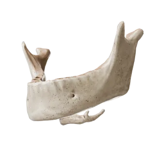
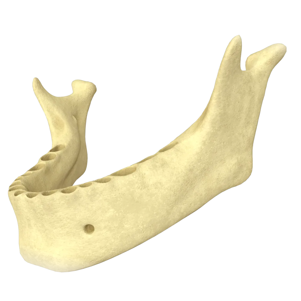
The mental foramen opens on the external surface of the mandibular body, usually below the interval between the first and second premolars.
-
Inferior
The mental foramen opens on the external surface of the mandibular body, usually below the interval between the first and second premolars.
-
External
The mental foramen opens on the external surface of the mandibular body, usually below the interval between the first and second premolars.
-
On the alveolar border
The mental foramen opens on the external surface of the mandibular body, usually below the interval between the first and second premolars.
-
I find it difficult to answer
The mental foramen opens on the external surface of the mandibular body, usually below the interval between the first and second premolars.
3. Which of the listed formations is located on the internal surface of the mandibular body and serves as the attachment site for the digastric muscle?
-
Mental spine
The digastric fossa is located on the internal surface at the base of the mandible on both sides of the median plane and is the site of origin of the anterior belly of the digastric muscle.
-
Digastric fossa
The digastric fossa is located on the internal surface at the base of the mandible on both sides of the median plane and is the site of origin of the anterior belly of the digastric muscle.
-
Mylohyoid line
The digastric fossa is located on the internal surface at the base of the mandible on both sides of the median plane and is the site of origin of the anterior belly of the digastric muscle.
-
Sublingual fossa
The digastric fossa is located on the internal surface at the base of the mandible on both sides of the median plane and is the site of origin of the anterior belly of the digastric muscle.
-
I find it difficult to answer
The digastric fossa is located on the internal surface at the base of the mandible on both sides of the median plane and is the site of origin of the anterior belly of the digastric muscle.
4. Which bone of the human skeleton is the only one that does not directly articulate (does not form joints or continuous connections) with any other bone?
-
Lacrimal bone
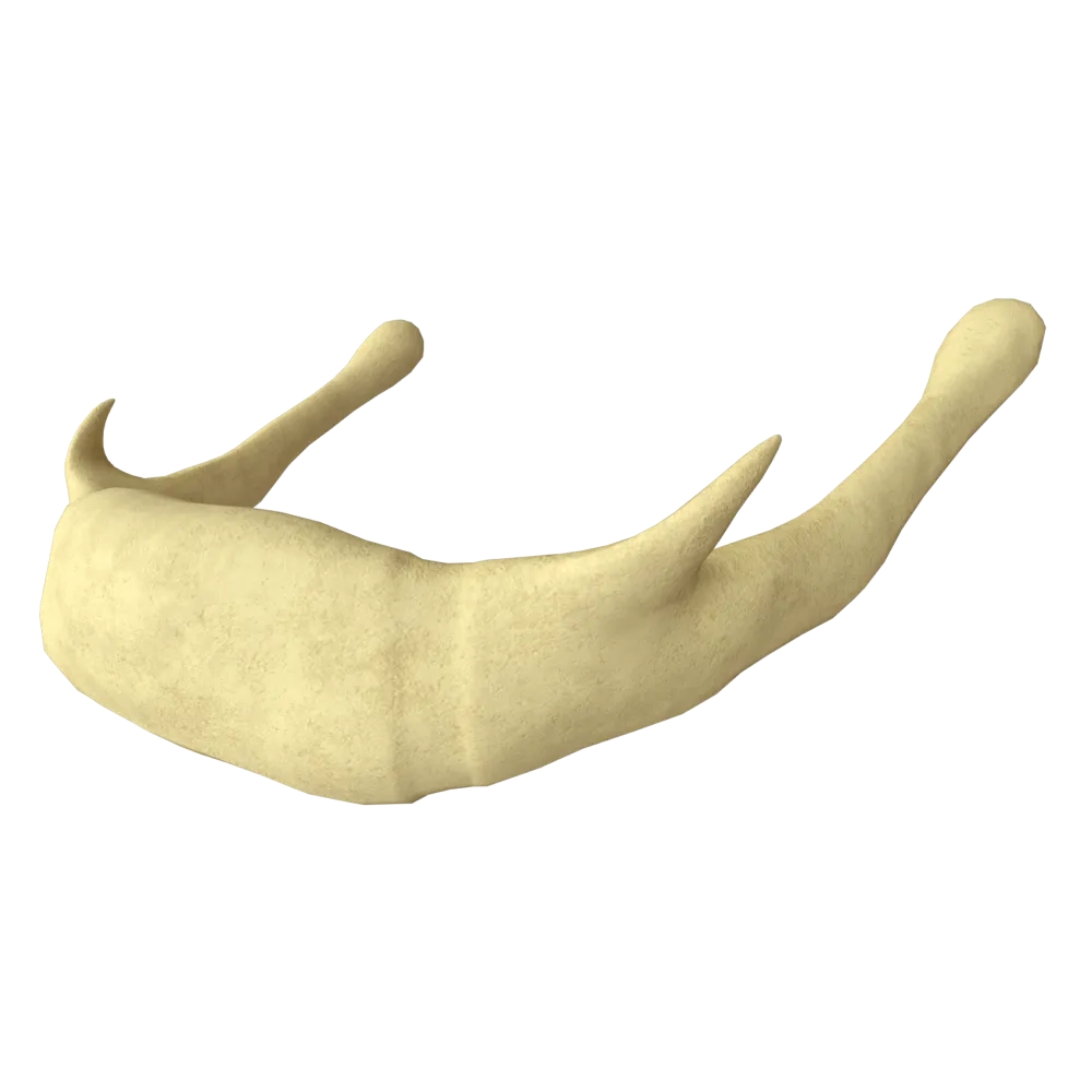
The hyoid bone is a unique element of the skeleton since it does not connect with any other bone.
-
Nasal bone
The hyoid bone is a unique element of the skeleton since it does not connect with any other bone.
-
Hyoid bone
The hyoid bone is a unique element of the skeleton since it does not connect with any other bone.
-
Inferior nasal concha
The hyoid bone is a unique element of the skeleton since it does not connect with any other bone.
-
I find it difficult to answer
The hyoid bone is a unique element of the skeleton since it does not connect with any other bone.
5. How many horns (paired processes) are present in the hyoid bone?
-
2 horns (1 pair)
The hyoid bone consists of an unpaired body (corpus) and 4 horns (2 pairs).
-
4 horns (2 pairs)
The hyoid bone consists of an unpaired body (corpus) and 4 horns (2 pairs).
-
3 horns
The hyoid bone consists of an unpaired body (corpus) and 4 horns (2 pairs).
-
6 horns (3 pairs)
The hyoid bone consists of an unpaired body (corpus) and 4 horns (2 pairs).
-
I find it difficult to answer
The hyoid bone consists of an unpaired body (corpus) and 4 horns (2 pairs).
6. Which of the listed bones is extracranial (does not belong to the bones of the skull, though topographically located in the region of the head and neck)?
-
Vomer
The hyoid bone is classified as an extracranial bone: it is located in the neck region, under the tongue, and is isolated from the rest of the skeleton (held only by muscles and ligaments).
-
Ethmoid bone
The hyoid bone is classified as an extracranial bone: it is located in the neck region, under the tongue, and is isolated from the rest of the skeleton (held only by muscles and ligaments).
-
Hyoid bone
The hyoid bone is classified as an extracranial bone: it is located in the neck region, under the tongue, and is isolated from the rest of the skeleton (held only by muscles and ligaments).
-
Lacrimal bone
The hyoid bone is classified as an extracranial bone: it is located in the neck region, under the tongue, and is isolated from the rest of the skeleton (held only by muscles and ligaments).
-
I find it difficult to answer
The hyoid bone is classified as an extracranial bone: it is located in the neck region, under the tongue, and is isolated from the rest of the skeleton (held only by muscles and ligaments).
7. Which foramen of the mandible is the entry point for the inferior alveolar nerve and the eponymous vessels into the mandible canal?
-
Mental foramen
The mandibular foramen is located on the internal surface of the mandibular ramus. Through it, the inferior alveolar nerve and vessels enter the mandibular canal.
-
Mandibular foramen
The mandibular foramen is located on the internal surface of the mandibular ramus. Through it, the inferior alveolar nerve and vessels enter the mandibular canal.
-
Incisive canal
The mandibular foramen is located on the internal surface of the mandibular ramus. Through it, the inferior alveolar nerve and vessels enter the mandibular canal.
-
Mandibular notch
The mandibular foramen is located on the internal surface of the mandibular ramus. Through it, the inferior alveolar nerve and vessels enter the mandibular canal.
-
I find it difficult to answer
The mandibular foramen is located on the internal surface of the mandibular ramus. Through it, the inferior alveolar nerve and vessels enter the mandibular canal.
8. Which tuberosity is located on the external surface of the angle of the mandible?
-
Pharyngeal tubercle
The masseteric tuberosity is located on the external (lateral) surface of the angle of the mandible. The masseter muscle attaches to it.
-
Pterygoid tuberosity
The masseteric tuberosity is located on the external (lateral) surface of the angle of the mandible. The masseter muscle attaches to it.
-
Masseteric tuberosity
The masseteric tuberosity is located on the external (lateral) surface of the angle of the mandible. The masseter muscle attaches to it.
-
Digastric fossa
The masseteric tuberosity is located on the external (lateral) surface of the angle of the mandible. The masseter muscle attaches to it.
-
I find it difficult to answer
The masseteric tuberosity is located on the external (lateral) surface of the angle of the mandible. The masseter muscle attaches to it.
9. How many processes does the mandibular ramus have along its upper margin?
-
1
The mandibular ramus ends with two processes at its upper margin, divided by the mandibular notch: the coronoid process and the condylar (articular) process.
-
2
The mandibular ramus ends with two processes at its upper margin, divided by the mandibular notch: the coronoid process and the condylar (articular) process.
-
3
The mandibular ramus ends with two processes at its upper margin, divided by the mandibular notch: the coronoid process and the condylar (articular) process.
-
4
The mandibular ramus ends with two processes at its upper margin, divided by the mandibular notch: the coronoid process and the condylar (articular) process.
-
I find it difficult to answer
The mandibular ramus ends with two processes at its upper margin, divided by the mandibular notch: the coronoid process and the condylar (articular) process.
10. Which fossa is located on the internal surface of the mandibular body above the mylohyoid line?
-
Submandibular fossa
The sublingual fossa is located above the mylohyoid line (closer to the chin). It houses the sublingual salivary gland.
-
Maxillary process
The sublingual fossa is located above the mylohyoid line (closer to the chin). It houses the sublingual salivary gland.
-
Sublingual fossa
The sublingual fossa is located above the mylohyoid line (closer to the chin). It houses the sublingual salivary gland.
-
Digastric fossa
The sublingual fossa is located above the mylohyoid line (closer to the chin). It houses the sublingual salivary gland.
-
I find it difficult to answer
The sublingual fossa is located above the mylohyoid line (closer to the chin). It houses the sublingual salivary gland.
11. Which fossa is located on the internal surface of the mandibular body below the mylohyoid line?
-
Sublingual fossa
The submandibular fossa is located below the mylohyoid line and houses the submandibular salivary gland.
-
Pterygoid fossa
The submandibular fossa is located below the mylohyoid line and houses the submandibular salivary gland.
-
Buccal fossa
The submandibular fossa is located below the mylohyoid line and houses the submandibular salivary gland.
-
Submandibular fossa
The submandibular fossa is located below the mylohyoid line and houses the submandibular salivary gland.
-
I find it difficult to answer
The submandibular fossa is located below the mylohyoid line and houses the submandibular salivary gland.
12. Which mandibular structure is located between the coronoid and condylar processes?
-
Mandibular notch
The mandibular notch is a semilunar depression located between the coronoid and condylar processes of the mandibular ramus.
-
Lingula of the mandible
The mandibular notch is a semilunar depression located between the coronoid and condylar processes of the mandibular ramus.
-
Masseteric tuberosity
The mandibular notch is a semilunar depression located between the coronoid and condylar processes of the mandibular ramus.
-
Pterygoid tuberosity
The mandibular notch is a semilunar depression located between the coronoid and condylar processes of the mandibular ramus.
-
I find it difficult to answer
The mandibular notch is a semilunar depression located between the coronoid and condylar processes of the mandibular ramus.
13. What is the lingula of the mandible?
-
Process to which the temporalis muscle attaches
Lingula of the mandible is a bony projection on the internal surface of the mandibular ramus. It covers the mandibular foramen.
-
Prominence above the mandibular foramen, covering the entrance to the mandibular canal
Lingula of the mandible is a bony projection on the internal surface of the mandibular ramus. It covers the mandibular foramen.
-
Prominence on the mental symphysis for the attachment of the tongue muscles
Lingula of the mandible is a bony projection on the internal surface of the mandibular ramus. It covers the mandibular foramen.
-
Cartilaginous plate in the composition of the temporomandibular joint
Lingula of the mandible is a bony projection on the internal surface of the mandibular ramus. It covers the mandibular foramen.
-
I find it difficult to answer
Lingula of the mandible is a bony projection on the internal surface of the mandibular ramus. It covers the mandibular foramen.
14. Which anatomical structure is located along the midline on the internal surface of the mandibular body?
-
Mylohyoid line
In the center (along the midline) of the internal surface of the mandibular body the mental spine is located. It is a solitary bony prominence to which the genioglossus and geniohyoid muscles attach.
-
Mental tubercle
In the center (along the midline) of the internal surface of the mandibular body the mental spine is located. It is a solitary bony prominence to which the genioglossus and geniohyoid muscles attach.
-
Mental spine
In the center (along the midline) of the internal surface of the mandibular body the mental spine is located. It is a solitary bony prominence to which the genioglossus and geniohyoid muscles attach.
-
Lingula of the mandible
In the center (along the midline) of the internal surface of the mandibular body the mental spine is located. It is a solitary bony prominence to which the genioglossus and geniohyoid muscles attach.
-
I find it difficult to answer
In the center (along the midline) of the internal surface of the mandibular body the mental spine is located. It is a solitary bony prominence to which the genioglossus and geniohyoid muscles attach.
15. Which tuberosity is located on the internal surface of the angle of the mandible?
-
Masseteric tuberosity
On the internal (medial) surface of the angle of the mandible the pterygoid tuberosity is located, to which the medial pterygoid muscle attaches.
-
Pterygoid tuberosity
On the internal (medial) surface of the angle of the mandible the pterygoid tuberosity is located, to which the medial pterygoid muscle attaches.
-
Pharyngeal tubercle
On the internal (medial) surface of the angle of the mandible the pterygoid tuberosity is located, to which the medial pterygoid muscle attaches.
-
Sphenoidal tubercle
On the internal (medial) surface of the angle of the mandible the pterygoid tuberosity is located, to which the medial pterygoid muscle attaches.
-
I find it difficult to answer
On the internal (medial) surface of the angle of the mandible the pterygoid tuberosity is located, to which the medial pterygoid muscle attaches.
16. Where is the lingula of the mandible located?
-
On the external surface of the ramus
Lingula of the mandible is a bony prominence on the internal surface of the ramus at the anteromedial edge of the mandibular foramen.
-
On the internal surface of the ramus at the anterior edge of the mandibular foramen
Lingula of the mandible is a bony prominence on the internal surface of the ramus at the anteromedial edge of the mandibular foramen.
-
On the body of the mandible
Lingula of the mandible is a bony prominence on the internal surface of the ramus at the anteromedial edge of the mandibular foramen.
-
On the mandibular notch
Lingula of the mandible is a bony prominence on the internal surface of the ramus at the anteromedial edge of the mandibular foramen.
-
I find it difficult to answer
Lingula of the mandible is a bony prominence on the internal surface of the ramus at the anteromedial edge of the mandibular foramen.
17. Which part of the hyoid bone connects with the greater horns?
-
Lesser horns
The greater horns of the hyoid bone extend posteriorly and laterally directly from the body, forming a single arch-like structure with it.
-
Body
The greater horns of the hyoid bone extend posteriorly and laterally directly from the body, forming a single arch-like structure with it.
-
Arch
The greater horns of the hyoid bone extend posteriorly and laterally directly from the body, forming a single arch-like structure with it.
-
Stylohyoid ligament
The greater horns of the hyoid bone extend posteriorly and laterally directly from the body, forming a single arch-like structure with it.
-
I find it difficult to answer
The greater horns of the hyoid bone extend posteriorly and laterally directly from the body, forming a single arch-like structure with it.
18. Does the mandible have a neck, and where exactly is it located?
-
Yes, on the coronoid process of the mandibular ramus
The mandible has a neck. It is located on the condylar process of the mandibular ramus, directly below its head.
-
No, the mandible does not have a neck
The mandible has a neck. It is located on the condylar process of the mandibular ramus, directly below its head.
-
Yes, on the condylar process below its head
The mandible has a neck. It is located on the condylar process of the mandibular ramus, directly below its head.
-
Yes, at the transition from the body to the ramus of the mandible
The mandible has a neck. It is located on the condylar process of the mandibular ramus, directly below its head.
-
I find it difficult to answer
The mandible has a neck. It is located on the condylar process of the mandibular ramus, directly below its head.
19. Is the mandible a paired bone in adults?
-
Yes, it is a paired bone
In adults, the mandible is an unpaired bone of the viscerocranium.
-
Paired only in women, unpaired in men
In adults, the mandible is an unpaired bone of the viscerocranium.
-
No, it is an unpaired bone
In adults, the mandible is an unpaired bone of the viscerocranium.
-
Paired, as it forms two isolated joints
In adults, the mandible is an unpaired bone of the viscerocranium.
-
I find it difficult to answer
In adults, the mandible is an unpaired bone of the viscerocranium.
20. Is the hyoid bone a paired anatomical structure?
-
Yes, it is a paired bone
The hyoid bone is an unpaired bone, located on the midline of the neck beneath the tongue.
-
Paired in children, but fuses into an unpaired bone in adults
The hyoid bone is an unpaired bone, located on the midline of the neck beneath the tongue.
-
No, it is an unpaired bone
The hyoid bone is an unpaired bone, located on the midline of the neck beneath the tongue.
-
Paired, as it divides into right and left halves
The hyoid bone is an unpaired bone, located on the midline of the neck beneath the tongue.
-
I find it difficult to answer
The hyoid bone is an unpaired bone, located on the midline of the neck beneath the tongue.
Retake this quiz?
Your current progress will be reset.

Cookie Consent
We use cookies to enhance your browsing experience, analyze site traffic, and deliver content. Please choose whether you accept all cookies or wish to reject non-essential tracking.
Cookie Preferences
Manage your cookie preferences below:
Essential cookies enable basic functions and are necessary for the proper function of the website.
Name
Description
Duration
Geolocation Config
This cookie is used to store the consent settings based on the visitor's location.
30 days
Cookie Preferences
This cookie is used to store the user's cookie consent preferences.
30 days
Google reCAPTCHA helps protect websites from spam and abuse by verifying user interactions through challenges.
Name
Description
Duration
_GRECAPTCHA
Google reCAPTCHA sets a necessary cookie (_GRECAPTCHA) when executed for the purpose of providing its risk analysis.
179 days
Statistics cookies collect information anonymously. This information helps us understand how visitors use our website.
Google Analytics is a powerful tool that tracks and analyzes website traffic for informed marketing decisions.
Service URL: policies.google.com (opens in a new window)
Name
Description
Duration
__utma
ID used to identify users and sessions
2 years after last activity
__utmt
Used to monitor number of Google Analytics server requests
10 minutes
__utmb
Used to distinguish new sessions and visits. This cookie is set when the GA.js javascript library is loaded and there is no existing __utmb cookie. The cookie is updated every time data is sent to the Google Analytics server.
30 minutes after last activity
__utmc
Used only with old Urchin versions of Google Analytics and not with GA.js. Was used to distinguish between new sessions and visits at the end of a session.
End of session (browser)
__utmz
Contains information about the traffic source or campaign that directed user to the website. The cookie is set when the GA.js javascript is loaded and updated when data is sent to the Google Anaytics server
6 months after last activity
__utmv
Contains custom information set by the web developer via the _setCustomVar method in Google Analytics. This cookie is updated every time new data is sent to the Google Analytics server.
2 years after last activity
__utmx
Used to determine whether a user is included in an A / B or Multivariate test.
18 months
_ga
ID used to identify users
2 years
_gali
Used by Google Analytics to determine which links on a page are being clicked
30 seconds
_ga_
ID used to identify users
2 years
_gid
ID used to identify users for 24 hours after last activity
24 hours
_gat
Used to monitor number of Google Analytics server requests when using Google Tag Manager
1 minute
_gac_
Contains information related to marketing campaigns of the user. These are shared with Google AdWords / Google Ads when the Google Ads and Google Analytics accounts are linked together.
90 days
Clarity is a web analytics service that tracks and reports website traffic.
Service URL: clarity.microsoft.com (opens in a new window)
Name
Description
Duration
_clsk
Connects multiple page views by a user into a single Clarity session recording.
1 day
_clck
Stores a unique user identifier to analyze visitor behavior through heatmaps and session recordings.
1 year
ANONCHK
Indicates whether MUID is transferred to ANID, a cookie used for advertising. Clarity doesn't use ANID and so this is always set to 0.
Session
CLID
Identifies the first-time Clarity saw this user on any site using Clarity.
12 months
_clsk
Connects multiple page views by a user into a single Clarity session recording.
12 months
_clck
Persists the Clarity User ID and preferences, unique to that site is attributed to the same user ID.
12 months