Aortic Stenosis: Etiology, Pathophysiology, Symptoms, Severity, Diagnosis, and Treatment
Oleg K.Cardiovascular surgeon, MD
15 min read·April 14, 2025
This article is for informational purposes only
The content on this website, including text, graphics, and other materials, is provided for informational purposes only. It is not intended as advice or guidance. Regarding your specific medical condition or treatment, please consult your healthcare provider.
Aortic stenosis (AS) is a morphological narrowing of the aortic valve (AV) orifice that obstructs the left ventricle (LV) outflow.
3D Animation: Rheumatic Aortic Stenosis (with Transformation)
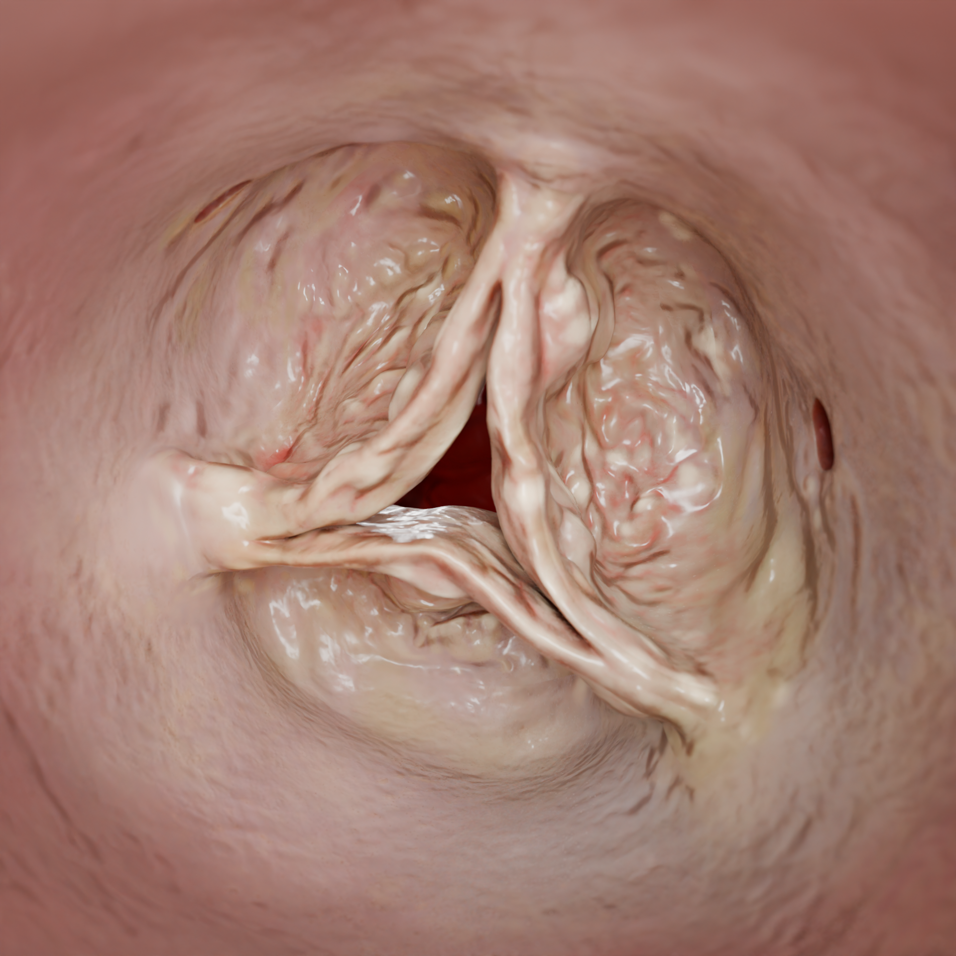
3D Animation: Degenerative Aortic Stenosis
Epidemiology
AS is the prevailing cause of morbidity among all valvular heart conditions. Its incidence is strongly age-related: AS may be identified in 12–13 %of the population over the age of 75, while its critical stage is generally observed in 3–4 % of patients.
Moreover, males are more prone to AS than females.
The condition is detected more frequently in high-income countries with developed healthcare systems. This trend is generally attributed to higher-quality medical services and better access to diagnostic tools. Developing countries, on the other hand, can provide limited data on AS prevalence, which may be linked to underdiagnosis and the lower life expectancy of the population.
Historically, AS was primarily associated with rheumatic fever, in which the valvular leaflets become deformed and fused. Over time, medical advances and lower rates of rheumatic disease in developed countries have altered the etiology of the disease. Currently, the major cause of AS is age-related degenerative changes in the valve, such as calcification and leaflet fibrosis. Furthermore, atherosclerosis has become a more prominent AS cause.
Today, the increasing number of AS patients is primarily linked to an aging population and lower mortality rates from other conditions. Improved diagnostic means and greater accessibility to medical services have also made it possible to detect more AS cases worldwide.
Rheumatic Aortic Stenosis – 3D ModelDegenerative Aortic Stenosis – 3D Model
Etiology
Degenerative changes: Age-related calcinosis of AV leaflets leading to thickening and reduced motility.
Congenital abnormalities: Bicuspid aortic valve (BAV), a predisposing factor for early calcification and eventual development of AS.
Rheumatic fever: A condition that causes the valvular leaflets to fuse and deform, contributing to AS.
Although rare, AS may also be associated with conditions such as chronic renal failure, carcinoid syndrome, Paget’s disease, and systemic lupus erythematosus.
3D Animation: Aortic Stenosis Development
Pathophysiology
When the valvular orifice narrows, resistance to blood ejection increases. This, in turn, raises systolic pressure in the left ventricle (LV) and leads to concentric LV hypertrophy. This is when the ventricular walls thicken without a proportional expansion of the cavity.
Once hypertrophy has developed, reduction of LV compliance (stretching) ensues. This impairs its ability to relax during diastole. An increase in LV diastolic blood pressure affects the left atrium (LA) which gives rise to hypertrophy and dilation. However, such compensatory mechanisms are insufficient to counteract the ever-increasing pressure in the pulmonary vessels. Eventually, this leads to blood congestion in the pulmonary circulation, which may trigger signs of cardiac failure.
In the early stages of AS, cardiac output is maintained via Frank — Starling mechanism. According to this principle, the force of contractions is directly related to the severity of myocardial dilation. However, prolonged pressure overload and progressive hypertrophy deplete the myocardial reserve, leading to reduced contractile function and systolic dysfunction.
Hypertrophied LV walls increase myocardial oxygen demand, while elevated diastolic blood pressure restricts blood supply to the coronary arteries. Consequently, arterial perfusion is reduced, potentially provoking myocardial ischemia even if no coronary atherosclerosis is observed.
Due to AS decompensation, the LV cavity and the left atrioventricular orifice become enlarged, leading to mitral regurgitation (MR).
Clinical Manifestations
The disease may exist for many years without producing any symptoms. Over time, the pathological changes described above collectively lead to symptoms characteristic of AS, such as dyspnea, angina pectoris, and syncope.
Dyspnea, for instance, may occur due to blood congestion in the pulmonary veins.
When a hypertrophied myocardium develops ischemia, a patient may experience angina pectoris.
Syncope is attributed to reduced cardiac output and insufficient cerebral blood flow.
Once observed, these symptoms may indicate an unfavorable prognosis and should prompt healthcare professionals to consider surgery. Cardiac asthma, lower limb swelling, and hepatomegaly (enlarged liver) are signs of disease decompensation that require immediate medical intervention.
Diagnosis
The primary tool for diagnosing AS is echocardiogram (ECG). It helps evaluate AV anatomy, AV calcification degree, AV effective orifice area (EOA), pressure gradient, LV function and size. For initial assessment, transthoracic echocardiogram (TTE) is generally recommended. If not informative, transesophageal echocardiogram (TEE) may be indicated.
Echocardiographic (ECG) Assessment of AS Severity
Parameter
Mild AS
Moderate AS
Severe AS
Critical САК
Maximal velocity (m/s)
<3.0
3.0-3.9
≥4.0
≥5.0
Mean pressure gradient(mmHg)
<20
20-39
≥40
≥60
EOA (cm²)
>1.5
1.0-1.5
≤1.0
≤0.6
Indexed EOA(cm²/m²)
>0.85
0.6-0.85
≤0.6
≤0.4
The following parameters are used to evaluate AS (the figures given below indicate cutoffs for severe AS):
Maximal velocity of AV blood flow (Vmax): ≥ 4.0 m/s;
AV mean pressure gradient (Δpm): ≥ 40 mmHg indicates severe SAC;
AV EOA: ≤ 1.0 cm² or ≤ 0.6 cm²/m².
Low cardiac output and a reduced LV ejection fraction (EF) (< 50 %) may require a dobutamine stress ECG. The results help distinguish between true severe AS and pseudo-severe AS.
MRI / CT: These imaging modalities may be used as supplementary diagnostic tools that may assist in determining the appropriate surgical approach.
CT angiography is the gold standard prior to TAVI (see below). This imaging technique provides data including anatomy of the aortic root and descending aorta, severity and extent of valvular and vascular calcification, risk factors for coronary artery obstruction, feasibility of vascular access.
MRI may help identify and assess myocardial fibrosis, which is an indication of decompensated AS.
Brain natriuretic peptide (BNP) or its precursor, N-terminal pro-brain natriuretic peptide (NT-proBNP), may be instrumental in risk stratification for AS patients. If elevated, these markers are associated with a worse prognosis and may serve as an additional rationale for intervention in asymptomatic patients.
AS Treatment
Risk factor modification helps slow the progression of AS and reduce potential complications. These measures include blood pressure control, a specialized diet and lipid-lowering medications to maintain normal serum lipid levels, total abstinence from smoking, strict blood glucose control, a balanced diet and regular physical activity to maintain a healthy body weight.
Medical therapy
Currently, there are no specific medications to slow the progression of AS. However, medical therapy may help alleviate symptoms and manage concomitant conditions: diuretics address pulmonary congestion in cases of cardiac failure; beta-blockers help control heat rate and blood pressure, especially in patients with concomitant coronary artery disease (CAD); angiotensin converting enzyme inhibitors or angiotensin II receptor blockers may be used in cardiac failure or hypertension. Note that these medications should be used with caution, as excessive blood pressure decrease may affect organ perfusion in patients with marked AS.
Aortic valve replacement (AVR) may be indicated in the following cases:
Symptomatic severe AS;
Asymptomatic severe AS accompanied by LV systolic dysfunction (EF < 50 %); and a positive stress test;
Very severe AS (mean pressure gradient ≥ 60 mmHg or maximal velocity > 5.0 m/s);
Rapid deterioration of the patient’s condition or rapid rise in serum BNP.
Transcatheter aortic valve implantation (TAVI) is a minimally invasive procedure to replace the AV. The most common access route is via the femoral artery. Alternatively, when the femoral arteries are too narrow, tortuous, or affected by atherosclerosis, a catheter may be inserted through: transapical access — via the LV apex; transaortic access — via the ascending aorta; transsubclavian access — via the subclavian artery. Both balloon-expandable and self-expanding valves may be employed.
TAVI should be preferred in patients ≥ 75 years of age; and/or patients with a high surgical risk (EuroSCORE II >8 %); and/or situations when open-heart surgery is contraindicated due to concomitant conditions.
Contraindications for TAVI:
Vascular access to the AV is not feasible (in marked atherosclerosis or narrow arteries);
Unfavorable valve or aorta anatomical features that render the procedure unfeasible;
Life expectancy is estimated to be less than 1 year or no improvement in the quality of life is expected following the procedure.
3D Animation: Transcatheter Aortic Valve Implantation
Find more scientifically accurate content on our social media
Subscribe and don’t miss out the latest resources
Surgical Aortic Valve Replacement (SAVR)
For most patients, open-heart surgery to replace the AV remains the standard treatment option. Both mechanical and biological valve prostheses may be employed. The procedure is performed under cardiopulmonary bypass. Individual anatomical features determine the surgical approach for AVR — via complete sternotomy, ministernotomy, or right anterior minithoracotomy.
Contraindications for SAVR: A high surgical risk or concomitant conditions that make surgery unsafe for a patient.
3D Animation: Aortic Valve Replacement
FAQ
1. Why does AS develop?
The most common causes are: • Age-related degenerative changes. • Congenital bicuspid aortic valve. • Complications of rheumatic fever. Although rare, AS may also be associated with conditions such as chronic renal failure, carcinoid syndrome, Paget’s disease, and systemic lupus erythematosus.
2. What auscultation findings are typical of AS?
Auscultation may reveal: • Rough systolic ejection murmur (most prominent in intercostal space 2, with radiation to the cervical vessels). • Diminished second heart sound (S2) (due to reduced leaflet motility). • Paradoxically split second heart sound (S2) (in severe AS).
• Further progression → Systolic dysfunction → LV dilation and MR → Cardiac failure.
4. What do patients with AS typically complain about?
Typical complaints include: • “Triad of symptoms”: Dyspnea — caused by pulmonary congestion. Angina pectoris — resulting from ischemia in the hypertrophied myocardium. Syncope — often triggered by physical activity. • Fatigue, dizziness (linked to reduced cardiac output).
5. What is the difference between TAVI and standard open-heart surgery?
TAVI does not imply thoracic incision but has a limited number of indications. Open aortic valve replacement (AVR) surgery is the standard procedure when patient’s surgical risks are low.
List of Sources
1.
VOKA 3D Anatomy & Pathology – Complete Anatomy and Pathology 3D Atlas [Internet]. VOKA 3D Anatomy & Pathology.
Available from: https://catalog.voka.io/
2.
Vahanian A, Beyersdorf F, Praz F, Milojevic M, Baldus S, Bauersachs J, Capodanno D, Conradi L, De Bonis M, De Paulis R, Delgado V, Freemantle N, Gilard M, Haugaa KH, Jeppsson A, Jüni P, Pierard L, Prendergast BD, Sádaba JR, Tribouilloy C, Wojakowski W; ESC/EACTS Scientific Document Group. 2021 ESC/EACTS Guidelines for the management of valvular heart disease. Eur Heart J. 2022 Feb 12;43(7):561–632. doi:10.1093/eurheartj/ehab395.
3.
Chen J, et al. Burden of valvular heart disease, 1990–2017: Results from the Global Burden of Disease Study 2017. J Glob Health. 2020 Sep 8;10(2):020404. doi:10.7189/jogh.10.020404.
4.
Joseph J, Naqvi SY, Giri J, Goldberg S. Aortic stenosis: Pathophysiology, diagnosis, and therapy. Am J Med. 2017 Mar;130(3):253–263. doi:10.1016/j.amjmed.2016.10.005.
5.
Zheng KH, Tzolos E, Dweck MR. Pathophysiology of aortic stenosis and future perspectives for medical therapy. Cardiol Clin. 2020 Feb;38(1):1–12. doi:10.1016/j.ccl.2019.09.010.
6.
Kanwar A, Thaden JJ, Nkomo VT. Management of patients with aortic valve stenosis. Mayo Clin Proc. 2018 Apr;93(4):488–508. doi:10.1016/j.mayocp.2018.01.020.
7.
Howard C, Jullian L, Joshi M, Noshirwani A, Bashir M, Harky A. TAVI and the future of aortic valve replacement. J Card Surg. 2019 Dec;34(12):1577–1590. doi:10.1111/jocs.14226.