1/20
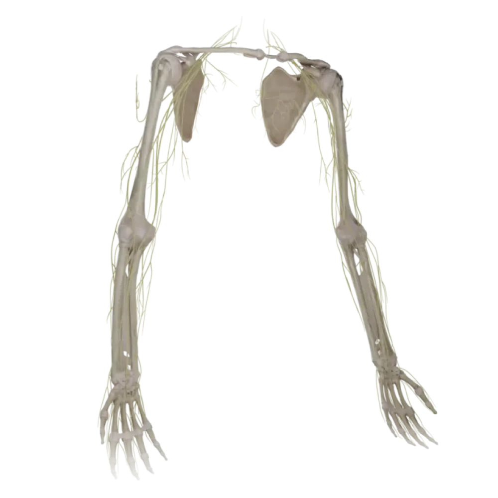

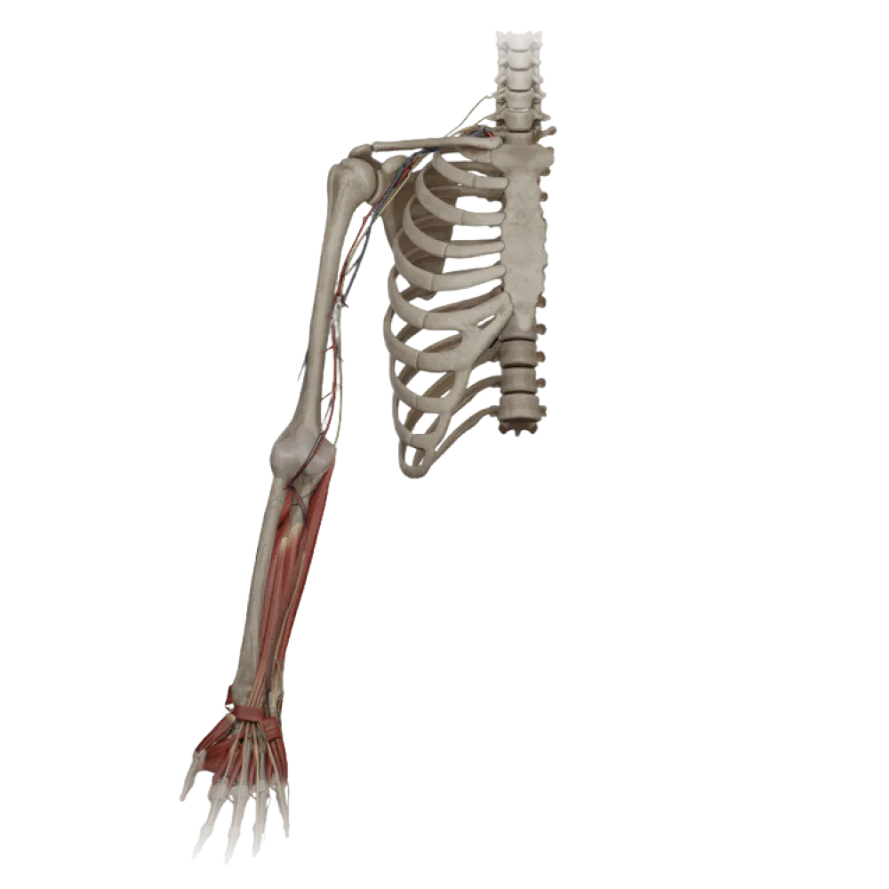
Test on the anatomy of the upper limb nerves
Assess knowledge of the anatomy of upper limb nerves. The test evaluates the topography, innervation zones, branching, and syngenesis of plexus structures.
1. Which bundles of the brachial plexus contribute to the formation of the median nerve?
-
Lateral and posterior
The median nerve is formed by the fusion of roots from the lateral (C5-C7) and medial (C8-T1) cords of the brachial plexus.
-
Only medial
The median nerve is formed by the fusion of roots from the lateral (C5-C7) and medial (C8-T1) cords of the brachial plexus.
-
Lateral and medial
The median nerve is formed by the fusion of roots from the lateral (C5-C7) and medial (C8-T1) cords of the brachial plexus.
-
Medial and posterior
The median nerve is formed by the fusion of roots from the lateral (C5-C7) and medial (C8-T1) cords of the brachial plexus.
-
I find it difficult to answer
The median nerve is formed by the fusion of roots from the lateral (C5-C7) and medial (C8-T1) cords of the brachial plexus.
2. Through which anatomical structure does the axillary nerve pass?
-
Quadrangular space
The axillary nerve leaves the axillary fossa through the quadrangular space with the posterior circumflex humeral artery.
-
Triangular foramen
The axillary nerve leaves the axillary fossa through the quadrangular space with the posterior circumflex humeral artery.
-
Spiral canal
The axillary nerve leaves the axillary fossa through the quadrangular space with the posterior circumflex humeral artery.
-
Subscapular space
The axillary nerve leaves the axillary fossa through the quadrangular space with the posterior circumflex humeral artery.
-
I find it difficult to answer
The axillary nerve leaves the axillary fossa through the quadrangular space with the posterior circumflex humeral artery.
3. Which muscle is pierced by the musculocutaneous nerve in the arm?
-
Biceps brachii
The musculocutaneous nerve emerges from the lateral cord, pierces the coracobrachialis muscle, and lies between the biceps and the brachialis muscles.
-
Triceps brachii muscle
The musculocutaneous nerve emerges from the lateral cord, pierces the coracobrachialis muscle, and lies between the biceps and the brachialis muscles.
-
Brachialis muscle
The musculocutaneous nerve emerges from the lateral cord, pierces the coracobrachialis muscle, and lies between the biceps and the brachialis muscles.
-
Coracobrachialis muscle
The musculocutaneous nerve emerges from the lateral cord, pierces the coracobrachialis muscle, and lies between the biceps and the brachialis muscles.
-
I find it difficult to answer
The musculocutaneous nerve emerges from the lateral cord, pierces the coracobrachialis muscle, and lies between the biceps and the brachialis muscles.
4. With which blood vessel does the radial nerve travel in the radial groove?
-
Brachial artery
The radial nerve descends within the radial groove on the posterior aspect of the humerus, accompanied by the deep brachial artery.
-
Deep brachial artery
The radial nerve descends within the radial groove on the posterior aspect of the humerus, accompanied by the deep brachial artery.
-
Superior ulnar collateral artery
The radial nerve descends within the radial groove on the posterior aspect of the humerus, accompanied by the deep brachial artery.
-
Radial collateral artery
The radial nerve descends within the radial groove on the posterior aspect of the humerus, accompanied by the deep brachial artery.
-
I find it difficult to answer
The radial nerve descends within the radial groove on the posterior aspect of the humerus, accompanied by the deep brachial artery.
5. Which muscle is innervated by the deep branch of the ulnar nerve?
-
Adductor pollicis muscle
The deep branch of the ulnar nerve innervates the hypothenar muscles, interossei, the 3rd and 4th lumbricals, and the adductor pollicis.
-
First lumbrical muscle
The deep branch of the ulnar nerve innervates the hypothenar muscles, interossei, the 3rd and 4th lumbricals, and the adductor pollicis.
-
Opponens pollicis muscle
The deep branch of the ulnar nerve innervates the hypothenar muscles, interossei, the 3rd and 4th lumbricals, and the adductor pollicis.
-
Abductor pollicis brevis
The deep branch of the ulnar nerve innervates the hypothenar muscles, interossei, the 3rd and 4th lumbricals, and the adductor pollicis.
-
I find it difficult to answer
The deep branch of the ulnar nerve innervates the hypothenar muscles, interossei, the 3rd and 4th lumbricals, and the adductor pollicis.
6. Where does the ulnar nerve lie at the level of the medial epicondyle?
-
In the cubital fossa, anterior to the epicondyle
At the level of the elbow joint, the ulnar nerve passes superficially in the groove for the ulnar nerve on the posterior surface of the medial epicondyle.
-
Within the medial intermuscular septum
At the level of the elbow joint, the ulnar nerve passes superficially in the groove for the ulnar nerve on the posterior surface of the medial epicondyle.
-
In the groove for the ulnar nerve behind the epicondyle
At the level of the elbow joint, the ulnar nerve passes superficially in the groove for the ulnar nerve on the posterior surface of the medial epicondyle.
-
Between the heads of the pronator teres
At the level of the elbow joint, the ulnar nerve passes superficially in the groove for the ulnar nerve on the posterior surface of the medial epicondyle.
-
I find it difficult to answer
At the level of the elbow joint, the ulnar nerve passes superficially in the groove for the ulnar nerve on the posterior surface of the medial epicondyle.
7. Between which structures does the median nerve pass in the forearm?
-
Between the brachioradialis and the pronator teres
In the forearm, the median nerve passes between the superficial and deep flexors of the fingers, providing them with branches.
-
Under the pronator quadratus
In the forearm, the median nerve passes between the superficial and deep flexors of the fingers, providing them with branches.
-
Between the flexor carpi ulnaris and the superficial flexor
In the forearm, the median nerve passes between the superficial and deep flexors of the fingers, providing them with branches.
-
Between the superficial and deep flexors of the fingers
In the forearm, the median nerve passes between the superficial and deep flexors of the fingers, providing them with branches.
-
I find it difficult to answer
In the forearm, the median nerve passes between the superficial and deep flexors of the fingers, providing them with branches.
8. Which area is innervated by the superficial branch of the radial nerve?
-
Skin of the palmar surface of the lateral 3.5 digits
The superficial branch of the radial nerve provides sensory innervation to the radial half of the dorsum of the hand and the dorsal surface of the proximal phalanges of digits 1, 2, and the lateral half of digit 3.
-
Skin of the dorsal surface of the lateral 2.5 digits
The superficial branch of the radial nerve provides sensory innervation to the radial half of the dorsum of the hand and the dorsal surface of the proximal phalanges of digits 1, 2, and the lateral half of digit 3.
-
Skin of the dorsal surface of the medial 1.5 digits
The superficial branch of the radial nerve provides sensory innervation to the radial half of the dorsum of the hand and the dorsal surface of the proximal phalanges of digits 1, 2, and the lateral half of digit 3.
-
Skin of the medial surface of the forearm
The superficial branch of the radial nerve provides sensory innervation to the radial half of the dorsum of the hand and the dorsal surface of the proximal phalanges of digits 1, 2, and the lateral half of digit 3.
-
I find it difficult to answer
The superficial branch of the radial nerve provides sensory innervation to the radial half of the dorsum of the hand and the dorsal surface of the proximal phalanges of digits 1, 2, and the lateral half of digit 3.
9. Which structure forms the anterior (superficial) wall of Guyon's canal (ulnar canal of the wrist)?
-
Palmar carpal ligament
Guyon's canal is bordered anteriorly by the palmar carpal ligament (an extension of the forearm fascia) and posteriorly by the flexor retinaculum.
-
Flexor retinaculum
Guyon's canal is bordered anteriorly by the palmar carpal ligament (an extension of the forearm fascia) and posteriorly by the flexor retinaculum.
-
Pisiform bone
Guyon's canal is bordered anteriorly by the palmar carpal ligament (an extension of the forearm fascia) and posteriorly by the flexor retinaculum.
-
Hook of hamate
Guyon's canal is bordered anteriorly by the palmar carpal ligament (an extension of the forearm fascia) and posteriorly by the flexor retinaculum.
-
I find it difficult to answer
Guyon's canal is bordered anteriorly by the palmar carpal ligament (an extension of the forearm fascia) and posteriorly by the flexor retinaculum.
10. The anterior interosseous nerve branches from which nerve in the forearm?
-
Ulnar nerve
The anterior interosseous nerve arises from the median nerve, innervating the deep muscles of the anterior compartment of the forearm (except the medial half of the flexor digitorum profundus).
-
Musculocutaneous nerve
The anterior interosseous nerve arises from the median nerve, innervating the deep muscles of the anterior compartment of the forearm (except the medial half of the flexor digitorum profundus).
-
Radial nerve
The anterior interosseous nerve arises from the median nerve, innervating the deep muscles of the anterior compartment of the forearm (except the medial half of the flexor digitorum profundus).
-
Median nerve
The anterior interosseous nerve arises from the median nerve, innervating the deep muscles of the anterior compartment of the forearm (except the medial half of the flexor digitorum profundus).
-
I find it difficult to answer
The anterior interosseous nerve arises from the median nerve, innervating the deep muscles of the anterior compartment of the forearm (except the medial half of the flexor digitorum profundus).
11. Which nerve passes through the scapular notch beneath the superior transverse ligament?
-
Subscapular nerve
The suprascapular nerve passes through the scapular notch into the supraspinous fossa, located below the superior transverse scapular ligament.
-
Thoracodorsal nerve
The suprascapular nerve passes through the scapular notch into the supraspinous fossa, located below the superior transverse scapular ligament.
-
Suprascapular nerve
The suprascapular nerve passes through the scapular notch into the supraspinous fossa, located below the superior transverse scapular ligament.
-
Axillary nerve
The suprascapular nerve passes through the scapular notch into the supraspinous fossa, located below the superior transverse scapular ligament.
-
I find it difficult to answer
The suprascapular nerve passes through the scapular notch into the supraspinous fossa, located below the superior transverse scapular ligament.
12. From which cord of the brachial plexus does the medial cutaneous nerve of the forearm arise?
-
Lateral cord
The medial cutaneous nerve of the forearm is a direct sensory branch of the medial cord of the brachial plexus.
-
Medial cord
The medial cutaneous nerve of the forearm is a direct sensory branch of the medial cord of the brachial plexus.
-
Posterior cord
The medial cutaneous nerve of the forearm is a direct sensory branch of the medial cord of the brachial plexus.
-
Supraclavicular part of the plexus
The medial cutaneous nerve of the forearm is a direct sensory branch of the medial cord of the brachial plexus.
-
I find it difficult to answer
The medial cutaneous nerve of the forearm is a direct sensory branch of the medial cord of the brachial plexus.
13. Which muscle is innervated by the long thoracic nerve?
-
Serratus anterior muscle
The long thoracic nerve descends along the outer surface of the serratus anterior muscle, providing motor innervation to it.
-
Pectoralis major muscle
The long thoracic nerve descends along the outer surface of the serratus anterior muscle, providing motor innervation to it.
-
Latissimus dorsi muscle
The long thoracic nerve descends along the outer surface of the serratus anterior muscle, providing motor innervation to it.
-
Pectoralis minor muscle
The long thoracic nerve descends along the outer surface of the serratus anterior muscle, providing motor innervation to it.
-
I find it difficult to answer
The long thoracic nerve descends along the outer surface of the serratus anterior muscle, providing motor innervation to it.
14. Through which muscle does the deep branch of the radial nerve pass when transitioning to the posterior surface of the forearm?
-
Pronator teres
The deep branch of the radial nerve pierces the supinator muscle (passing through the arcade of Frohse), directing itself to the posterior compartment of the forearm.
-
Brachioradialis muscle
The deep branch of the radial nerve pierces the supinator muscle (passing through the arcade of Frohse), directing itself to the posterior compartment of the forearm.
-
Extensor digitorum
The deep branch of the radial nerve pierces the supinator muscle (passing through the arcade of Frohse), directing itself to the posterior compartment of the forearm.
-
Supinator
The deep branch of the radial nerve pierces the supinator muscle (passing through the arcade of Frohse), directing itself to the posterior compartment of the forearm.
-
I find it difficult to answer
The deep branch of the radial nerve pierces the supinator muscle (passing through the arcade of Frohse), directing itself to the posterior compartment of the forearm.
15. Which nerve accompanies the lateral cutaneous vein of the arm (v. cephalica)?
-
Medial cutaneous nerve of the arm
The lateral cutaneous nerve of the forearm (ultimate extension of the musculocutaneous nerve) accompanies the v. cephalica in the forearm. cephalica.
-
Lateral cutaneous nerve of the forearm
The lateral cutaneous nerve of the forearm (ultimate extension of the musculocutaneous nerve) accompanies the v. cephalica in the forearm. cephalica.
-
Posterior cutaneous nerve of the forearm
The lateral cutaneous nerve of the forearm (ultimate extension of the musculocutaneous nerve) accompanies the v. cephalica in the forearm. cephalica.
-
Medial antebrachial cutaneous nerve
The lateral cutaneous nerve of the forearm (ultimate extension of the musculocutaneous nerve) accompanies the v. cephalica in the forearm. cephalica.
-
I find it difficult to answer
The lateral cutaneous nerve of the forearm (ultimate extension of the musculocutaneous nerve) accompanies the v. cephalica in the forearm. cephalica.
16. What is the topographical relationship of the median nerve to the brachial artery in the cubital fossa?
-
The nerve is lateral to the artery
In the cubital fossa, the median nerve lies medial to the brachial artery, with both structures covered by the bicipital aponeurosis (Pirogov's aponeurosis).
-
The nerve is posterior to the artery
In the cubital fossa, the median nerve lies medial to the brachial artery, with both structures covered by the bicipital aponeurosis (Pirogov's aponeurosis).
-
The nerve is medial to the artery
In the cubital fossa, the median nerve lies medial to the brachial artery, with both structures covered by the bicipital aponeurosis (Pirogov's aponeurosis).
-
The nerve is anterior to the artery
In the cubital fossa, the median nerve lies medial to the brachial artery, with both structures covered by the bicipital aponeurosis (Pirogov's aponeurosis).
-
I find it difficult to answer
In the cubital fossa, the median nerve lies medial to the brachial artery, with both structures covered by the bicipital aponeurosis (Pirogov's aponeurosis).
17. Which group of hand muscles is innervated by the recurrent branch of the median nerve?
-
Thenar muscles
The recurrent (motor) branch of the median nerve innervates the thenar muscles: the short abductor, opponens, and the superficial head of the short flexor of the thumb.
-
Interossei muscles
The recurrent (motor) branch of the median nerve innervates the thenar muscles: the short abductor, opponens, and the superficial head of the short flexor of the thumb.
-
Hypothenar muscles
The recurrent (motor) branch of the median nerve innervates the thenar muscles: the short abductor, opponens, and the superficial head of the short flexor of the thumb.
-
Lumbrical muscles (all)
The recurrent (motor) branch of the median nerve innervates the thenar muscles: the short abductor, opponens, and the superficial head of the short flexor of the thumb.
-
I find it difficult to answer
The recurrent (motor) branch of the median nerve innervates the thenar muscles: the short abductor, opponens, and the superficial head of the short flexor of the thumb.
18. From which segments of the spinal cord is the radial nerve predominantly formed?
-
C8-T1
The radial nerve is a continuation of the posterior cord of the brachial plexus and contains fibers from all roots forming the plexus (C5-T1).
-
C5-C7
The radial nerve is a continuation of the posterior cord of the brachial plexus and contains fibers from all roots forming the plexus (C5-T1).
-
C7-T1
The radial nerve is a continuation of the posterior cord of the brachial plexus and contains fibers from all roots forming the plexus (C5-T1).
-
C5-T1
The radial nerve is a continuation of the posterior cord of the brachial plexus and contains fibers from all roots forming the plexus (C5-T1).
-
I find it difficult to answer
The radial nerve is a continuation of the posterior cord of the brachial plexus and contains fibers from all roots forming the plexus (C5-T1).
19. Which muscle receives dual innervation (from the median and ulnar nerves)?
-
Flexor digitorum superficialis
The flexor digitorum profundus is innervated by two nerves: the medial part (for the 4th-5th fingers) by the ulnar nerve, and the lateral part (for the 2nd-3rd fingers) by the anterior interosseous nerve (from the median nerve).
-
Flexor pollicis longus
The flexor digitorum profundus is innervated by two nerves: the medial part (for the 4th-5th fingers) by the ulnar nerve, and the lateral part (for the 2nd-3rd fingers) by the anterior interosseous nerve (from the median nerve).
-
Flexor digitorum profundus
The flexor digitorum profundus is innervated by two nerves: the medial part (for the 4th-5th fingers) by the ulnar nerve, and the lateral part (for the 2nd-3rd fingers) by the anterior interosseous nerve (from the median nerve).
-
Flexor carpi radialis
The flexor digitorum profundus is innervated by two nerves: the medial part (for the 4th-5th fingers) by the ulnar nerve, and the lateral part (for the 2nd-3rd fingers) by the anterior interosseous nerve (from the median nerve).
-
I find it difficult to answer
The flexor digitorum profundus is innervated by two nerves: the medial part (for the 4th-5th fingers) by the ulnar nerve, and the lateral part (for the 2nd-3rd fingers) by the anterior interosseous nerve (from the median nerve).
20. Which nerve crosses the tendon of the flexor digitorum longus in the area of the carpal tunnel?
-
Median nerve
The median nerve passes through the carpal tunnel along with the flexor tendons, positioned most superficially.
-
Deep branch of the ulnar nerve
The median nerve passes through the carpal tunnel along with the flexor tendons, positioned most superficially.
-
Superficial branch of radial nerve
The median nerve passes through the carpal tunnel along with the flexor tendons, positioned most superficially.
-
Dorsal branch of ulnar nerve
The median nerve passes through the carpal tunnel along with the flexor tendons, positioned most superficially.
-
I find it difficult to answer
The median nerve passes through the carpal tunnel along with the flexor tendons, positioned most superficially.
Retake this quiz?
Your current progress will be reset.
Nerves of the upper limb
Ulnar nerve
0/20