
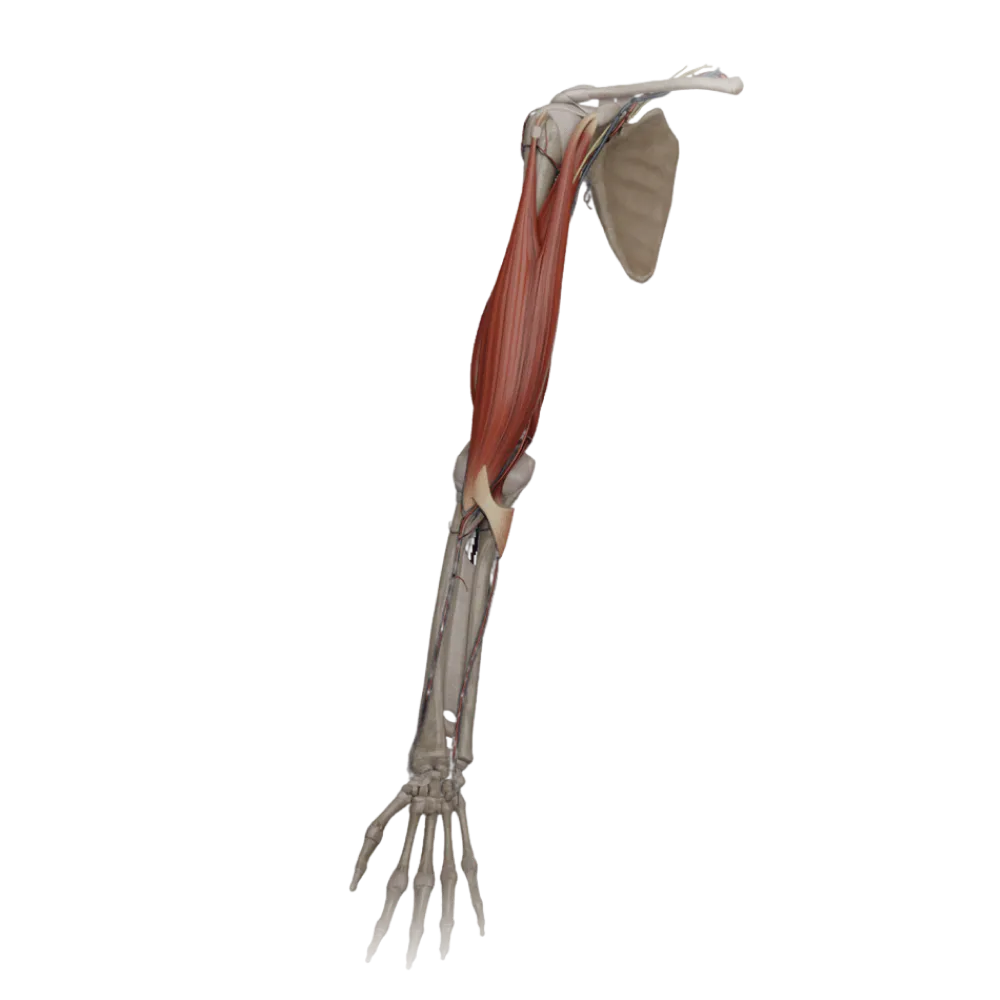

Forearm muscle anatomy test
Evaluate your knowledge of forearm muscle anatomy. The test assesses their innervation, blood supply, and the topography of neurovascular bundles.
1/20
bold
text
1. Which nerve innervates the flexor carpi ulnaris (m. flexor carpi ulnaris)?
-
Median nerve
The flexor carpi ulnaris is one of the muscles on the anterior surface of the forearm and is innervated by the ulnar nerve, not the median nerve.
-
Ulnar nerve
The flexor carpi ulnaris is one of the muscles on the anterior surface of the forearm and is innervated by the ulnar nerve, not the median nerve.
-
Radial nerve
The flexor carpi ulnaris is one of the muscles on the anterior surface of the forearm and is innervated by the ulnar nerve, not the median nerve.
-
Musculocutaneous nerve
The flexor carpi ulnaris is one of the muscles on the anterior surface of the forearm and is innervated by the ulnar nerve, not the median nerve.
-
I find it difficult to answer
The flexor carpi ulnaris is one of the muscles on the anterior surface of the forearm and is innervated by the ulnar nerve, not the median nerve.
2. Which muscle of the anterior forearm group is innervated by the radial nerve?
-
Pronator teres
Despite its location in the anterior region of the forearm, the brachioradialis is innervated by radial nerve branches.
-
Flexor carpi radialis
Despite its location in the anterior region of the forearm, the brachioradialis is innervated by radial nerve branches.
-
Palmaris longus muscle
Despite its location in the anterior region of the forearm, the brachioradialis is innervated by radial nerve branches.
-
Brachioradialis muscle
Despite its location in the anterior region of the forearm, the brachioradialis is innervated by radial nerve branches.
-
I find it difficult to answer
Despite its location in the anterior region of the forearm, the brachioradialis is innervated by radial nerve branches.
3. Which sections of the deep flexor of the fingers (m. flexor digitorum profundus) are innervated by the ulnar nerve?
-
Sections for fingers IV and V
The flexor digitorum profundus has dual innervation: the medial portion (fingers IV, V) is innervated by the ulnar nerve, and the lateral by the median.
-
Sections for fingers II and III
The flexor digitorum profundus has dual innervation: the medial portion (fingers IV, V) is innervated by the ulnar nerve, and the lateral by the median.
-
Sections for fingers I and II
The flexor digitorum profundus has dual innervation: the medial portion (fingers IV, V) is innervated by the ulnar nerve, and the lateral by the median.
-
All sections are innervated by the median nerve
The flexor digitorum profundus has dual innervation: the medial portion (fingers IV, V) is innervated by the ulnar nerve, and the lateral by the median.
-
I find it difficult to answer
The flexor digitorum profundus has dual innervation: the medial portion (fingers IV, V) is innervated by the ulnar nerve, and the lateral by the median.
4. From which artery is the supinator (m. supinator) primarily supplied?
-
Anterior interosseous artery
The supinator is supplied by branches of the radial artery (a. recurrens radialis) and the recurrent interosseous artery.
-
Superficial palmar arch
The supinator is supplied by branches of the radial artery (a. recurrens radialis) and the recurrent interosseous artery.
-
Recurrent radial artery
The supinator is supplied by branches of the radial artery (a. recurrens radialis) and the recurrent interosseous artery.
-
Ulnar artery
The supinator is supplied by branches of the radial artery (a. recurrens radialis) and the recurrent interosseous artery.
-
I find it difficult to answer
The supinator is supplied by branches of the radial artery (a. recurrens radialis) and the recurrent interosseous artery.
5. Which nerve innervates the pronator quadratus (m. pronator quadratus)?
-
Ulnar nerve
The pronator quadratus is located in the deep layer of the anterior group and is innervated by the anterior interosseous nerve (a branch of the median nerve).
-
Anterior interosseous nerve
The pronator quadratus is located in the deep layer of the anterior group and is innervated by the anterior interosseous nerve (a branch of the median nerve).
-
Deep branch of radial nerve
The pronator quadratus is located in the deep layer of the anterior group and is innervated by the anterior interosseous nerve (a branch of the median nerve).
-
Superficial branch of radial nerve
The pronator quadratus is located in the deep layer of the anterior group and is innervated by the anterior interosseous nerve (a branch of the median nerve).
-
I find it difficult to answer
The pronator quadratus is located in the deep layer of the anterior group and is innervated by the anterior interosseous nerve (a branch of the median nerve).
6. Which artery runs in the radial groove (sulcus radialis) of the forearm along with the superficial branch of the radial nerve?
-
Anterior interosseous artery
In the radial groove formed by the brachioradialis and the radial flexor of the wrist, the radial artery passes.
-
Ulnar artery
In the radial groove formed by the brachioradialis and the radial flexor of the wrist, the radial artery passes.
-
Common interosseous artery
In the radial groove formed by the brachioradialis and the radial flexor of the wrist, the radial artery passes.
-
Radial artery
In the radial groove formed by the brachioradialis and the radial flexor of the wrist, the radial artery passes.
-
I find it difficult to answer
In the radial groove formed by the brachioradialis and the radial flexor of the wrist, the radial artery passes.
7. Identify the nerve innervating the extensor digitorum (m. extensor digitorum):
-
Deep branch of radial nerve
All muscles of the posterior forearm group, including the extensor digitorum, are innervated by branches of the radial nerve (primarily the deep branch).
-
Median nerve
All muscles of the posterior forearm group, including the extensor digitorum, are innervated by branches of the radial nerve (primarily the deep branch).
-
Musculocutaneous nerve
All muscles of the posterior forearm group, including the extensor digitorum, are innervated by branches of the radial nerve (primarily the deep branch).
-
Ulnar nerve
All muscles of the posterior forearm group, including the extensor digitorum, are innervated by branches of the radial nerve (primarily the deep branch).
-
I find it difficult to answer
All muscles of the posterior forearm group, including the extensor digitorum, are innervated by branches of the radial nerve (primarily the deep branch).
8. The blood supply to the superficial flexor of the fingers (m. flexor digitorum superficialis) is primarily from:
-
Posterior interosseous artery
The flexor digitorum superficialis receives branches from both the radial and ulnar arteries of the forearm.
-
Deep brachial artery
The flexor digitorum superficialis receives branches from both the radial and ulnar arteries of the forearm.
-
Radial and ulnar arteries
The flexor digitorum superficialis receives branches from both the radial and ulnar arteries of the forearm.
-
Axillary artery
The flexor digitorum superficialis receives branches from both the radial and ulnar arteries of the forearm.
-
I find it difficult to answer
The flexor digitorum superficialis receives branches from both the radial and ulnar arteries of the forearm.
9. Which forearm muscle is innervated by the superficial branch of the radial nerve?
-
Extensor carpi radialis longus
The superficial branch of the radial nerve is entirely sensory and does not innervate forearm muscles.
-
None of the forearm muscles
The superficial branch of the radial nerve is entirely sensory and does not innervate forearm muscles.
-
Extensor carpi radialis brevis
The superficial branch of the radial nerve is entirely sensory and does not innervate forearm muscles.
-
Extensor carpi ulnaris
The superficial branch of the radial nerve is entirely sensory and does not innervate forearm muscles.
-
I find it difficult to answer
The superficial branch of the radial nerve is entirely sensory and does not innervate forearm muscles.
10. From which vessel does the common interosseous artery (a. interossea communis) originate?
-
Radial artery
The common interosseous artery is a large, short branch of the ulnar artery, which then divides into the anterior and posterior interosseous arteries.
-
Brachial artery
The common interosseous artery is a large, short branch of the ulnar artery, which then divides into the anterior and posterior interosseous arteries.
-
Subclavian artery
The common interosseous artery is a large, short branch of the ulnar artery, which then divides into the anterior and posterior interosseous arteries.
-
Ulnar artery
The common interosseous artery is a large, short branch of the ulnar artery, which then divides into the anterior and posterior interosseous arteries.
-
I find it difficult to answer
The common interosseous artery is a large, short branch of the ulnar artery, which then divides into the anterior and posterior interosseous arteries.
11. Which nerve pierces the supinator, passing through the supinator canal (canalis supinatorius)?
-
Deep branch of radial nerve
The deep branch of the radial nerve penetrates the posterior forearm region, piercing the m. supinator.
-
Anterior interosseous nerve
The deep branch of the radial nerve penetrates the posterior forearm region, piercing the m. supinator.
-
Median nerve
The deep branch of the radial nerve penetrates the posterior forearm region, piercing the m. supinator.
-
Ulnar nerve
The deep branch of the radial nerve penetrates the posterior forearm region, piercing the m. supinator.
-
I find it difficult to answer
The deep branch of the radial nerve penetrates the posterior forearm region, piercing the m. supinator.
12. Which forearm muscle is innervated directly by the trunk of the radial nerve before its division into superficial and deep branches?
-
Extensor digiti minimi
M. extensor carpi radialis longus and m. brachioradialis are innervated by the radial nerve before it enters the supinator canal.
-
Extensor indicis
M. extensor carpi radialis longus and m. brachioradialis are innervated by the radial nerve before it enters the supinator canal.
-
Extensor carpi radialis longus
M. extensor carpi radialis longus and m. brachioradialis are innervated by the radial nerve before it enters the supinator canal.
-
Extensor carpi radialis brevis
M. extensor carpi radialis longus and m. brachioradialis are innervated by the radial nerve before it enters the supinator canal.
-
I find it difficult to answer
M. extensor carpi radialis longus and m. brachioradialis are innervated by the radial nerve before it enters the supinator canal.
13. The anterior interosseous artery (a. interossea anterior) primarily supplies:
-
The superficial muscles of the posterior group
The anterior interosseous artery runs along the anterior surface of the interosseous membrane supplying the deep flexors and the pronator quadratus.
-
The deep muscles of the anterior group
The anterior interosseous artery runs along the anterior surface of the interosseous membrane supplying the deep flexors and the pronator quadratus.
-
The lateral group muscles
The anterior interosseous artery runs along the anterior surface of the interosseous membrane supplying the deep flexors and the pronator quadratus.
-
The skin of the posterior aspect of the forearm
The anterior interosseous artery runs along the anterior surface of the interosseous membrane supplying the deep flexors and the pronator quadratus.
-
I find it difficult to answer
The anterior interosseous artery runs along the anterior surface of the interosseous membrane supplying the deep flexors and the pronator quadratus.
14. Through which heads does the median nerve pass on the forearm?
-
Flexor carpi ulnaris
The median nerve exits the cubital fossa, passing between the humeral and ulnar heads of the pronator teres.
-
Brachioradialis muscle
The median nerve exits the cubital fossa, passing between the humeral and ulnar heads of the pronator teres.
-
Supinator
The median nerve exits the cubital fossa, passing between the humeral and ulnar heads of the pronator teres.
-
Pronator teres
The median nerve exits the cubital fossa, passing between the humeral and ulnar heads of the pronator teres.
-
I find it difficult to answer
The median nerve exits the cubital fossa, passing between the humeral and ulnar heads of the pronator teres.
15. The ulnar nerve on the forearm follows the ulnar groove (sulcus ulnaris) accompanied by:
-
Ulnar artery
Within the ulnar groove, formed by the flexor carpi ulnaris and the flexor digitorum superficialis, lie the ulnar artery and nerve.
-
Radial artery
Within the ulnar groove, formed by the flexor carpi ulnaris and the flexor digitorum superficialis, lie the ulnar artery and nerve.
-
Anterior interosseous artery
Within the ulnar groove, formed by the flexor carpi ulnaris and the flexor digitorum superficialis, lie the ulnar artery and nerve.
-
Deep vein of the arm
Within the ulnar groove, formed by the flexor carpi ulnaris and the flexor digitorum superficialis, lie the ulnar artery and nerve.
-
I find it difficult to answer
Within the ulnar groove, formed by the flexor carpi ulnaris and the flexor digitorum superficialis, lie the ulnar artery and nerve.
16. Which artery participates in supplying the brachioradialis muscle?
-
Posterior interosseous artery
The brachioradialis muscle receives supply from the radial artery as well as the radial collateral and recurrent radial arteries.
-
Ulnar artery
The brachioradialis muscle receives supply from the radial artery as well as the radial collateral and recurrent radial arteries.
-
Radial artery
The brachioradialis muscle receives supply from the radial artery as well as the radial collateral and recurrent radial arteries.
-
Common interosseous artery
The brachioradialis muscle receives supply from the radial artery as well as the radial collateral and recurrent radial arteries.
-
I find it difficult to answer
The brachioradialis muscle receives supply from the radial artery as well as the radial collateral and recurrent radial arteries.
17. The posterior interosseous artery (a. interossea posterior) exits onto the posterior surface of the forearm through:
-
Supinator canal
The posterior interosseous artery pierces the interosseous membrane in its upper region, exiting to the posterior compartment of the forearm.
-
The opening in the proximal part of the interosseous membrane
The posterior interosseous artery pierces the interosseous membrane in its upper region, exiting to the posterior compartment of the forearm.
-
Wrist canal
The posterior interosseous artery pierces the interosseous membrane in its upper region, exiting to the posterior compartment of the forearm.
-
Guyon's canal
The posterior interosseous artery pierces the interosseous membrane in its upper region, exiting to the posterior compartment of the forearm.
-
I find it difficult to answer
The posterior interosseous artery pierces the interosseous membrane in its upper region, exiting to the posterior compartment of the forearm.
18. The muscles of the lateral group of the forearm (brachioradialis, long and short radial extensors of the wrist) are innervated by:
-
Musculocutaneous nerve
The entire lateral and posterior groups of forearm muscles receive innervation from the radial nerve and its deep branch.
-
Median nerve
The entire lateral and posterior groups of forearm muscles receive innervation from the radial nerve and its deep branch.
-
Axillary nerve
The entire lateral and posterior groups of forearm muscles receive innervation from the radial nerve and its deep branch.
-
Radial nerve
The entire lateral and posterior groups of forearm muscles receive innervation from the radial nerve and its deep branch.
-
I find it difficult to answer
The entire lateral and posterior groups of forearm muscles receive innervation from the radial nerve and its deep branch.
19. The flexor carpi ulnaris (m. flexor carpi ulnaris) is predominantly supplied by:
-
Ulnar artery
The flexor carpi ulnaris is positioned medially and receives branches from the ulnar artery, superior and inferior ulnar collateral arteries.
-
Radial artery
The flexor carpi ulnaris is positioned medially and receives branches from the ulnar artery, superior and inferior ulnar collateral arteries.
-
Anterior interosseous artery
The flexor carpi ulnaris is positioned medially and receives branches from the ulnar artery, superior and inferior ulnar collateral arteries.
-
Deep brachial artery
The flexor carpi ulnaris is positioned medially and receives branches from the ulnar artery, superior and inferior ulnar collateral arteries.
-
I find it difficult to answer
The flexor carpi ulnaris is positioned medially and receives branches from the ulnar artery, superior and inferior ulnar collateral arteries.
20. Which nerve innervates the abductor pollicis longus muscle (m. abductor pollicis longus)?
-
Anterior interosseous nerve
The abductor pollicis longus belongs to the deep layer of the posterior group and is innervated by the posterior interosseous nerve.
-
Superficial branch of radial nerve
The abductor pollicis longus belongs to the deep layer of the posterior group and is innervated by the posterior interosseous nerve.
-
Posterior interosseous nerve
The abductor pollicis longus belongs to the deep layer of the posterior group and is innervated by the posterior interosseous nerve.
-
Ulnar nerve
The abductor pollicis longus belongs to the deep layer of the posterior group and is innervated by the posterior interosseous nerve.
-
I find it difficult to answer
The abductor pollicis longus belongs to the deep layer of the posterior group and is innervated by the posterior interosseous nerve.
Retake this quiz?
Your current progress will be reset.