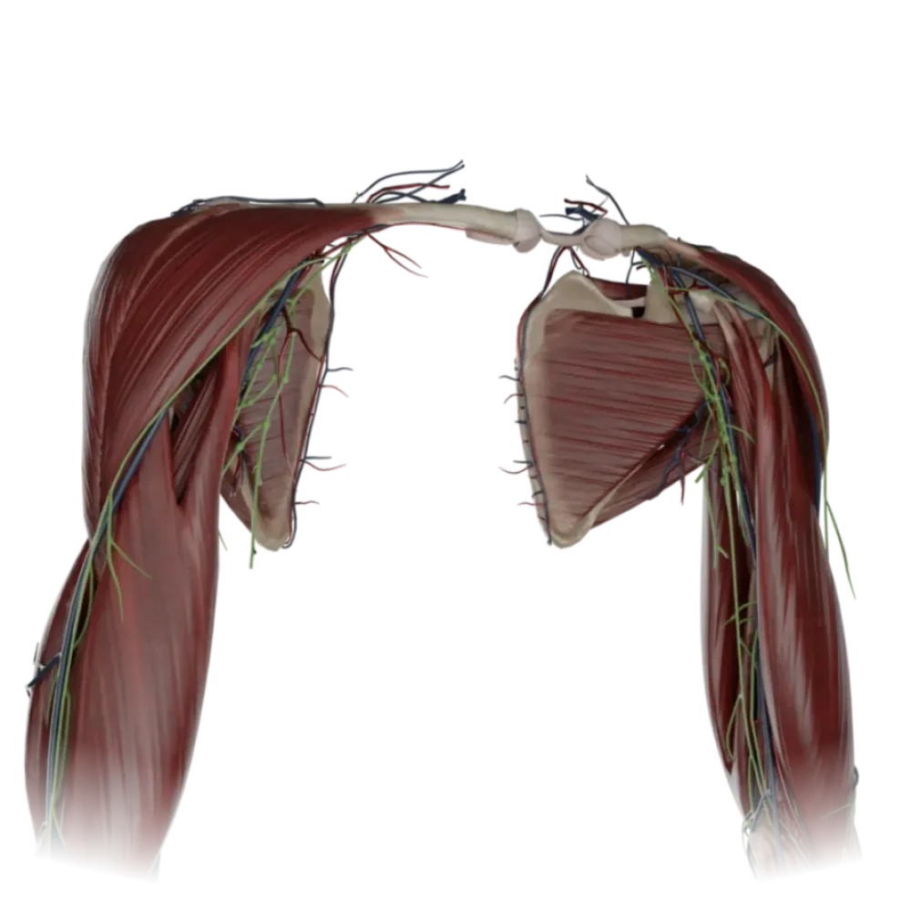


Anatomy test of the axillary cavity
Check the knowledge of axillary cavity anatomy. The test covers the topography, walls, triangles, openings and their neurovascular content.
1/20
bold
text
1. What forms the anterior wall of the axillary cavity?
-
Pectoralis major and minor muscles
The anterior wall is formed by the pectoralis major and minor muscles.
-
Serratus anterior muscle
The anterior wall is formed by the pectoralis major and minor muscles.
-
Subscapularis and teres major muscles
The anterior wall is formed by the pectoralis major and minor muscles.
-
Coracobrachialis muscle
The anterior wall is formed by the pectoralis major and minor muscles.
-
I find it difficult to answer
The anterior wall is formed by the pectoralis major and minor muscles.
2. What forms the medial wall of the axillary cavity?
-
Latissimus dorsi muscle
The medial wall is formed by the serratus anterior muscle and the lateral surface of the thoracic cage.
-
Pectoralis major muscle
The medial wall is formed by the serratus anterior muscle and the lateral surface of the thoracic cage.
-
Serratus anterior muscle
The medial wall is formed by the serratus anterior muscle and the lateral surface of the thoracic cage.
-
Biceps brachii
The medial wall is formed by the serratus anterior muscle and the lateral surface of the thoracic cage.
-
I find it difficult to answer
The medial wall is formed by the serratus anterior muscle and the lateral surface of the thoracic cage.
3. Which muscle contributes to the formation of the posterior wall of the axillary cavity?
-
Coracobrachialis muscle
The posterior wall is formed by the subscapularis, teres major, and the latissimus dorsi muscles.
-
Pectoralis minor muscle
The posterior wall is formed by the subscapularis, teres major, and the latissimus dorsi muscles.
-
Serratus anterior muscle
The posterior wall is formed by the subscapularis, teres major, and the latissimus dorsi muscles.
-
Latissimus dorsi muscle
The posterior wall is formed by the subscapularis, teres major, and the latissimus dorsi muscles.
-
I find it difficult to answer
The posterior wall is formed by the subscapularis, teres major, and the latissimus dorsi muscles.
4. Which structure limits the axillary cavity laterally?
-
The thoracic cage and the intercostal muscles
The lateral wall is formed by the medial surface of the humerus, the short head of the biceps, and the coracobrachialis muscle.
-
Humerus and coracobrachialis muscle
The lateral wall is formed by the medial surface of the humerus, the short head of the biceps, and the coracobrachialis muscle.
-
Clavicle and subclavius muscle
The lateral wall is formed by the medial surface of the humerus, the short head of the biceps, and the coracobrachialis muscle.
-
Scapula and subscapularis muscle
The lateral wall is formed by the medial surface of the humerus, the short head of the biceps, and the coracobrachialis muscle.
-
I find it difficult to answer
The lateral wall is formed by the medial surface of the humerus, the short head of the biceps, and the coracobrachialis muscle.
5. Which triangles are identified on the anterior wall of the axillary cavity?
-
Carotid, scapulotracheal, submandibular
Three topographic triangles are identified on the anterior wall: clavipectoral, pectoral, and subpectoral.
-
Clavipectoral, pectoral, subpectoral
Three topographic triangles are identified on the anterior wall: clavipectoral, pectoral, and subpectoral.
-
Lumbar, femoral, popliteal
Three topographic triangles are identified on the anterior wall: clavipectoral, pectoral, and subpectoral.
-
Scapuloclavicular, scapulotrapezius
Three topographic triangles are identified on the anterior wall: clavipectoral, pectoral, and subpectoral.
-
I find it difficult to answer
Three topographic triangles are identified on the anterior wall: clavipectoral, pectoral, and subpectoral.
6. From which triangle of the anterior wall does the axillary artery give off a branch. thoracoacromial?
-
In the subpectoral triangle
The thoracoacromial artery (a. thoracoacromialis) arises from the axillary artery within the clavipectoral triangle.
-
In the pectoral triangle
The thoracoacromial artery (a. thoracoacromialis) arises from the axillary artery within the clavipectoral triangle.
-
In the clavipectoral triangle
The thoracoacromial artery (a. thoracoacromialis) arises from the axillary artery within the clavipectoral triangle.
-
In the scapular-pectoral triangle
The thoracoacromial artery (a. thoracoacromialis) arises from the axillary artery within the clavipectoral triangle.
-
I find it difficult to answer
The thoracoacromial artery (a. thoracoacromialis) arises from the axillary artery within the clavipectoral triangle.
7. What limits the superior aspect of the quadrilateral foramen (foramen quadrilaterum)?
-
By the teres major muscle
The superior aspect of the foramen is limited by the lower edge of the subscapularis muscle (anteriorly) and the teres minor muscle (posteriorly).
-
The long head of the triceps brachii muscle
The superior aspect of the foramen is limited by the lower edge of the subscapularis muscle (anteriorly) and the teres minor muscle (posteriorly).
-
By the surgical neck of the humerus
The superior aspect of the foramen is limited by the lower edge of the subscapularis muscle (anteriorly) and the teres minor muscle (posteriorly).
-
Subscapularis and teres minor muscles
The superior aspect of the foramen is limited by the lower edge of the subscapularis muscle (anteriorly) and the teres minor muscle (posteriorly).
-
I find it difficult to answer
The superior aspect of the foramen is limited by the lower edge of the subscapularis muscle (anteriorly) and the teres minor muscle (posteriorly).
8. Which structure passes through the quadrilateral foramen?
-
Radial nerve
Through the foramen quadrilaterum pass the axillary nerve and the posterior circumflex humeral artery. axillary and a. circumflex humeral posterior.
-
Axillary nerve
Through the foramen quadrilaterum pass the axillary nerve and the posterior circumflex humeral artery. axillary and a. circumflex humeral posterior.
-
Median nerve
Through the foramen quadrilaterum pass the axillary nerve and the posterior circumflex humeral artery. axillary and a. circumflex humeral posterior.
-
Ulnar nerve
Through the foramen quadrilaterum pass the axillary nerve and the posterior circumflex humeral artery. axillary and a. circumflex humeral posterior.
-
I find it difficult to answer
Through the foramen quadrilaterum pass the axillary nerve and the posterior circumflex humeral artery. axillary and a. circumflex humeral posterior.
9. What limits the lateral aspect of the trilateral foramen (foramen trilaterum)?
-
The surgical neck of the humerus
The lateral boundary of the trilateral foramen is the long head of the triceps brachii muscle.
-
Short head of the biceps brachii
The lateral boundary of the trilateral foramen is the long head of the triceps brachii muscle.
-
Coracobrachialis muscle
The lateral boundary of the trilateral foramen is the long head of the triceps brachii muscle.
-
Long head of the triceps brachii
The lateral boundary of the trilateral foramen is the long head of the triceps brachii muscle.
-
I find it difficult to answer
The lateral boundary of the trilateral foramen is the long head of the triceps brachii muscle.
10. What passes through the triangular space (foramen trilaterum)?
-
Circumflex scapular artery
Through the trilateral foramen passes the circumflex scapular artery. circumflex scapulae.
-
Posterior circumflex humeral artery
Through the trilateral foramen passes the circumflex scapular artery. circumflex scapulae.
-
Thoracodorsal artery
Through the trilateral foramen passes the circumflex scapular artery. circumflex scapulae.
-
Subscapular artery
Through the trilateral foramen passes the circumflex scapular artery. circumflex scapulae.
-
I find it difficult to answer
Through the trilateral foramen passes the circumflex scapular artery. circumflex scapulae.
11. Which vein, piercing the clavipectoral fascia, drains into the axillary vein in the clavipectoral triangle?
-
Vena basilica
The lateral subcutaneous vein of the arm (v. cephalica) pierces the clavipectoral fascia and drains into the axillary vein.
-
Vena brachialis
The lateral subcutaneous vein of the arm (v. cephalica) pierces the clavipectoral fascia and drains into the axillary vein.
-
Vena cephalica
The lateral subcutaneous vein of the arm (v. cephalica) pierces the clavipectoral fascia and drains into the axillary vein.
-
Vena mediana cubiti
The lateral subcutaneous vein of the arm (v. cephalica) pierces the clavipectoral fascia and drains into the axillary vein.
-
I find it difficult to answer
The lateral subcutaneous vein of the arm (v. cephalica) pierces the clavipectoral fascia and drains into the axillary vein.
12. The topography of the pectoral triangle (trigonum pectorale) corresponds to the contours of which muscle?
-
Minor pectoral muscle
The pectoral triangle is strictly projected according to the contours of the minor pectoral muscle.
-
Major pectoral muscle
The pectoral triangle is strictly projected according to the contours of the minor pectoral muscle.
-
Subclavius muscle
The pectoral triangle is strictly projected according to the contours of the minor pectoral muscle.
-
Serratus anterior muscle
The pectoral triangle is strictly projected according to the contours of the minor pectoral muscle.
-
I find it difficult to answer
The pectoral triangle is strictly projected according to the contours of the minor pectoral muscle.
13. What forms the inferior boundary of the subpectoral triangle (trigonum subpectorale)?
-
Clavicle
The subpectoral triangle is located between the lower edges of the minor and major pectoral muscles.
-
Lower edge of the minor pectoral muscle
The subpectoral triangle is located between the lower edges of the minor and major pectoral muscles.
-
Upper edge of the major pectoral muscle
The subpectoral triangle is located between the lower edges of the minor and major pectoral muscles.
-
Lower edge of the major pectoral muscle
The subpectoral triangle is located between the lower edges of the minor and major pectoral muscles.
-
I find it difficult to answer
The subpectoral triangle is located between the lower edges of the minor and major pectoral muscles.
14. Which structure forms the medial wall of the quadrilateral foramen?
-
Surgical neck of the arm
The medial boundary of the quadrilateral foramen is formed by the long head of the triceps brachii muscle.
-
Long head of the triceps brachii
The medial boundary of the quadrilateral foramen is formed by the long head of the triceps brachii muscle.
-
Teres major muscle
The medial boundary of the quadrilateral foramen is formed by the long head of the triceps brachii muscle.
-
Subscapular muscle
The medial boundary of the quadrilateral foramen is formed by the long head of the triceps brachii muscle.
-
I find it difficult to answer
The medial boundary of the quadrilateral foramen is formed by the long head of the triceps brachii muscle.
15. Which muscle forms the inferior boundary of both the trilateral and quadrilateral foramina?
-
Subscapular muscle
The inferior boundary of both openings is formed by the teres major muscle and the tendon of the latissimus dorsi muscle.
-
Teres minor muscle
The inferior boundary of both openings is formed by the teres major muscle and the tendon of the latissimus dorsi muscle.
-
Teres major muscle
The inferior boundary of both openings is formed by the teres major muscle and the tendon of the latissimus dorsi muscle.
-
Infraspinatus muscle
The inferior boundary of both openings is formed by the teres major muscle and the tendon of the latissimus dorsi muscle.
-
I find it difficult to answer
The inferior boundary of both openings is formed by the teres major muscle and the tendon of the latissimus dorsi muscle.
16. Which large branch arises from the axillary artery in the subpectoral triangle?
-
Superior thoracic artery
In the subpectoral triangle, the subscapular artery branches from the axillary artery. Subscapularis.
-
Thoracoacromial artery
In the subpectoral triangle, the subscapular artery branches from the axillary artery. Subscapularis.
-
Lateral thoracic artery
In the subpectoral triangle, the subscapular artery branches from the axillary artery. Subscapularis.
-
Subscapular artery
In the subpectoral triangle, the subscapular artery branches from the axillary artery. Subscapularis.
-
I find it difficult to answer
In the subpectoral triangle, the subscapular artery branches from the axillary artery. Subscapularis.
17. Which fascia forms the floor of the axillary cavity (fossa axillaris)?
-
Fascia clavipectoralis
The floor of the axillary cavity is closed by the axillary fascia (fascia axillaris).
-
Fascia axillaris
The floor of the axillary cavity is closed by the axillary fascia (fascia axillaris).
-
Fascia pectoralis
The floor of the axillary cavity is closed by the axillary fascia (fascia axillaris).
-
Fascia endothoracica
The floor of the axillary cavity is closed by the axillary fascia (fascia axillaris).
-
I find it difficult to answer
The floor of the axillary cavity is closed by the axillary fascia (fascia axillaris).
18. In which triangle does the axillary artery give off the lateral thoracic artery (a. thoracica lateralis)?
-
In the pectoral
The lateral thoracic artery branches from the main trunk within the pectoral triangle.
-
In the clavipectoral
The lateral thoracic artery branches from the main trunk within the pectoral triangle.
-
In the subpectoral
The lateral thoracic artery branches from the main trunk within the pectoral triangle.
-
In the deltopectoral
The lateral thoracic artery branches from the main trunk within the pectoral triangle.
-
I find it difficult to answer
The lateral thoracic artery branches from the main trunk within the pectoral triangle.
19. With which space does the axillary cavity communicate through the quadrilateral foramen?
-
With the cranial cavity
Through the quadrilateral foramen, the axillary cavity communicates with the subdeltoid cellular space.
-
With the posterior scapular space
Through the quadrilateral foramen, the axillary cavity communicates with the subdeltoid cellular space.
-
With the subdeltoid space
Through the quadrilateral foramen, the axillary cavity communicates with the subdeltoid cellular space.
-
With the anterior mediastinum
Through the quadrilateral foramen, the axillary cavity communicates with the subdeltoid cellular space.
-
I find it difficult to answer
Through the quadrilateral foramen, the axillary cavity communicates with the subdeltoid cellular space.
20. How is the axillary vein positioned in relation to the axillary artery in the cavity?
-
Laterally and posteriorly
Within the neurovascular bundle, the axillary vein lies medially and slightly anterior to the artery.
-
Surrounds the artery on all sides
Within the neurovascular bundle, the axillary vein lies medially and slightly anterior to the artery.
-
Strictly posterior
Within the neurovascular bundle, the axillary vein lies medially and slightly anterior to the artery.
-
Medially and anteriorly
Within the neurovascular bundle, the axillary vein lies medially and slightly anterior to the artery.
-
I find it difficult to answer
Within the neurovascular bundle, the axillary vein lies medially and slightly anterior to the artery.
Retake this quiz?
Your current progress will be reset.