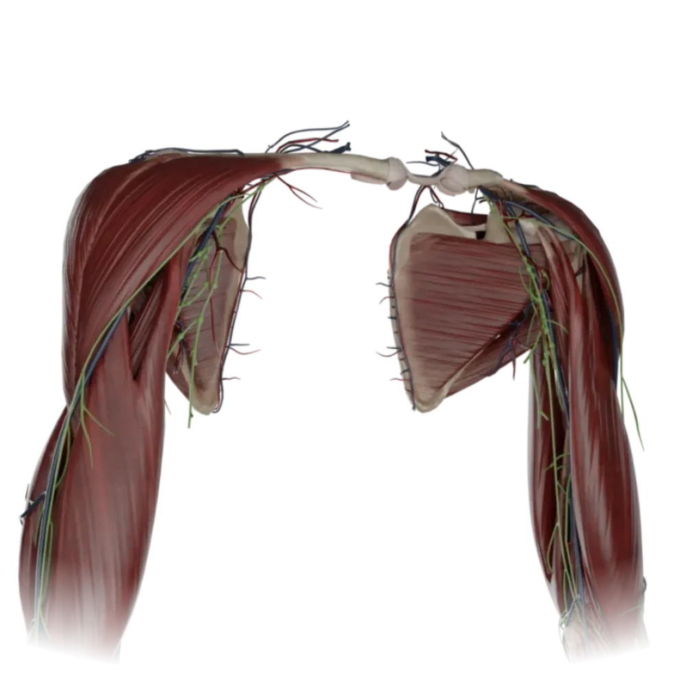
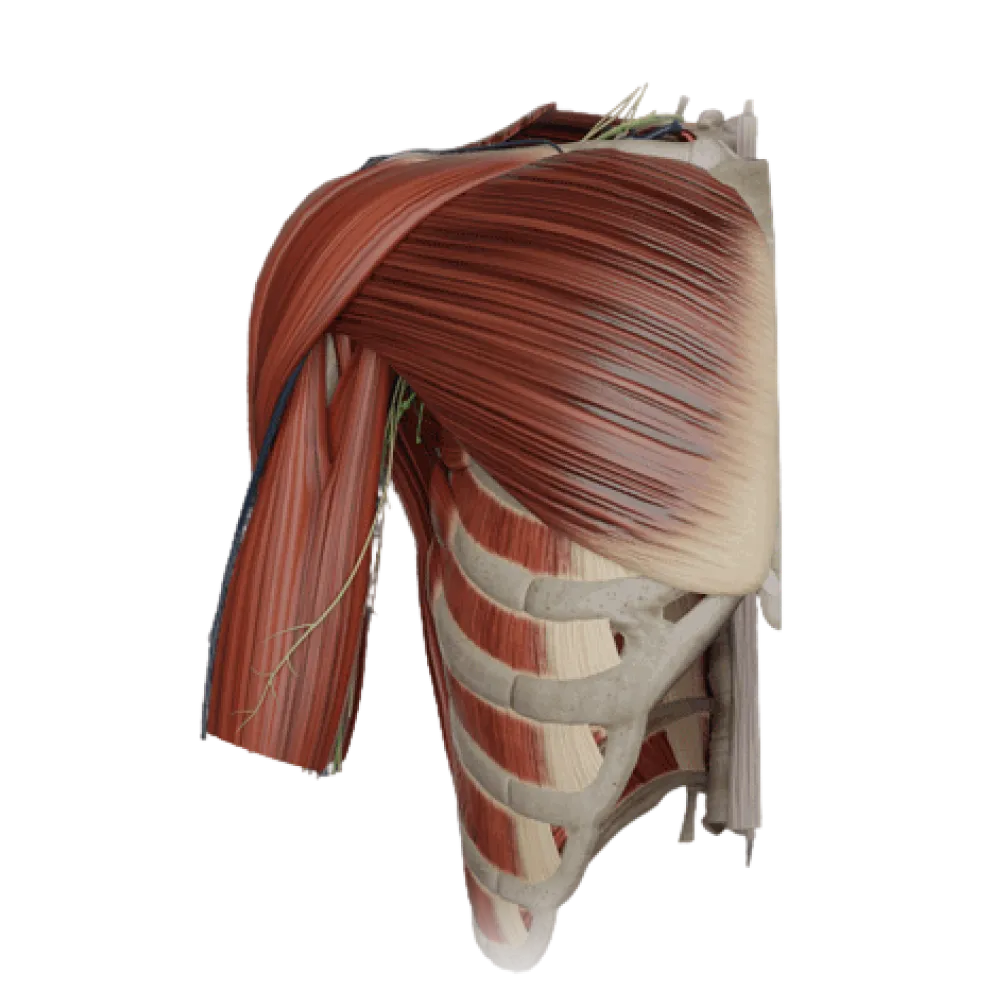
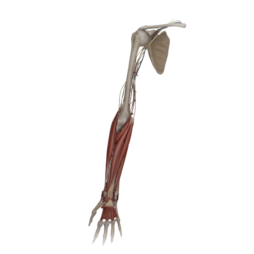
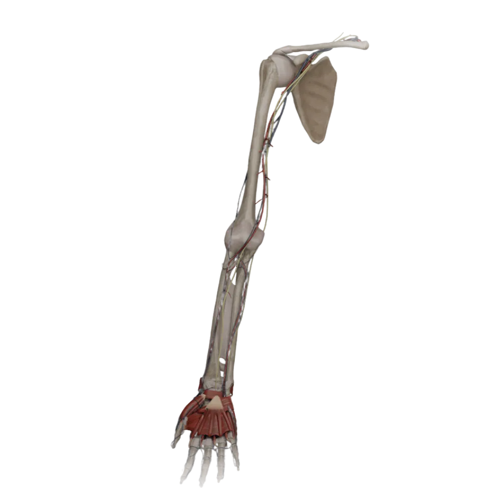


Anatomy test of the arm
Evaluate the knowledge of the upper limb topography. The test verifies the anatomy of the canals, fossae, and grooves of the arm, forearm, and hand.
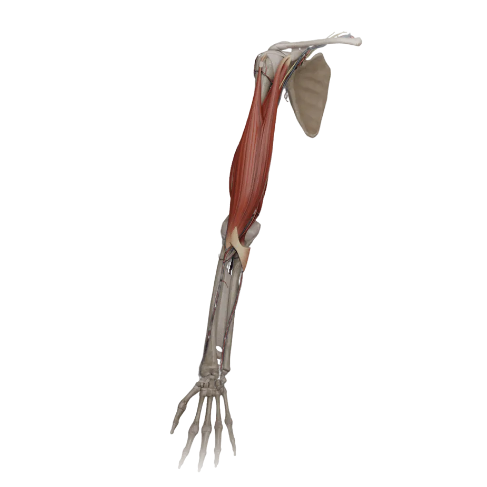
1/20
bold
text
1. The radial nerve canal (canalis nervi radialis) is located:
-
Between the humerus and the medial head of the triceps brachii muscle
The radial nerve canal is formed by the groove of the radial nerve on the humerus and the adjoining medial and lateral heads of the triceps brachii muscle.
-
Within the thickness of the biceps brachii muscle
The radial nerve canal is formed by the groove of the radial nerve on the humerus and the adjoining medial and lateral heads of the triceps brachii muscle.
-
Between the humerus, the lateral and medial heads of the triceps brachii muscle
The radial nerve canal is formed by the groove of the radial nerve on the humerus and the adjoining medial and lateral heads of the triceps brachii muscle.
-
Between the long and medial heads of the triceps brachii muscle
The radial nerve canal is formed by the groove of the radial nerve on the humerus and the adjoining medial and lateral heads of the triceps brachii muscle.
-
I find it difficult to answer
The radial nerve canal is formed by the groove of the radial nerve on the humerus and the adjoining medial and lateral heads of the triceps brachii muscle.
2. The quadrangular space (foramen quadrilaterum) is bounded superiorly by:
-
By the teres major muscle
The quadrangular space on the posterior wall of the axillary cavity is bounded superiorly by the teres minor muscle (and the lower border of the subscapularis muscle).
-
The long head of the triceps brachii muscle
The quadrangular space on the posterior wall of the axillary cavity is bounded superiorly by the teres minor muscle (and the lower border of the subscapularis muscle).
-
The surgical neck of the humerus
The quadrangular space on the posterior wall of the axillary cavity is bounded superiorly by the teres minor muscle (and the lower border of the subscapularis muscle).
-
By the teres minor muscle
The quadrangular space on the posterior wall of the axillary cavity is bounded superiorly by the teres minor muscle (and the lower border of the subscapularis muscle).
-
I find it difficult to answer
The quadrangular space on the posterior wall of the axillary cavity is bounded superiorly by the teres minor muscle (and the lower border of the subscapularis muscle).
3. What passes through the triangular space (foramen trilaterum)?
-
Deep brachial artery
The circumflex scapular artery (a. circumflexa scapulae), a branch of the subscapular artery, passes through the triangular space.
-
Circumflex scapular artery
The circumflex scapular artery (a. circumflexa scapulae), a branch of the subscapular artery, passes through the triangular space.
-
Axillary nerve
The circumflex scapular artery (a. circumflexa scapulae), a branch of the subscapular artery, passes through the triangular space.
-
Posterior circumflex humeral artery
The circumflex scapular artery (a. circumflexa scapulae), a branch of the subscapular artery, passes through the triangular space.
-
I find it difficult to answer
The circumflex scapular artery (a. circumflexa scapulae), a branch of the subscapular artery, passes through the triangular space.
4. The cubital fossa (fossa cubitalis) is bounded laterally by:
-
By the brachioradialis muscle
The cubital fossa is laterally bounded by the brachioradialis muscle (m. brachioradialis), and medially by the pronator teres muscle.
-
Pronator teres
The cubital fossa is laterally bounded by the brachioradialis muscle (m. brachioradialis), and medially by the pronator teres muscle.
-
By the brachialis muscle
The cubital fossa is laterally bounded by the brachioradialis muscle (m. brachioradialis), and medially by the pronator teres muscle.
-
The supinator muscle
The cubital fossa is laterally bounded by the brachioradialis muscle (m. brachioradialis), and medially by the pronator teres muscle.
-
I find it difficult to answer
The cubital fossa is laterally bounded by the brachioradialis muscle (m. brachioradialis), and medially by the pronator teres muscle.
5. The medial bicipital groove (sulcus bicipitalis medialis) contains:
-
The radial nerve and deep brachial artery
In the medial bicipital groove, the main neurovascular bundle of the arm, including the brachial artery, accompanying veins, and the median nerve, passes.
-
The ulnar nerve and the superior ulnar collateral artery
In the medial bicipital groove, the main neurovascular bundle of the arm, including the brachial artery, accompanying veins, and the median nerve, passes.
-
Musculocutaneous nerve
In the medial bicipital groove, the main neurovascular bundle of the arm, including the brachial artery, accompanying veins, and the median nerve, passes.
-
The median nerve and brachial artery
In the medial bicipital groove, the main neurovascular bundle of the arm, including the brachial artery, accompanying veins, and the median nerve, passes.
-
I find it difficult to answer
In the medial bicipital groove, the main neurovascular bundle of the arm, including the brachial artery, accompanying veins, and the median nerve, passes.
6. The carpal tunnel (canalis carpi) is formed by:
-
The metacarpal bones and palmar aponeurosis
The carpal tunnel is formed by the carpal groove, constituted by the carpal bones, and closed anteriorly by the flexor retinaculum (retinaculum flexorum).
-
The carpal bones and flexor retinaculum
The carpal tunnel is formed by the carpal groove, constituted by the carpal bones, and closed anteriorly by the flexor retinaculum (retinaculum flexorum).
-
The carpal bones and extensor retinaculum
The carpal tunnel is formed by the carpal groove, constituted by the carpal bones, and closed anteriorly by the flexor retinaculum (retinaculum flexorum).
-
The phalanges and flexor tendons
The carpal tunnel is formed by the carpal groove, constituted by the carpal bones, and closed anteriorly by the flexor retinaculum (retinaculum flexorum).
-
I find it difficult to answer
The carpal tunnel is formed by the carpal groove, constituted by the carpal bones, and closed anteriorly by the flexor retinaculum (retinaculum flexorum).
7. The carpal canal (canalis carpi) does NOT contain:
-
Ulnar nerve
The ulnar nerve passes outside the carpal tunnel, in its own ulnar canal (Guyon's canal).
-
Median nerve
The ulnar nerve passes outside the carpal tunnel, in its own ulnar canal (Guyon's canal).
-
Tendons of the flexor digitorum superficialis muscle
The ulnar nerve passes outside the carpal tunnel, in its own ulnar canal (Guyon's canal).
-
The tendon of the flexor pollicis longus muscle
The ulnar nerve passes outside the carpal tunnel, in its own ulnar canal (Guyon's canal).
-
I find it difficult to answer
The ulnar nerve passes outside the carpal tunnel, in its own ulnar canal (Guyon's canal).
8. The ulnar groove of the forearm (sulcus ulnaris) is located between:
-
The pronator teres and the flexor carpi radialis
The ulnar groove is located in the lower half of the forearm between the muscles. flexor carpi ulnaris (medially) and m. flexor digitorum superficialis (laterally).
-
The brachioradialis muscle and the flexor carpi radialis muscle
The ulnar groove is located in the lower half of the forearm between the muscles. flexor carpi ulnaris (medially) and m. flexor digitorum superficialis (laterally).
-
The flexor carpi radialis muscle and the flexor digitorum superficialis muscle
The ulnar groove is located in the lower half of the forearm between the muscles. flexor carpi ulnaris (medially) and m. flexor digitorum superficialis (laterally).
-
The flexor digitorum superficialis muscle and the flexor carpi ulnaris
The ulnar groove is located in the lower half of the forearm between the muscles. flexor carpi ulnaris (medially) and m. flexor digitorum superficialis (laterally).
-
I find it difficult to answer
The ulnar groove is located in the lower half of the forearm between the muscles. flexor carpi ulnaris (medially) and m. flexor digitorum superficialis (laterally).
9. The radial groove of the forearm (sulcus radialis) is bounded by:
-
The flexor carpi radialis and the palmar longus muscle
The radial groove is laterally bounded by the brachioradialis muscle and medially by the flexor carpi radialis muscle.
-
The brachioradialis muscle and the flexor carpi radialis muscle
The radial groove is laterally bounded by the brachioradialis muscle and medially by the flexor carpi radialis muscle.
-
The brachioradialis muscle and the supinator
The radial groove is laterally bounded by the brachioradialis muscle and medially by the flexor carpi radialis muscle.
-
By the extensor carpi radialis longus and brevis
The radial groove is laterally bounded by the brachioradialis muscle and medially by the flexor carpi radialis muscle.
-
I find it difficult to answer
The radial groove is laterally bounded by the brachioradialis muscle and medially by the flexor carpi radialis muscle.
10. In the radial groove of the forearm (sulcus radialis) passes:
-
Median nerve
In the radial groove is located the radial neurovascular bundle: the radial artery with veins and the superficial branch of the radial nerve.
-
Deep branch of radial nerve
In the radial groove is located the radial neurovascular bundle: the radial artery with veins and the superficial branch of the radial nerve.
-
The superficial branch of the radial nerve and the radial artery
In the radial groove is located the radial neurovascular bundle: the radial artery with veins and the superficial branch of the radial nerve.
-
Ulnar artery
In the radial groove is located the radial neurovascular bundle: the radial artery with veins and the superficial branch of the radial nerve.
-
I find it difficult to answer
In the radial groove is located the radial neurovascular bundle: the radial artery with veins and the superficial branch of the radial nerve.
11. Guyon's canal (canalis carpi ulnaris) contains:
-
Median nerve
The ulnar canal of the wrist (Guyon's canal) serves as a passage for the ulnar nerve and vessels to the hand.
-
The ulnar nerve and ulnar artery
The ulnar canal of the wrist (Guyon's canal) serves as a passage for the ulnar nerve and vessels to the hand.
-
The radial artery and the superficial branch of the radial nerve
The ulnar canal of the wrist (Guyon's canal) serves as a passage for the ulnar nerve and vessels to the hand.
-
The tendon of the flexor carpi ulnaris muscle
The ulnar canal of the wrist (Guyon's canal) serves as a passage for the ulnar nerve and vessels to the hand.
-
I find it difficult to answer
The ulnar canal of the wrist (Guyon's canal) serves as a passage for the ulnar nerve and vessels to the hand.
12. The median groove of the forearm (sulcus medianus) is located between:
-
The flexor carpi radialis muscle and the flexor digitorum superficialis muscle
The median groove is located between the muscles. flexor carpi radialis (laterally) and m. flexor digitorum superficialis (medially).
-
The brachioradialis and the pronator teres
The median groove is located between the muscles. flexor carpi radialis (laterally) and m. flexor digitorum superficialis (medially).
-
The flexor digitorum superficialis muscle and the flexor carpi ulnaris
The median groove is located between the muscles. flexor carpi radialis (laterally) and m. flexor digitorum superficialis (medially).
-
The flexor digitorum profundus and the pronator quadratus
The median groove is located between the muscles. flexor carpi radialis (laterally) and m. flexor digitorum superficialis (medially).
-
I find it difficult to answer
The median groove is located between the muscles. flexor carpi radialis (laterally) and m. flexor digitorum superficialis (medially).
13. The anatomical snuffbox is medially (ulnar) bounded:
-
By the tendon of the abductor pollicis longus muscle
The ulnar (medial) boundary of the anatomical snuffbox is formed by the tendon of the extensor pollicis longus. extensor pollicis longus.
-
By the tendon of the extensor pollicis longus muscle
The ulnar (medial) boundary of the anatomical snuffbox is formed by the tendon of the extensor pollicis longus. extensor pollicis longus.
-
By the tendon of the extensor pollicis brevis muscle
The ulnar (medial) boundary of the anatomical snuffbox is formed by the tendon of the extensor pollicis longus. extensor pollicis longus.
-
By the tendon of the flexor carpi radialis muscle
The ulnar (medial) boundary of the anatomical snuffbox is formed by the tendon of the extensor pollicis longus. extensor pollicis longus.
-
I find it difficult to answer
The ulnar (medial) boundary of the anatomical snuffbox is formed by the tendon of the extensor pollicis longus. extensor pollicis longus.
14. The floor of the anatomical snuffbox is formed by:
-
The lunate and triquetral bones
The floor of the radial fossa (anatomical snuffbox) is formed by the scaphoid bone (os scaphoideum) and the trapezium bone (os trapezium).
-
The pisiform and hamate bones
The floor of the radial fossa (anatomical snuffbox) is formed by the scaphoid bone (os scaphoideum) and the trapezium bone (os trapezium).
-
The capitate and trapezoid bones
The floor of the radial fossa (anatomical snuffbox) is formed by the scaphoid bone (os scaphoideum) and the trapezium bone (os trapezium).
-
The scaphoid and trapezium bones
The floor of the radial fossa (anatomical snuffbox) is formed by the scaphoid bone (os scaphoideum) and the trapezium bone (os trapezium).
-
I find it difficult to answer
The floor of the radial fossa (anatomical snuffbox) is formed by the scaphoid bone (os scaphoideum) and the trapezium bone (os trapezium).
15. The supinator canal (canalis supinatorius) transmits:
-
Median nerve
The supinator canal is located between the superficial and deep fascicles of the supinator muscle, through which the deep branch of the radial nerve passes to the posterior surface of the forearm.
-
Musculocutaneous nerve
The supinator canal is located between the superficial and deep fascicles of the supinator muscle, through which the deep branch of the radial nerve passes to the posterior surface of the forearm.
-
The deep branch of the radial nerve
The supinator canal is located between the superficial and deep fascicles of the supinator muscle, through which the deep branch of the radial nerve passes to the posterior surface of the forearm.
-
Ulnar nerve
The supinator canal is located between the superficial and deep fascicles of the supinator muscle, through which the deep branch of the radial nerve passes to the posterior surface of the forearm.
-
I find it difficult to answer
The supinator canal is located between the superficial and deep fascicles of the supinator muscle, through which the deep branch of the radial nerve passes to the posterior surface of the forearm.
16. The canal through which the ulnar nerve transitions from the arm to the forearm is called:
-
Cubital canal
The ulnar nerve passes behind the medial epicondyle of the humerus through the cubital canal (Mushe canal), formed by the ulnar collateral ligament and the aponeurosis of the flexor carpi ulnaris.
-
Supinator canal
The ulnar nerve passes behind the medial epicondyle of the humerus through the cubital canal (Mushe canal), formed by the ulnar collateral ligament and the aponeurosis of the flexor carpi ulnaris.
-
Guyon's canal
The ulnar nerve passes behind the medial epicondyle of the humerus through the cubital canal (Mushe canal), formed by the ulnar collateral ligament and the aponeurosis of the flexor carpi ulnaris.
-
The pronator teres canal
The ulnar nerve passes behind the medial epicondyle of the humerus through the cubital canal (Mushe canal), formed by the ulnar collateral ligament and the aponeurosis of the flexor carpi ulnaris.
-
I find it difficult to answer
The ulnar nerve passes behind the medial epicondyle of the humerus through the cubital canal (Mushe canal), formed by the ulnar collateral ligament and the aponeurosis of the flexor carpi ulnaris.
17. The median nerve in the forearm pierces the muscle:
-
Pronator quadratus
The median nerve exits the cubital fossa, passing between the two heads of the pronator teres muscle (m. pronator teres).
-
Pronator teres
The median nerve exits the cubital fossa, passing between the two heads of the pronator teres muscle (m. pronator teres).
-
Supinator
The median nerve exits the cubital fossa, passing between the two heads of the pronator teres muscle (m. pronator teres).
-
Brachioradialis muscle
The median nerve exits the cubital fossa, passing between the two heads of the pronator teres muscle (m. pronator teres).
-
I find it difficult to answer
The median nerve exits the cubital fossa, passing between the two heads of the pronator teres muscle (m. pronator teres).
18. In the radial nerve canal (humeromuscular canal), along with the nerve passes:
-
Deep brachial artery
The radial nerve passes in the canalis humeromuscularis along with the deep brachial artery (a. profunda brachii) and its accompanying veins.
-
Brachial artery
The radial nerve passes in the canalis humeromuscularis along with the deep brachial artery (a. profunda brachii) and its accompanying veins.
-
The superior ulnar collateral artery
The radial nerve passes in the canalis humeromuscularis along with the deep brachial artery (a. profunda brachii) and its accompanying veins.
-
The inferior ulnar collateral artery
The radial nerve passes in the canalis humeromuscularis along with the deep brachial artery (a. profunda brachii) and its accompanying veins.
-
I find it difficult to answer
The radial nerve passes in the canalis humeromuscularis along with the deep brachial artery (a. profunda brachii) and its accompanying veins.
19. The inferior border of the cubital fossa (fossa cubitalis) is formed by:
-
A line connecting the epicondyles of the humerus
The cubital fossa has the shape of an inverted triangle, with its apex (inferior border) formed by the point of convergence of the brachioradialis muscle and pronator teres.
-
The tendon of the biceps brachii muscle
The cubital fossa has the shape of an inverted triangle, with its apex (inferior border) formed by the point of convergence of the brachioradialis muscle and pronator teres.
-
The convergence of the brachioradialis muscle and pronator teres
The cubital fossa has the shape of an inverted triangle, with its apex (inferior border) formed by the point of convergence of the brachioradialis muscle and pronator teres.
-
The bicipital aponeurosis (Pirogoff's)
The cubital fossa has the shape of an inverted triangle, with its apex (inferior border) formed by the point of convergence of the brachioradialis muscle and pronator teres.
-
I find it difficult to answer
The cubital fossa has the shape of an inverted triangle, with its apex (inferior border) formed by the point of convergence of the brachioradialis muscle and pronator teres.
20. The lateral bicipital groove (sulcus bicipitalis lateralis) contains:
-
Brachial artery
In the lateral bicipital groove of the arm runs a large subcutaneous vein — the lateral subcutaneous vein of the arm (vena cephalica).
-
Medial subcutaneous vein of the arm (v. basilica)
In the lateral bicipital groove of the arm runs a large subcutaneous vein — the lateral subcutaneous vein of the arm (vena cephalica).
-
Radial nerve
In the lateral bicipital groove of the arm runs a large subcutaneous vein — the lateral subcutaneous vein of the arm (vena cephalica).
-
Lateral subcutaneous vein of the arm (v. cephalica)
In the lateral bicipital groove of the arm runs a large subcutaneous vein — the lateral subcutaneous vein of the arm (vena cephalica).
-
I find it difficult to answer
In the lateral bicipital groove of the arm runs a large subcutaneous vein — the lateral subcutaneous vein of the arm (vena cephalica).
Retake this quiz?
Your current progress will be reset.