1/20


Upper limb muscles anatomy test
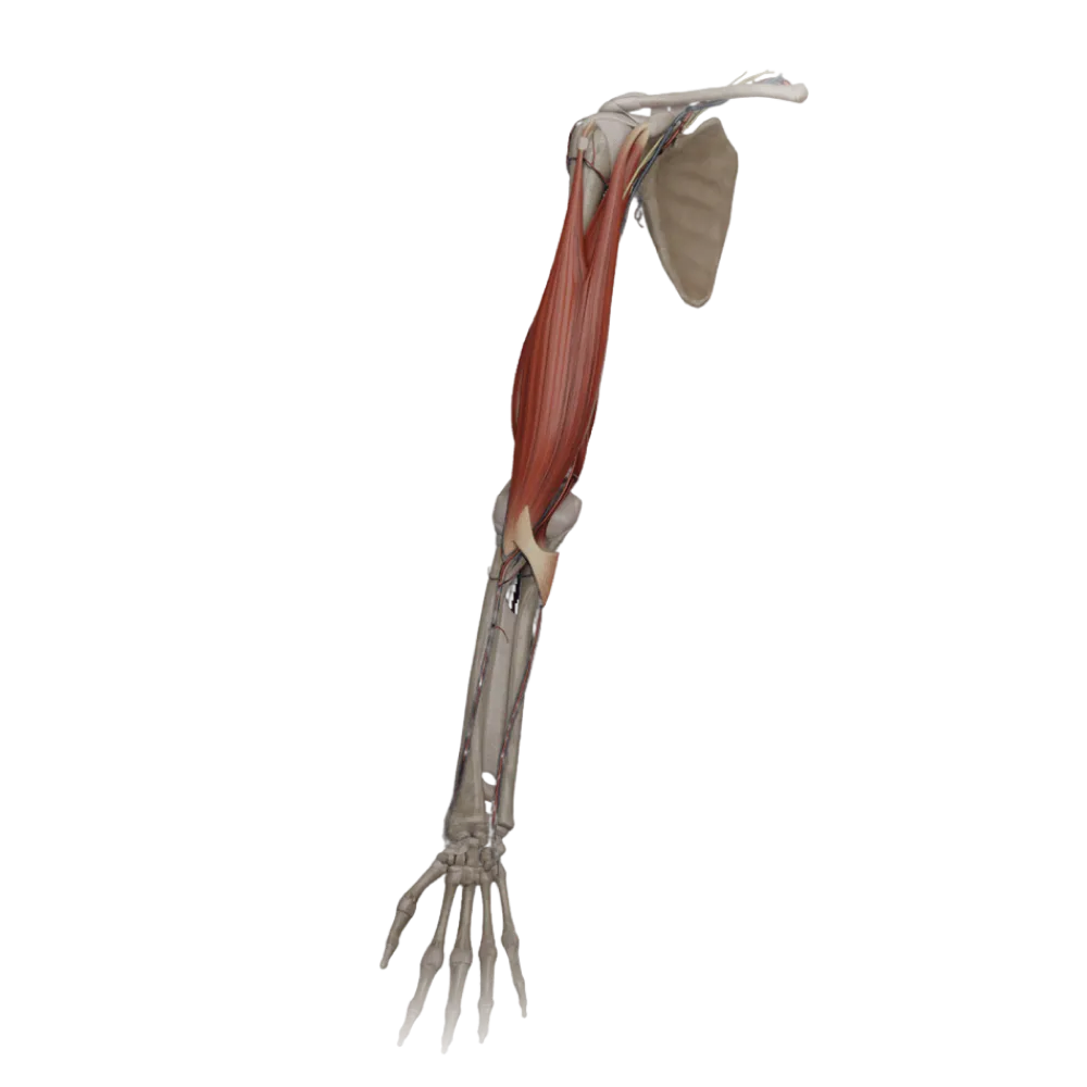
Evaluate the knowledge of upper limb muscle anatomy. The test assesses the topography, functions, innervation features, and boundaries of muscle channels.
1. Which muscle is not part of the rotator cuff of the shoulder?
-
Supraspinatus muscle
The teres major muscle is not part of the rotator cuff, which includes the supraspinatus, infraspinatus, teres minor, and subscapularis muscles.
-
Infraspinatus muscle
The teres major muscle is not part of the rotator cuff, which includes the supraspinatus, infraspinatus, teres minor, and subscapularis muscles.
-
Teres major muscle
The teres major muscle is not part of the rotator cuff, which includes the supraspinatus, infraspinatus, teres minor, and subscapularis muscles.
-
Subscapular muscle
The teres major muscle is not part of the rotator cuff, which includes the supraspinatus, infraspinatus, teres minor, and subscapularis muscles.
-
I find it difficult to answer
The teres major muscle is not part of the rotator cuff, which includes the supraspinatus, infraspinatus, teres minor, and subscapularis muscles.
2. Which nerve innervates the deltoid muscle?
-
Musculocutaneous nerve
The deltoid muscle is innervated by the axillary nerve (n. axillaris), which passes through the quadrangular space.
-
Axillary nerve
The deltoid muscle is innervated by the axillary nerve (n. axillaris), which passes through the quadrangular space.
-
Radial nerve
The deltoid muscle is innervated by the axillary nerve (n. axillaris), which passes through the quadrangular space.
-
Median nerve
The deltoid muscle is innervated by the axillary nerve (n. axillaris), which passes through the quadrangular space.
-
I find it difficult to answer
The deltoid muscle is innervated by the axillary nerve (n. axillaris), which passes through the quadrangular space.
3. Which muscle attaches to the coracoid process of the scapula?
-
Pectoralis minor muscle
Pectoralis minor muscle (m. pectoralis minor) originates from the 3rd to the 5th ribs and attaches to the coracoid process of the scapula.
-
Pectoralis major muscle
Pectoralis minor muscle (m. pectoralis minor) originates from the 3rd to the 5th ribs and attaches to the coracoid process of the scapula.
-
Subclavius muscle
Pectoralis minor muscle (m. pectoralis minor) originates from the 3rd to the 5th ribs and attaches to the coracoid process of the scapula.
-
Serratus anterior muscle
Pectoralis minor muscle (m. pectoralis minor) originates from the 3rd to the 5th ribs and attaches to the coracoid process of the scapula.
-
I find it difficult to answer
Pectoralis minor muscle (m. pectoralis minor) originates from the 3rd to the 5th ribs and attaches to the coracoid process of the scapula.
4. What forms the lateral boundary of the cubital fossa?
-
Pronator teres
The lateral border of the cubital fossa is formed by the brachioradialis muscle (m. brachioradialis), while the medial border is formed by the pronator teres.
-
By the brachialis muscle
The lateral border of the cubital fossa is formed by the brachioradialis muscle (m. brachioradialis), while the medial border is formed by the pronator teres.
-
By the brachioradialis muscle
The lateral border of the cubital fossa is formed by the brachioradialis muscle (m. brachioradialis), while the medial border is formed by the pronator teres.
-
By the biceps brachii muscle
The lateral border of the cubital fossa is formed by the brachioradialis muscle (m. brachioradialis), while the medial border is formed by the pronator teres.
-
I find it difficult to answer
The lateral border of the cubital fossa is formed by the brachioradialis muscle (m. brachioradialis), while the medial border is formed by the pronator teres.
5. Which muscle is the main supinator of the forearm when the elbow is extended?
-
Biceps brachii
When the elbow is extended, the primary supinator is the supinator muscle, whereas the biceps brachii is most effective when the elbow is flexed.
-
Supinator muscle
When the elbow is extended, the primary supinator is the supinator muscle, whereas the biceps brachii is most effective when the elbow is flexed.
-
Brachioradialis muscle
When the elbow is extended, the primary supinator is the supinator muscle, whereas the biceps brachii is most effective when the elbow is flexed.
-
Pronator teres
When the elbow is extended, the primary supinator is the supinator muscle, whereas the biceps brachii is most effective when the elbow is flexed.
-
I find it difficult to answer
When the elbow is extended, the primary supinator is the supinator muscle, whereas the biceps brachii is most effective when the elbow is flexed.
6. Which nerve passes between the heads of the flexor carpi ulnaris?
-
Median nerve
The ulnar nerve (n. ulnaris) passes on the forearm between the humeral and ulnar heads of the flexor carpi ulnaris.
-
Ulnar nerve
The ulnar nerve (n. ulnaris) passes on the forearm between the humeral and ulnar heads of the flexor carpi ulnaris.
-
Radial nerve
The ulnar nerve (n. ulnaris) passes on the forearm between the humeral and ulnar heads of the flexor carpi ulnaris.
-
Musculocutaneous nerve
The ulnar nerve (n. ulnaris) passes on the forearm between the humeral and ulnar heads of the flexor carpi ulnaris.
-
I find it difficult to answer
The ulnar nerve (n. ulnaris) passes on the forearm between the humeral and ulnar heads of the flexor carpi ulnaris.
7. Which muscle flexes the distal phalanges of the II-V fingers?
-
Flexor digitorum superficialis
The flexor digitorum profundus (m. flexor digitorum profundus) attaches to the distal phalanges, facilitating their flexion.
-
Flexor digitorum profundus
The flexor digitorum profundus (m. flexor digitorum profundus) attaches to the distal phalanges, facilitating their flexion.
-
Lumbrical muscles
The flexor digitorum profundus (m. flexor digitorum profundus) attaches to the distal phalanges, facilitating their flexion.
-
Palmar interossei muscles
The flexor digitorum profundus (m. flexor digitorum profundus) attaches to the distal phalanges, facilitating their flexion.
-
I find it difficult to answer
The flexor digitorum profundus (m. flexor digitorum profundus) attaches to the distal phalanges, facilitating their flexion.
8. Where is the attachment site of the latissimus dorsi muscle on the humerus?
-
Crest of the greater tubercle
The latissimus dorsi muscle attaches to the crest of the lesser tubercle (crista tuberculi minoris) of the humerus.
-
Crest of the lesser tubercle
The latissimus dorsi muscle attaches to the crest of the lesser tubercle (crista tuberculi minoris) of the humerus.
-
Deltoid tuberosity
The latissimus dorsi muscle attaches to the crest of the lesser tubercle (crista tuberculi minoris) of the humerus.
-
Coronoid fossa
The latissimus dorsi muscle attaches to the crest of the lesser tubercle (crista tuberculi minoris) of the humerus.
-
I find it difficult to answer
The latissimus dorsi muscle attaches to the crest of the lesser tubercle (crista tuberculi minoris) of the humerus.
9. Which of the listed muscles in the hypothenar eminence is innervated by the median nerve?
-
Abductor digiti minimi muscle
All hypothenar muscles are innervated by the deep branch of the ulnar nerve.
-
Flexor digiti minimi brevis
All hypothenar muscles are innervated by the deep branch of the ulnar nerve.
-
Opponens digiti minimi muscle
All hypothenar muscles are innervated by the deep branch of the ulnar nerve.
-
None of the above
All hypothenar muscles are innervated by the deep branch of the ulnar nerve.
-
I find it difficult to answer
All hypothenar muscles are innervated by the deep branch of the ulnar nerve.
10. How many palmar interossei muscles are found in the human hand?
-
Two.
There are three palmar interossei muscles in the hand that adduct the II, IV, and V fingers toward the middle (III) finger.
-
Three.
There are three palmar interossei muscles in the hand that adduct the II, IV, and V fingers toward the middle (III) finger.
-
Four.
There are three palmar interossei muscles in the hand that adduct the II, IV, and V fingers toward the middle (III) finger.
-
Five
There are three palmar interossei muscles in the hand that adduct the II, IV, and V fingers toward the middle (III) finger.
-
I find it difficult to answer
There are three palmar interossei muscles in the hand that adduct the II, IV, and V fingers toward the middle (III) finger.
11. What limits the upper part of the three-sided opening of the axillary space?
-
By the teres major muscle
The upper aspect of the triangle is bounded by the teres minor (conceptual boundary) or the subscapularis muscle.
-
By the teres minor muscle
The upper aspect of the triangle is bounded by the teres minor (conceptual boundary) or the subscapularis muscle.
-
By the long head of the triceps
The upper aspect of the triangle is bounded by the teres minor (conceptual boundary) or the subscapularis muscle.
-
By the surgical neck of the humerus
The upper aspect of the triangle is bounded by the teres minor (conceptual boundary) or the subscapularis muscle.
-
I find it difficult to answer
The upper aspect of the triangle is bounded by the teres minor (conceptual boundary) or the subscapularis muscle.
12. Which muscle does not originate from the lateral epicondyle of the humerus?
-
Extensor carpi radialis longus
The pronator teres originates from the medial epicondyle of the humerus, whereas the other listed muscles originate from the lateral epicondyle.
-
Extensor carpi radialis brevis
The pronator teres originates from the medial epicondyle of the humerus, whereas the other listed muscles originate from the lateral epicondyle.
-
Extensor digitorum
The pronator teres originates from the medial epicondyle of the humerus, whereas the other listed muscles originate from the lateral epicondyle.
-
Pronator teres
The pronator teres originates from the medial epicondyle of the humerus, whereas the other listed muscles originate from the lateral epicondyle.
-
I find it difficult to answer
The pronator teres originates from the medial epicondyle of the humerus, whereas the other listed muscles originate from the lateral epicondyle.
13. What structure forms the medial border of the anatomical snuffbox?
-
Tendon of the abductor pollicis longus muscle
The medial (ulnar) border of the anatomical snuffbox is formed by the tendon of the extensor pollicis longus (m. extensor pollicis longus).
-
Tendon of the extensor pollicis brevis muscle
The medial (ulnar) border of the anatomical snuffbox is formed by the tendon of the extensor pollicis longus (m. extensor pollicis longus).
-
Tendon of the extensor pollicis longus muscle
The medial (ulnar) border of the anatomical snuffbox is formed by the tendon of the extensor pollicis longus (m. extensor pollicis longus).
-
Styloid process of the radius
The medial (ulnar) border of the anatomical snuffbox is formed by the tendon of the extensor pollicis longus (m. extensor pollicis longus).
-
I find it difficult to answer
The medial (ulnar) border of the anatomical snuffbox is formed by the tendon of the extensor pollicis longus (m. extensor pollicis longus).
14. What function do the dorsal interossei of the hand perform?
-
Flexion in the interphalangeal joints
The dorsal interossei muscles (mm. interossei dorsales) abduct (spread) the II and IV fingers from the middle (III) finger.
-
Finger adduction
The dorsal interossei muscles (mm. interossei dorsales) abduct (spread) the II and IV fingers from the middle (III) finger.
-
Finger abduction
The dorsal interossei muscles (mm. interossei dorsales) abduct (spread) the II and IV fingers from the middle (III) finger.
-
Thumb opposition
The dorsal interossei muscles (mm. interossei dorsales) abduct (spread) the II and IV fingers from the middle (III) finger.
-
I find it difficult to answer
The dorsal interossei muscles (mm. interossei dorsales) abduct (spread) the II and IV fingers from the middle (III) finger.
15. Which nerve pierces the coracobrachialis muscle?
-
Axillary nerve
The musculocutaneous nerve (n. musculocutaneus) typically pierces the coracobrachialis muscle (m. coracobrachialis) in the arm.
-
Musculocutaneous nerve
The musculocutaneous nerve (n. musculocutaneus) typically pierces the coracobrachialis muscle (m. coracobrachialis) in the arm.
-
Median nerve
The musculocutaneous nerve (n. musculocutaneus) typically pierces the coracobrachialis muscle (m. coracobrachialis) in the arm.
-
Ulnar nerve
The musculocutaneous nerve (n. musculocutaneus) typically pierces the coracobrachialis muscle (m. coracobrachialis) in the arm.
-
I find it difficult to answer
The musculocutaneous nerve (n. musculocutaneus) typically pierces the coracobrachialis muscle (m. coracobrachialis) in the arm.
16. Where is Guyon's canal located?
-
In the area of the elbow joint
The ulnar canal (Guyon's canal) is located medially, between the pisiform and the hook of the hamate bone; it contains the ulnar neurovascular bundle.
-
At the level of the carpal ligament on the radial side
The ulnar canal (Guyon's canal) is located medially, between the pisiform and the hook of the hamate bone; it contains the ulnar neurovascular bundle.
-
Between the pisiform and the hook of the hamate bone
The ulnar canal (Guyon's canal) is located medially, between the pisiform and the hook of the hamate bone; it contains the ulnar neurovascular bundle.
-
In the area of the surgical neck of the humerus
The ulnar canal (Guyon's canal) is located medially, between the pisiform and the hook of the hamate bone; it contains the ulnar neurovascular bundle.
-
I find it difficult to answer
The ulnar canal (Guyon's canal) is located medially, between the pisiform and the hook of the hamate bone; it contains the ulnar neurovascular bundle.
17. Which forearm muscle is not innervated by the radial nerve?
-
Brachioradialis muscle
The flexor carpi ulnaris (m. flexor carpi ulnaris) is innervated by the ulnar nerve, whereas the muscles of the posterior forearm group and the brachioradialis muscle are innervated by the radial nerve.
-
Extensor carpi radialis longus
The flexor carpi ulnaris (m. flexor carpi ulnaris) is innervated by the ulnar nerve, whereas the muscles of the posterior forearm group and the brachioradialis muscle are innervated by the radial nerve.
-
Flexor carpi ulnaris
The flexor carpi ulnaris (m. flexor carpi ulnaris) is innervated by the ulnar nerve, whereas the muscles of the posterior forearm group and the brachioradialis muscle are innervated by the radial nerve.
-
Supinator muscle
The flexor carpi ulnaris (m. flexor carpi ulnaris) is innervated by the ulnar nerve, whereas the muscles of the posterior forearm group and the brachioradialis muscle are innervated by the radial nerve.
-
I find it difficult to answer
The flexor carpi ulnaris (m. flexor carpi ulnaris) is innervated by the ulnar nerve, whereas the muscles of the posterior forearm group and the brachioradialis muscle are innervated by the radial nerve.
18. To which finger does the tendon of the extensor carpi ulnaris attach?
-
To the base of the second metacarpal bone
The tendon of the extensor carpi ulnaris (m. extensor carpi ulnaris) attaches to the base of the fifth metacarpal bone.
-
To the base of the third metacarpal bone
The tendon of the extensor carpi ulnaris (m. extensor carpi ulnaris) attaches to the base of the fifth metacarpal bone.
-
To the base of the fourth metacarpal bone
The tendon of the extensor carpi ulnaris (m. extensor carpi ulnaris) attaches to the base of the fifth metacarpal bone.
-
To the base of the fifth metacarpal bone
The tendon of the extensor carpi ulnaris (m. extensor carpi ulnaris) attaches to the base of the fifth metacarpal bone.
-
I find it difficult to answer
The tendon of the extensor carpi ulnaris (m. extensor carpi ulnaris) attaches to the base of the fifth metacarpal bone.
19. Which tendon passes through the glenohumeral joint cavity?
-
Short head of the biceps brachii
The tendon of the long head of the biceps brachii (caput longum m. bicipitis brachii) passes within the glenohumeral joint cavity, above the head of the humerus.
-
Long head of the biceps brachii
The tendon of the long head of the biceps brachii (caput longum m. bicipitis brachii) passes within the glenohumeral joint cavity, above the head of the humerus.
-
Coracobrachialis muscle
The tendon of the long head of the biceps brachii (caput longum m. bicipitis brachii) passes within the glenohumeral joint cavity, above the head of the humerus.
-
Subscapularis muscle
The tendon of the long head of the biceps brachii (caput longum m. bicipitis brachii) passes within the glenohumeral joint cavity, above the head of the humerus.
-
I find it difficult to answer
The tendon of the long head of the biceps brachii (caput longum m. bicipitis brachii) passes within the glenohumeral joint cavity, above the head of the humerus.
20. What is the main source of blood supply to the superficial palmar arch?
-
Radial artery
The superficial palmar arch (arcus palmaris superficialis) is primarily formed by the ulnar artery with participation from the superficial palmar branch of the radial artery.
-
Ulnar artery
The superficial palmar arch (arcus palmaris superficialis) is primarily formed by the ulnar artery with participation from the superficial palmar branch of the radial artery.
-
Anterior interosseous artery
The superficial palmar arch (arcus palmaris superficialis) is primarily formed by the ulnar artery with participation from the superficial palmar branch of the radial artery.
-
Deep brachial artery
The superficial palmar arch (arcus palmaris superficialis) is primarily formed by the ulnar artery with participation from the superficial palmar branch of the radial artery.
-
I find it difficult to answer
The superficial palmar arch (arcus palmaris superficialis) is primarily formed by the ulnar artery with participation from the superficial palmar branch of the radial artery.
Retake this quiz?
Your current progress will be reset.