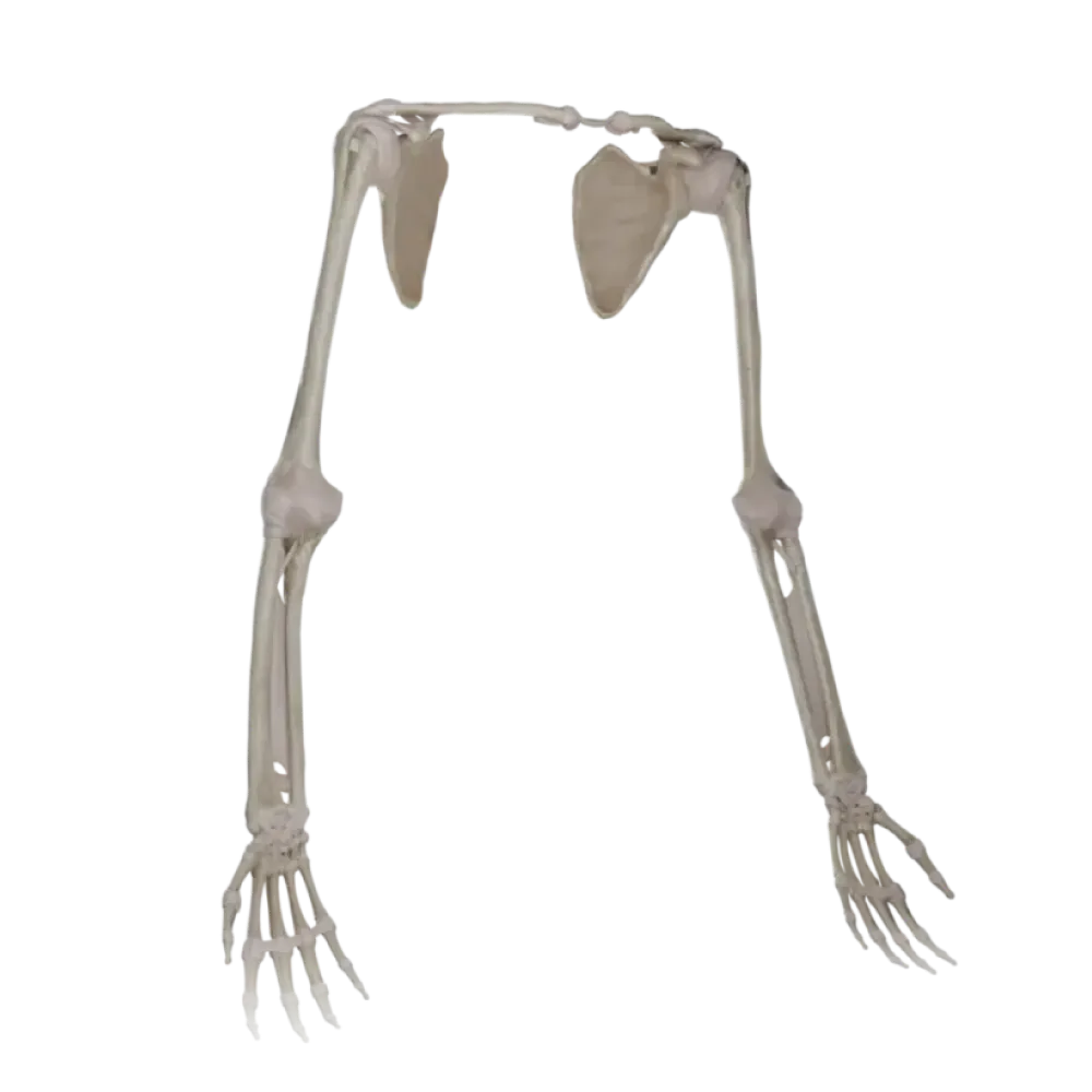

Hand joints anatomy test
Assess your knowledge of hand joints anatomy. The test examines their topography, morphology, antagonist muscles, blood supply sources, and innervation.
1/20
bold
text
1. Indicate the type of radiocarpal joint (articulatio radiocarpea) based on the articular surfaces' shape
-
Ball-and-socket
The radiocarpal joint is a complex elliptic biaxial joint.
-
Ellipsoidal
The radiocarpal joint is a complex elliptic biaxial joint.
-
Hinge
The radiocarpal joint is a complex elliptic biaxial joint.
-
Saddle-shaped
The radiocarpal joint is a complex elliptic biaxial joint.
-
I find it difficult to answer
The radiocarpal joint is a complex elliptic biaxial joint.
2. Which artery is the primary source of formation of the deep palmar arch (arcus palmaris profundus)?
-
Radial artery
The deep palmar arch is predominantly formed by the terminal portion of the radial artery.
-
Ulnar artery
The deep palmar arch is predominantly formed by the terminal portion of the radial artery.
-
A. interossea anterior
The deep palmar arch is predominantly formed by the terminal portion of the radial artery.
-
Brachial artery
The deep palmar arch is predominantly formed by the terminal portion of the radial artery.
-
I find it difficult to answer
The deep palmar arch is predominantly formed by the terminal portion of the radial artery.
3. Which nerve innervates all the palmar and dorsal interosseous muscles of the hand?
-
Median nerve
All the interosseous muscles of the hand receive innervation from the deep branch of the ulnar nerve.
-
Radial nerve
All the interosseous muscles of the hand receive innervation from the deep branch of the ulnar nerve.
-
Ulnar nerve
All the interosseous muscles of the hand receive innervation from the deep branch of the ulnar nerve.
-
Musculocutaneous nerve
All the interosseous muscles of the hand receive innervation from the deep branch of the ulnar nerve.
-
I find it difficult to answer
All the interosseous muscles of the hand receive innervation from the deep branch of the ulnar nerve.
4. Which joint of the hand is a typical saddle joint?
-
Art mediocarpal
The carpometacarpal joint of the thumb is a classic saddle joint.
-
Art metacarpophalangea pollicis
The carpometacarpal joint of the thumb is a classic saddle joint.
-
Art radiocarpal
The carpometacarpal joint of the thumb is a classic saddle joint.
-
Art carpometacarpea pollicis
The carpometacarpal joint of the thumb is a classic saddle joint.
-
I find it difficult to answer
The carpometacarpal joint of the thumb is a classic saddle joint.
5. Indicate the muscle responsible for flexion at the distal interphalangeal joints of the II-V fingers
-
M. flexor digitorum superficialis
The flexor digitorum profundus attaches to the distal phalanges, facilitating their flexion.
-
Mm lumbricals
The flexor digitorum profundus attaches to the distal phalanges, facilitating their flexion.
-
Flexor digitorum profundus
The flexor digitorum profundus attaches to the distal phalanges, facilitating their flexion.
-
Mm palmar interossei
The flexor digitorum profundus attaches to the distal phalanges, facilitating their flexion.
-
I find it difficult to answer
The flexor digitorum profundus attaches to the distal phalanges, facilitating their flexion.
6. Which structure forms the flexor retinaculum (retinaculum flexorum)?
-
Fascia dorsalis manus
The flexor retinaculum is a robust thickening of the fascia that retains the tendons in the carpal tunnel.
-
Aponeurosis palmaris
The flexor retinaculum is a robust thickening of the fascia that retains the tendons in the carpal tunnel.
-
Ligamentum carpi radiatum
The flexor retinaculum is a robust thickening of the fascia that retains the tendons in the carpal tunnel.
-
Thickening of the proper fascia of the forearm
The flexor retinaculum is a robust thickening of the fascia that retains the tendons in the carpal tunnel.
-
I find it difficult to answer
The flexor retinaculum is a robust thickening of the fascia that retains the tendons in the carpal tunnel.
7. Which muscles provide adduction (adductio) of the II, IV, and V fingers to the middle finger?
-
Mm dorsal interossei
The palmar interosseous muscles adduct the II, IV, and V fingers to the axial line of the hand.
-
Mm palmar interossei
The palmar interosseous muscles adduct the II, IV, and V fingers to the axial line of the hand.
-
Mm lumbricals
The palmar interosseous muscles adduct the II, IV, and V fingers to the axial line of the hand.
-
M. adductor pollicis
The palmar interosseous muscles adduct the II, IV, and V fingers to the axial line of the hand.
-
I find it difficult to answer
The palmar interosseous muscles adduct the II, IV, and V fingers to the axial line of the hand.
8. Through which structure does the median nerve (n. medianus) pass when transitioning to the palm?
-
Canalis ulnaris
The median nerve passes through the carpal canal beneath the flexor retinaculum.
-
Carpal canal
The median nerve passes through the carpal canal beneath the flexor retinaculum.
-
Canalis radialis
The median nerve passes through the carpal canal beneath the flexor retinaculum.
-
Spatium subaponeuroticum
The median nerve passes through the carpal canal beneath the flexor retinaculum.
-
I find it difficult to answer
The median nerve passes through the carpal canal beneath the flexor retinaculum.
9. Which carpal bone directly articulates with the radius?
-
Os hamatum
The scaphoid is part of the proximal row forming the head of the radiocarpal joint.
-
Os capitatum
The scaphoid is part of the proximal row forming the head of the radiocarpal joint.
-
Os scaphoideum
The scaphoid is part of the proximal row forming the head of the radiocarpal joint.
-
Os trapezoideum
The scaphoid is part of the proximal row forming the head of the radiocarpal joint.
-
I find it difficult to answer
The scaphoid is part of the proximal row forming the head of the radiocarpal joint.
10. Indicate the source of innervation of the lumbricals (mm. lumbricales) of the III and IV fingers
-
Ulnar nerve
Two lateral lumbricals are innervated by n. medianus, two medial ones by n. ulnaris
-
Radial nerve
Two lateral lumbricals are innervated by n. medianus, two medial ones by n. ulnaris
-
Musculocutaneous nerve
Two lateral lumbricals are innervated by n. medianus, two medial ones by n. ulnaris
-
Median nerve
Two lateral lumbricals are innervated by n. medianus, two medial ones by n. ulnaris
-
I find it difficult to answer
Two lateral lumbricals are innervated by n. medianus, two medial ones by n. ulnaris
11. Where is the dorsal carpal network (rete carpi dorsale) topographically located?
-
Under the skin of the dorsum of the hand
The dorsal carpal network lies on the ligaments beneath the tendons of the extensor muscles.
-
Under the extensor tendons
The dorsal carpal network lies on the ligaments beneath the tendons of the extensor muscles.
-
In the thickness of the interosseous muscles
The dorsal carpal network lies on the ligaments beneath the tendons of the extensor muscles.
-
On the palmar surface
The dorsal carpal network lies on the ligaments beneath the tendons of the extensor muscles.
-
I find it difficult to answer
The dorsal carpal network lies on the ligaments beneath the tendons of the extensor muscles.
12. Which artery primarily forms the superficial palmar arch (arcus palmaris superficialis)?
-
Radial artery
The ulnar artery, upon reaching the palm, anastomoses with a branch of the radial, forming the superficial arch.
-
Posterior interosseous artery
The ulnar artery, upon reaching the palm, anastomoses with a branch of the radial, forming the superficial arch.
-
A. princeps pollicis
The ulnar artery, upon reaching the palm, anastomoses with a branch of the radial, forming the superficial arch.
-
Ulnar artery
The ulnar artery, upon reaching the palm, anastomoses with a branch of the radial, forming the superficial arch.
-
I find it difficult to answer
The ulnar artery, upon reaching the palm, anastomoses with a branch of the radial, forming the superficial arch.
13. What movement is limited in a typical elliptical metacarpophalangeal joint?
-
Flexio
Rotational movements around the longitudinal axis in metacarpophalangeal joints are not possible.
-
Abductio
Rotational movements around the longitudinal axis in metacarpophalangeal joints are not possible.
-
Rotatio
Rotational movements around the longitudinal axis in metacarpophalangeal joints are not possible.
-
Circumductio
Rotational movements around the longitudinal axis in metacarpophalangeal joints are not possible.
-
I find it difficult to answer
Rotational movements around the longitudinal axis in metacarpophalangeal joints are not possible.
14. Which bone does the tendon of m. flexor carpi ulnaris?
-
Os scaphoideum
The tendon of the ulnar flexor of the wrist terminates on the pisiform bone.
-
Os pisiforme
The tendon of the ulnar flexor of the wrist terminates on the pisiform bone.
-
Os trapezoideum
The tendon of the ulnar flexor of the wrist terminates on the pisiform bone.
-
Os lunatum
The tendon of the ulnar flexor of the wrist terminates on the pisiform bone.
-
I find it difficult to answer
The tendon of the ulnar flexor of the wrist terminates on the pisiform bone.
15. Which nerve branches provide innervation to the skin of the palmar surface of the little finger?
-
Median nerve
The ulnar nerve is responsible for sensory innervation of the ulnar edge of the palm and 1.5 fingers.
-
Radial nerve
The ulnar nerve is responsible for sensory innervation of the ulnar edge of the palm and 1.5 fingers.
-
Ulnar nerve
The ulnar nerve is responsible for sensory innervation of the ulnar edge of the palm and 1.5 fingers.
-
N. cutaneus brachii
The ulnar nerve is responsible for sensory innervation of the ulnar edge of the palm and 1.5 fingers.
-
I find it difficult to answer
The ulnar nerve is responsible for sensory innervation of the ulnar edge of the palm and 1.5 fingers.
16. In which joint does opposition (oppositio) of the thumb occur?
-
Art carpometacarpea pollicis
The saddle shape of the carpometacarpal joint of the thumb allows for opposition.
-
Art mediocarpal
The saddle shape of the carpometacarpal joint of the thumb allows for opposition.
-
Art intercarpal
The saddle shape of the carpometacarpal joint of the thumb allows for opposition.
-
Art radiocarpal
The saddle shape of the carpometacarpal joint of the thumb allows for opposition.
-
I find it difficult to answer
The saddle shape of the carpometacarpal joint of the thumb allows for opposition.
17. Which ligaments restrict lateral movements (abduction/adduction) in interphalangeal joints?
-
Ligg palmar
Collateral ligaments fix interphalangeal joints, allowing only flexion and extension.
-
Ligg collateral
Collateral ligaments fix interphalangeal joints, allowing only flexion and extension.
-
Lig pisohamate
Collateral ligaments fix interphalangeal joints, allowing only flexion and extension.
-
Extensor retinaculum
Collateral ligaments fix interphalangeal joints, allowing only flexion and extension.
-
I find it difficult to answer
Collateral ligaments fix interphalangeal joints, allowing only flexion and extension.
18. Which arteries contribute to the formation of the palmar network of the wrist (rete carpi palmare)?
-
Only a. radialis
The palmar network is formed by the carpal branches of the radial, ulnar, and anterior interosseous arteries.
-
Only a. ulnaris
The palmar network is formed by the carpal branches of the radial, ulnar, and anterior interosseous arteries.
-
Aa. radial, ulnar, and anterior interosseous
The palmar network is formed by the carpal branches of the radial, ulnar, and anterior interosseous arteries.
-
Deep brachial artery
The palmar network is formed by the carpal branches of the radial, ulnar, and anterior interosseous arteries.
-
I find it difficult to answer
The palmar network is formed by the carpal branches of the radial, ulnar, and anterior interosseous arteries.
19. Which muscle group abducts fingers II, IV and V away from the middle finger
-
Mm palmar interossei
The dorsal interosseous muscles are abductors of the II-IV fingers of the hand.
-
Mm lumbricals
The dorsal interosseous muscles are abductors of the II-IV fingers of the hand.
-
M. opponens digiti minimi
The dorsal interosseous muscles are abductors of the II-IV fingers of the hand.
-
Mm dorsal interossei
The dorsal interosseous muscles are abductors of the II-IV fingers of the hand.
-
I find it difficult to answer
The dorsal interosseous muscles are abductors of the II-IV fingers of the hand.
20. Identify the central bone involved in the formation of the midcarpal joint (art. mediocarpea)
-
Os pisiforme
The capitate bone is the largest bone of the distal row, part of the midcarpal joint.
-
Os capitatum
The capitate bone is the largest bone of the distal row, part of the midcarpal joint.
-
Os hamatum
The capitate bone is the largest bone of the distal row, part of the midcarpal joint.
-
Os triquetrum
The capitate bone is the largest bone of the distal row, part of the midcarpal joint.
-
I find it difficult to answer
The capitate bone is the largest bone of the distal row, part of the midcarpal joint.
Retake this quiz?
Your current progress will be reset.