
Anatomy test of the radioulnar and radiocarpal joints
Assess the knowledge of the anatomy of the distal radioulnar and radiocarpal joints: their structure, ligaments, blood supply, innervation, and biomechanics.
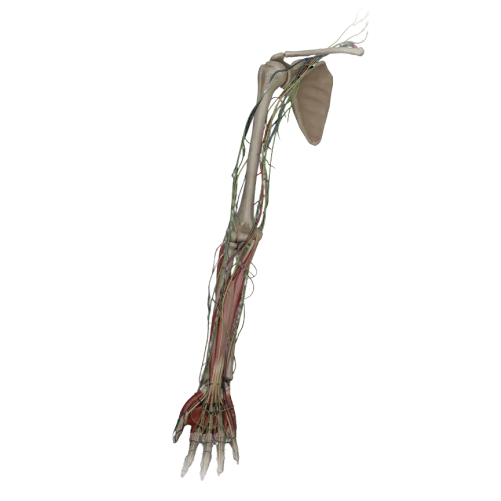
1/20
bold
text
1. Which structure supplements the articular fossa in the distal radioulnar joint?
-
Lateral meniscus
The distal radioulnar joint contains a triangular fibrocartilaginous disc, separating the ulnar head from the wrist bones.
-
Articular disc (discus articularis)
The distal radioulnar joint contains a triangular fibrocartilaginous disc, separating the ulnar head from the wrist bones.
-
Articular labrum (labrum articulare)
The distal radioulnar joint contains a triangular fibrocartilaginous disc, separating the ulnar head from the wrist bones.
-
Quadrate ligament
The distal radioulnar joint contains a triangular fibrocartilaginous disc, separating the ulnar head from the wrist bones.
-
I find it difficult to answer
The distal radioulnar joint contains a triangular fibrocartilaginous disc, separating the ulnar head from the wrist bones.
2. Identify the bone from the proximal row of the wrist that does not directly contribute to the formation of the radiocarpal joint.
-
Navicular
The pisiform bone is sesamoid and situated within the tendon of the flexor carpi ulnaris, not contacting the radius.
-
Lunate
The pisiform bone is sesamoid and situated within the tendon of the flexor carpi ulnaris, not contacting the radius.
-
Triquetral
The pisiform bone is sesamoid and situated within the tendon of the flexor carpi ulnaris, not contacting the radius.
-
Pisiform
The pisiform bone is sesamoid and situated within the tendon of the flexor carpi ulnaris, not contacting the radius.
-
I find it difficult to answer
The pisiform bone is sesamoid and situated within the tendon of the flexor carpi ulnaris, not contacting the radius.
3. Which muscle is the primary pronator in the distal radioulnar joint?
-
Quadratus pronator (m. pronator quadratus)
The quadratus pronator is located directly over the distal radioulnar joint and facilitates rotation of the radius around the ulna.
-
Teres pronator (m. pronator teres)
The quadratus pronator is located directly over the distal radioulnar joint and facilitates rotation of the radius around the ulna.
-
Brachioradialis muscle
The quadratus pronator is located directly over the distal radioulnar joint and facilitates rotation of the radius around the ulna.
-
Flexor carpi radialis
The quadratus pronator is located directly over the distal radioulnar joint and facilitates rotation of the radius around the ulna.
-
I find it difficult to answer
The quadratus pronator is located directly over the distal radioulnar joint and facilitates rotation of the radius around the ulna.
4. Through which canal does the radial artery pass when transitioning to the dorsal hand?
-
Wrist canal
The radial artery travels beneath the tendons of the abductor pollicis longus and the extensor pollicis brevis in the anatomical snuffbox region.
-
Guyon's canal
The radial artery travels beneath the tendons of the abductor pollicis longus and the extensor pollicis brevis in the anatomical snuffbox region.
-
Anatomical snuffbox
The radial artery travels beneath the tendons of the abductor pollicis longus and the extensor pollicis brevis in the anatomical snuffbox region.
-
Radial wrist canal
The radial artery travels beneath the tendons of the abductor pollicis longus and the extensor pollicis brevis in the anatomical snuffbox region.
-
I find it difficult to answer
The radial artery travels beneath the tendons of the abductor pollicis longus and the extensor pollicis brevis in the anatomical snuffbox region.
5. What type of joint by the shape of the articular surfaces is the radiocarpal joint?
-
Hinge
Articulatio radiocarpea is a typical ellipsoidal joint, allowing movement around two axes.
-
Ellipsoidal
Articulatio radiocarpea is a typical ellipsoidal joint, allowing movement around two axes.
-
Saddle-shaped
Articulatio radiocarpea is a typical ellipsoidal joint, allowing movement around two axes.
-
Ball-and-socket
Articulatio radiocarpea is a typical ellipsoidal joint, allowing movement around two axes.
-
I find it difficult to answer
Articulatio radiocarpea is a typical ellipsoidal joint, allowing movement around two axes.
6. Innervation of the m. originates from which nerve branch. extensor carpi ulnaris?
-
Deep branch of radial nerve
The extensor carpi ulnaris is innervated by the deep branch of the radial nerve (posterior interosseous nerve).
-
Median nerve
The extensor carpi ulnaris is innervated by the deep branch of the radial nerve (posterior interosseous nerve).
-
Ulnar nerve
The extensor carpi ulnaris is innervated by the deep branch of the radial nerve (posterior interosseous nerve).
-
Posterior cutaneous nerve of the forearm
The extensor carpi ulnaris is innervated by the deep branch of the radial nerve (posterior interosseous nerve).
-
I find it difficult to answer
The extensor carpi ulnaris is innervated by the deep branch of the radial nerve (posterior interosseous nerve).
7. Which ligament restricts excessive adduction (ulnar deviation) in the radiocarpal joint?
-
Palmar radiocarpal ligament
Ligamentum collaterale carpi radiale tightens with ulnar deviation of the hand, limiting this movement.
-
Dorsal radiocarpal ligament
Ligamentum collaterale carpi radiale tightens with ulnar deviation of the hand, limiting this movement.
-
Radial collateral ligament of the wrist
Ligamentum collaterale carpi radiale tightens with ulnar deviation of the hand, limiting this movement.
-
Ulnar collateral ligament of the wrist
Ligamentum collaterale carpi radiale tightens with ulnar deviation of the hand, limiting this movement.
-
I find it difficult to answer
Ligamentum collaterale carpi radiale tightens with ulnar deviation of the hand, limiting this movement.
8. Which of the vessels listed contributes to the formation of the palmar arterial carpal network (rete carpale palmare)?
-
Anterior interosseous artery
Rete carpale palmare is formed by anastomosis of the palmar carpal branches of the radial and ulnar arteries.
-
Superficial palmar arch
Rete carpale palmare is formed by anastomosis of the palmar carpal branches of the radial and ulnar arteries.
-
Dorsal interosseous artery
Rete carpale palmare is formed by anastomosis of the palmar carpal branches of the radial and ulnar arteries.
-
Palmar carpal branch of the radial artery
Rete carpale palmare is formed by anastomosis of the palmar carpal branches of the radial and ulnar arteries.
-
I find it difficult to answer
Rete carpale palmare is formed by anastomosis of the palmar carpal branches of the radial and ulnar arteries.
9. Which muscle performs simultaneous flexion and radial abduction of the hand?
-
Palmaris longus
The flexor carpi radialis causes flexion and radial deviation of the hand upon contraction.
-
M. flexor carpi radialis
The flexor carpi radialis causes flexion and radial deviation of the hand upon contraction.
-
M. flexor carpi ulnaris
The flexor carpi radialis causes flexion and radial deviation of the hand upon contraction.
-
M. abductor pollicis longus
The flexor carpi radialis causes flexion and radial deviation of the hand upon contraction.
-
I find it difficult to answer
The flexor carpi radialis causes flexion and radial deviation of the hand upon contraction.
10. Identify the source of innervation of the distal radioulnar joint capsule from the palmar aspect.
-
Anterior interosseous nerve, branch of the median nerve
The anterior interosseous nerve (branch of the median nerve) provides sensory innervation to the palmar section of the distal radioulnar joint capsule.
-
Posterior interosseous nerve
The anterior interosseous nerve (branch of the median nerve) provides sensory innervation to the palmar section of the distal radioulnar joint capsule.
-
Musculocutaneous nerve
The anterior interosseous nerve (branch of the median nerve) provides sensory innervation to the palmar section of the distal radioulnar joint capsule.
-
Ulnar nerve (dorsal ramus)
The anterior interosseous nerve (branch of the median nerve) provides sensory innervation to the palmar section of the distal radioulnar joint capsule.
-
I find it difficult to answer
The anterior interosseous nerve (branch of the median nerve) provides sensory innervation to the palmar section of the distal radioulnar joint capsule.
11. Which structures does the flexor retinaculum (retinaculum flexorum) stretch between?
-
Scaphoid and lunate bones
Medially, the retinaculum attaches to the pisiform and the hook of the hamate, forming the roof of the carpal canal.
-
Styloid processes of the radius and ulna
Medially, the retinaculum attaches to the pisiform and the hook of the hamate, forming the roof of the carpal canal.
-
Pisiform and hook of hamate (medially)
Medially, the retinaculum attaches to the pisiform and the hook of the hamate, forming the roof of the carpal canal.
-
The capitate and trapezoid bones
Medially, the retinaculum attaches to the pisiform and the hook of the hamate, forming the roof of the carpal canal.
-
I find it difficult to answer
Medially, the retinaculum attaches to the pisiform and the hook of the hamate, forming the roof of the carpal canal.
12. Which muscle is the antagonist to the flexor carpi ulnaris during sagittal plane movements?
-
M. extensor carpi ulnaris
The extensor carpi ulnaris acts as an antagonist to the flexor carpi ulnaris in flexion but as a synergist in adduction.
-
M. flexor carpi radialis
The extensor carpi ulnaris acts as an antagonist to the flexor carpi ulnaris in flexion but as a synergist in adduction.
-
Palmaris longus
The extensor carpi ulnaris acts as an antagonist to the flexor carpi ulnaris in flexion but as a synergist in adduction.
-
M. flexor digitorum superficialis
The extensor carpi ulnaris acts as an antagonist to the flexor carpi ulnaris in flexion but as a synergist in adduction.
-
I find it difficult to answer
The extensor carpi ulnaris acts as an antagonist to the flexor carpi ulnaris in flexion but as a synergist in adduction.
13. Which artery passes through Guyon's canal?
-
Radial artery
The ulnar artery, along with the ulnar nerve, traverses Guyon's canal, positioned laterally to the pisiform bone.
-
Anterior interosseous artery
The ulnar artery, along with the ulnar nerve, traverses Guyon's canal, positioned laterally to the pisiform bone.
-
Ulnar artery
The ulnar artery, along with the ulnar nerve, traverses Guyon's canal, positioned laterally to the pisiform bone.
-
Deep palmar arch
The ulnar artery, along with the ulnar nerve, traverses Guyon's canal, positioned laterally to the pisiform bone.
-
I find it difficult to answer
The ulnar artery, along with the ulnar nerve, traverses Guyon's canal, positioned laterally to the pisiform bone.
14. Which of the following is a characteristic movement in the distal radioulnar joint?
-
Flexion and extension
The distal radioulnar joint is functionally linked with the proximal one and facilitates rotational movements of the forearm.
-
Adduction and abduction
The distal radioulnar joint is functionally linked with the proximal one and facilitates rotational movements of the forearm.
-
Rotation (pronation and supination)
The distal radioulnar joint is functionally linked with the proximal one and facilitates rotational movements of the forearm.
-
Circumduction
The distal radioulnar joint is functionally linked with the proximal one and facilitates rotational movements of the forearm.
-
I find it difficult to answer
The distal radioulnar joint is functionally linked with the proximal one and facilitates rotational movements of the forearm.
15. Which ligament of the radiocarpal joint is the strongest?
-
Lig dorsal radiocarpal
The palmar radiocarpal ligament is the most robust, preventing excessive extension of the hand.
-
Lig palmar radiocarpal
The palmar radiocarpal ligament is the most robust, preventing excessive extension of the hand.
-
Lig palmar ulnocarpal
The palmar radiocarpal ligament is the most robust, preventing excessive extension of the hand.
-
Lig radiate carpus
The palmar radiocarpal ligament is the most robust, preventing excessive extension of the hand.
-
I find it difficult to answer
The palmar radiocarpal ligament is the most robust, preventing excessive extension of the hand.
16. Specify the attachment site of the extensor carpi radialis brevis tendon. To the base of the third metacarpal bone.
-
Base of the first metacarpal bone
The short radial wrist extensor attaches to the dorsal surface of the base of the third metacarpal bone.
-
Base of the second metacarpal bone
The short radial wrist extensor attaches to the dorsal surface of the base of the third metacarpal bone.
-
Base of the fourth metacarpal bone
The short radial wrist extensor attaches to the dorsal surface of the base of the third metacarpal bone.
-
Base of the third metacarpal bone
The short radial wrist extensor attaches to the dorsal surface of the base of the third metacarpal bone.
-
I find it difficult to answer
The short radial wrist extensor attaches to the dorsal surface of the base of the third metacarpal bone.
17. Which nerve innervates the m. palmaris longus?
-
Radial nerve
The long palmar muscle belongs to the superficial layer of the anterior group of forearm muscles and is innervated by the median nerve.
-
Ulnar nerve
The long palmar muscle belongs to the superficial layer of the anterior group of forearm muscles and is innervated by the median nerve.
-
Median nerve
The long palmar muscle belongs to the superficial layer of the anterior group of forearm muscles and is innervated by the median nerve.
-
Musculocutaneous nerve
The long palmar muscle belongs to the superficial layer of the anterior group of forearm muscles and is innervated by the median nerve.
-
I find it difficult to answer
The long palmar muscle belongs to the superficial layer of the anterior group of forearm muscles and is innervated by the median nerve.
18. Participating in the formation of the dorsal arterial wrist network (rete carpale dorsale) is:
-
Ramus carpeus dorsalis a. radialis
The dorsal carpal branch of the radial artery anastomoses with a similar branch of the ulnar artery and interosseous arteries.
-
A. princeps pollicis
The dorsal carpal branch of the radial artery anastomoses with a similar branch of the ulnar artery and interosseous arteries.
-
A. radialis indicis
The dorsal carpal branch of the radial artery anastomoses with a similar branch of the ulnar artery and interosseous arteries.
-
A. palmaris profunda
The dorsal carpal branch of the radial artery anastomoses with a similar branch of the ulnar artery and interosseous arteries.
-
I find it difficult to answer
The dorsal carpal branch of the radial artery anastomoses with a similar branch of the ulnar artery and interosseous arteries.
19. Around which axis does abduction and adduction of the hand occur in the radiocarpal joint?
-
Frontal
Abduction and adduction (radial and ulnar deviation) occur around the sagittal axis.
-
Vertical
Abduction and adduction (radial and ulnar deviation) occur around the sagittal axis.
-
Longitudinal
Abduction and adduction (radial and ulnar deviation) occur around the sagittal axis.
-
Sagittal
Abduction and adduction (radial and ulnar deviation) occur around the sagittal axis.
-
I find it difficult to answer
Abduction and adduction (radial and ulnar deviation) occur around the sagittal axis.
20. Specify the synergist m. flexor carpi ulnaris during wrist adduction.
-
M. flexor carpi radialis
The ulnar flexor and the ulnar extensor of the wrist act together as synergists during wrist adduction.
-
M. extensor carpi ulnaris
The ulnar flexor and the ulnar extensor of the wrist act together as synergists during wrist adduction.
-
M. extensor carpi radialis longus
The ulnar flexor and the ulnar extensor of the wrist act together as synergists during wrist adduction.
-
Pronator teres
The ulnar flexor and the ulnar extensor of the wrist act together as synergists during wrist adduction.
-
I find it difficult to answer
The ulnar flexor and the ulnar extensor of the wrist act together as synergists during wrist adduction.
Retake this quiz?
Your current progress will be reset.