
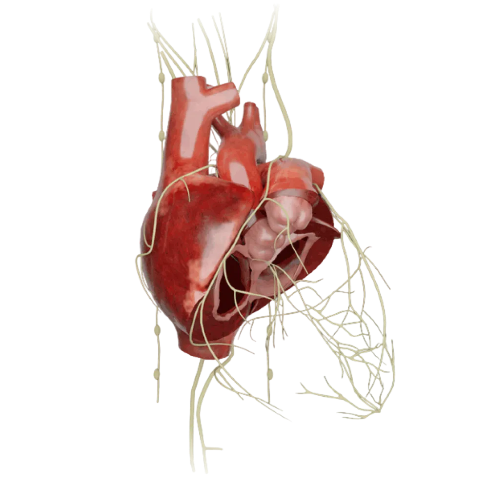
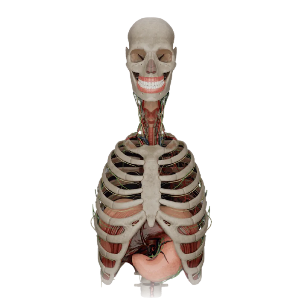

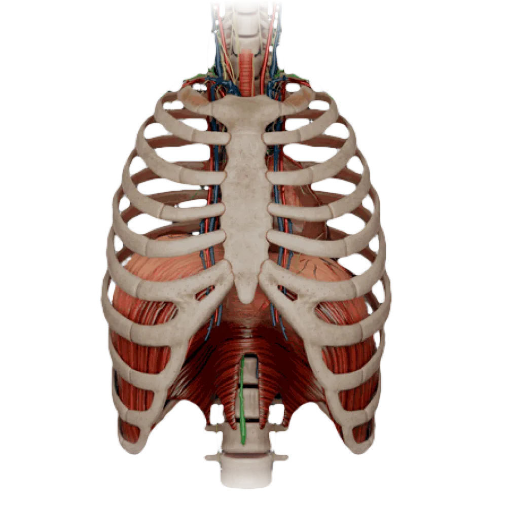

Anatomy test of the heart and pericardium
Evaluate the knowledge of heart and pericardium anatomy. The test assesses the topography, chambers, valve apparatus, blood supply, innervation, and sinuses.
1/20
bold
text
1. Where is the normal projection of the apex of the heart (apex cordis) in an adult?
-
In the fifth intercostal space, 1-1.5 cm lateral to the left midclavicular line
The apex of the heart is projected in the fifth left intercostal space, 1-1.5 cm medial to the left midclavicular line.
-
In the fifth intercostal space, 1-1.5 cm medial to the left midclavicular line
The apex of the heart is projected in the fifth left intercostal space, 1-1.5 cm medial to the left midclavicular line.
-
In the fourth intercostal space at the left parasternal line
The apex of the heart is projected in the fifth left intercostal space, 1-1.5 cm medial to the left midclavicular line.
-
In the sixth intercostal space at the left anterior axillary line
The apex of the heart is projected in the fifth left intercostal space, 1-1.5 cm medial to the left midclavicular line.
-
I find it difficult to answer
The apex of the heart is projected in the fifth left intercostal space, 1-1.5 cm medial to the left midclavicular line.
2. What blood vessels course through the anterior interventricular groove (sulcus interventricularis anterior)?
-
R. interventricularis posterior and v. cordis media
In the anterior interventricular groove run the anterior interventricular branch of the left coronary artery and the great cardiac vein.
-
A. coronaria dextra and v. cordis parva
In the anterior interventricular groove run the anterior interventricular branch of the left coronary artery and the great cardiac vein.
-
R. circumflexus and v. posterior ventriculi sinistri
In the anterior interventricular groove run the anterior interventricular branch of the left coronary artery and the great cardiac vein.
-
R. interventricularis anterior and v. cordis magna
In the anterior interventricular groove run the anterior interventricular branch of the left coronary artery and the great cardiac vein.
-
I find it difficult to answer
In the anterior interventricular groove run the anterior interventricular branch of the left coronary artery and the great cardiac vein.
3. From which artery does the branch supplying the sinuatrial node (r. nodi sinuatrialis) usually arise?
-
From the right coronary artery (a. coronaria dextra)
In 60-65% of people, the branch supplying the sinuatrial node originates from the initial portion of the right coronary artery.
-
From the circumflex branch of the left coronary artery (r. circumflexus)
In 60-65% of people, the branch supplying the sinuatrial node originates from the initial portion of the right coronary artery.
-
From the anterior interventricular branch (r. interventricularis anterior)
In 60-65% of people, the branch supplying the sinuatrial node originates from the initial portion of the right coronary artery.
-
From the posterior interventricular branch (r. interventricularis posterior)
In 60-65% of people, the branch supplying the sinuatrial node originates from the initial portion of the right coronary artery.
-
I find it difficult to answer
In 60-65% of people, the branch supplying the sinuatrial node originates from the initial portion of the right coronary artery.
4. Where does the opening of the coronary sinus (ostium sinus coronarii) open?
-
Into the superior vena cava
The coronary sinus, which collects venous blood from the majority of the heart, opens into the right atrium between the valve of the inferior vena cava and the interatrial septum.
-
Into the left atrium
The coronary sinus, which collects venous blood from the majority of the heart, opens into the right atrium between the valve of the inferior vena cava and the interatrial septum.
-
Into the right atrium
The coronary sinus, which collects venous blood from the majority of the heart, opens into the right atrium between the valve of the inferior vena cava and the interatrial septum.
-
Into the right ventricle
The coronary sinus, which collects venous blood from the majority of the heart, opens into the right atrium between the valve of the inferior vena cava and the interatrial septum.
-
I find it difficult to answer
The coronary sinus, which collects venous blood from the majority of the heart, opens into the right atrium between the valve of the inferior vena cava and the interatrial septum.
5. At what spinal level is the base of the heart (basis cordis) projected when lying down?
-
Th5-Th8
The base of the heart, mainly formed by the atria, is projected at the level of the Th5-Th8 vertebrae when lying down.
-
Th2-Th4
The base of the heart, mainly formed by the atria, is projected at the level of the Th5-Th8 vertebrae when lying down.
-
Th8-Th10
The base of the heart, mainly formed by the atria, is projected at the level of the Th5-Th8 vertebrae when lying down.
-
T1-T3
The base of the heart, mainly formed by the atria, is projected at the level of the Th5-Th8 vertebrae when lying down.
-
I find it difficult to answer
The base of the heart, mainly formed by the atria, is projected at the level of the Th5-Th8 vertebrae when lying down.
6. What is the transverse pericardial sinus (sinus transversus pericardii) bounded by anteriorly?
-
By the left atrium and the pulmonary veins
The transverse pericardial sinus is located between the ascending aorta and the pulmonary trunk anteriorly, and the anterior wall of the atria and the superior vena cava posteriorly.
-
By the ascending aorta and the pulmonary trunk
The transverse pericardial sinus is located between the ascending aorta and the pulmonary trunk anteriorly, and the anterior wall of the atria and the superior vena cava posteriorly.
-
By the superior and inferior vena cava
The transverse pericardial sinus is located between the ascending aorta and the pulmonary trunk anteriorly, and the anterior wall of the atria and the superior vena cava posteriorly.
-
By the anterior chest wall
The transverse pericardial sinus is located between the ascending aorta and the pulmonary trunk anteriorly, and the anterior wall of the atria and the superior vena cava posteriorly.
-
I find it difficult to answer
The transverse pericardial sinus is located between the ascending aorta and the pulmonary trunk anteriorly, and the anterior wall of the atria and the superior vena cava posteriorly.
7. In which anatomical structure is the His bundle trunk (fasciculus atrioventricularis) located?
-
In the wall of the right atrium between the openings of the vena cava
The trunk of the His bundle (fasciculus atrioventricularis) passes through the right fibrous triangle and is located in the membranous part of the interventricular septum.
-
In the muscular part of the interventricular septum
The trunk of the His bundle (fasciculus atrioventricularis) passes through the right fibrous triangle and is located in the membranous part of the interventricular septum.
-
In the membranous part of the interventricular septum
The trunk of the His bundle (fasciculus atrioventricularis) passes through the right fibrous triangle and is located in the membranous part of the interventricular septum.
-
At the base of the anterior papillary muscle of the right ventricle
The trunk of the His bundle (fasciculus atrioventricularis) passes through the right fibrous triangle and is located in the membranous part of the interventricular septum.
-
I find it difficult to answer
The trunk of the His bundle (fasciculus atrioventricularis) passes through the right fibrous triangle and is located in the membranous part of the interventricular septum.
8. Which artery is the primary source of blood supply to the fibrous pericardium?
-
A. musculophrenica
The primary blood supply to the pericardium is provided by the pericardiophrenic artery (a. pericardiophrenica), which is a branch of the internal thoracic artery.
-
A. thoracica interna
The primary blood supply to the pericardium is provided by the pericardiophrenic artery (a. pericardiophrenica), which is a branch of the internal thoracic artery.
-
A. epigastrica superior
The primary blood supply to the pericardium is provided by the pericardiophrenic artery (a. pericardiophrenica), which is a branch of the internal thoracic artery.
-
A. pericardiophrenica
The primary blood supply to the pericardium is provided by the pericardiophrenic artery (a. pericardiophrenica), which is a branch of the internal thoracic artery.
-
I find it difficult to answer
The primary blood supply to the pericardium is provided by the pericardiophrenic artery (a. pericardiophrenica), which is a branch of the internal thoracic artery.
9. Which artery supplies the anterior two-thirds of the interventricular septum?
-
A. coronaria dextra
The anterior two-thirds of the interventricular septum are vascularized by the branches of the anterior interventricular branch, arising from the left coronary artery.
-
R. interventricularis anterior (from a. coronaria sinistra)
The anterior two-thirds of the interventricular septum are vascularized by the branches of the anterior interventricular branch, arising from the left coronary artery.
-
R. interventricularis posterior
The anterior two-thirds of the interventricular septum are vascularized by the branches of the anterior interventricular branch, arising from the left coronary artery.
-
R. circumflexus
The anterior two-thirds of the interventricular septum are vascularized by the branches of the anterior interventricular branch, arising from the left coronary artery.
-
I find it difficult to answer
The anterior two-thirds of the interventricular septum are vascularized by the branches of the anterior interventricular branch, arising from the left coronary artery.
10. Where is the fossa ovalis located in the heart of an adult?
-
On the interatrial septum on the side of the right atrium
The fossa ovalis is a remnant of the fetal foramen ovale and is prominently expressed on the right surface of the interatrial septum (in the right atrium).
-
On the interventricular septum
The fossa ovalis is a remnant of the fetal foramen ovale and is prominently expressed on the right surface of the interatrial septum (in the right atrium).
-
In the wall of the left ventricle below the mitral valve
The fossa ovalis is a remnant of the fetal foramen ovale and is prominently expressed on the right surface of the interatrial septum (in the right atrium).
-
On the interatrial septum on the side of the left atrium
The fossa ovalis is a remnant of the fetal foramen ovale and is prominently expressed on the right surface of the interatrial septum (in the right atrium).
-
I find it difficult to answer
The fossa ovalis is a remnant of the fetal foramen ovale and is prominently expressed on the right surface of the interatrial septum (in the right atrium).
11. Which structure forms the right border of the cardiac silhouette on a chest X-ray in the anteroposterior view?
-
Right निलय
The right border of the heart, from the third to the fifth costal cartilage to the right of the sternum, is formed by the lateral edge of the right atrium.
-
Pulmonary trunk
The right border of the heart, from the third to the fifth costal cartilage to the right of the sternum, is formed by the lateral edge of the right atrium.
-
Ascending aorta
The right border of the heart, from the third to the fifth costal cartilage to the right of the sternum, is formed by the lateral edge of the right atrium.
-
Right atrium
The right border of the heart, from the third to the fifth costal cartilage to the right of the sternum, is formed by the lateral edge of the right atrium.
-
I find it difficult to answer
The right border of the heart, from the third to the fifth costal cartilage to the right of the sternum, is formed by the lateral edge of the right atrium.
12. What is understood by the topographic term 'crux cordis'?
-
The intersection of the aortic arch and the pulmonary trunk
The crux cordis is an area on the posterior (diaphragmatic) surface of the heart, where the posterior interventricular groove intersects with the coronary groove.
-
The point where the left and right coronary arteries converge
The crux cordis is an area on the posterior (diaphragmatic) surface of the heart, where the posterior interventricular groove intersects with the coronary groove.
-
The intersection of the coronary and interventricular grooves on the diaphragmatic surface
The crux cordis is an area on the posterior (diaphragmatic) surface of the heart, where the posterior interventricular groove intersects with the coronary groove.
-
Projection of the four valves of the heart onto the chest wall
The crux cordis is an area on the posterior (diaphragmatic) surface of the heart, where the posterior interventricular groove intersects with the coronary groove.
-
I find it difficult to answer
The crux cordis is an area on the posterior (diaphragmatic) surface of the heart, where the posterior interventricular groove intersects with the coronary groove.
13. Where do the postganglionic sympathetic fibers that innervate the heart (nn. cardiaci) originate?
-
From the vagus nerve
The cardiac sympathetic nerves (nn. cardiaci cervicales et thoracici) are formed by the axons of neurons of the cervical and upper thoracic ganglia of the sympathetic trunk.
-
From the celiac plexus
The cardiac sympathetic nerves (nn. cardiaci cervicales et thoracici) are formed by the axons of neurons of the cervical and upper thoracic ganglia of the sympathetic trunk.
-
From the cervical and upper thoracic ganglia of the sympathetic trunk
The cardiac sympathetic nerves (nn. cardiaci cervicales et thoracici) are formed by the axons of neurons of the cervical and upper thoracic ganglia of the sympathetic trunk.
-
Directly from the spinal nerves T1-T4
The cardiac sympathetic nerves (nn. cardiaci cervicales et thoracici) are formed by the axons of neurons of the cervical and upper thoracic ganglia of the sympathetic trunk.
-
I find it difficult to answer
The cardiac sympathetic nerves (nn. cardiaci cervicales et thoracici) are formed by the axons of neurons of the cervical and upper thoracic ganglia of the sympathetic trunk.
14. Where is the oblique pericardial sinus (sinus obliquus pericardii) located?
-
Between the ascending aorta and the pulmonary trunk
The oblique pericardial sinus is located on the posterior wall of the atria, bounded by the left and right pulmonary veins and the inferior vena cava.
-
Between the inferior vena cava and the pulmonary veins on the posterior wall of the atria
The oblique pericardial sinus is located on the posterior wall of the atria, bounded by the left and right pulmonary veins and the inferior vena cava.
-
At the transition of the parietal pericardial leaflet to the diaphragm
The oblique pericardial sinus is located on the posterior wall of the atria, bounded by the left and right pulmonary veins and the inferior vena cava.
-
Behind the right atrium along the terminal groove
The oblique pericardial sinus is located on the posterior wall of the atria, bounded by the left and right pulmonary veins and the inferior vena cava.
-
I find it difficult to answer
The oblique pericardial sinus is located on the posterior wall of the atria, bounded by the left and right pulmonary veins and the inferior vena cava.
15. What is the name of the valve located at the opening of the coronary sinus?
-
Thebesian valve (valvula sinus coronarii)
The orifice of the coronary sinus is bounded by a thin fold of the endocardium — the valve of the coronary sinus (Thebesian valve).
-
Eustachian valve (valvula venae cavae inferioris)
The orifice of the coronary sinus is bounded by a thin fold of the endocardium — the valve of the coronary sinus (Thebesian valve).
-
Arantius valve
The orifice of the coronary sinus is bounded by a thin fold of the endocardium — the valve of the coronary sinus (Thebesian valve).
-
Gürtler's valve
The orifice of the coronary sinus is bounded by a thin fold of the endocardium — the valve of the coronary sinus (Thebesian valve).
-
I find it difficult to answer
The orifice of the coronary sinus is bounded by a thin fold of the endocardium — the valve of the coronary sinus (Thebesian valve).
16. To which regional lymph nodes does lymph from the heart primarily drain?
-
Nodi lymphatici coeliaci et gastrici
Lymphatic drainage from the heart primarily occurs to the inferior tracheobronchial (bifurcation) and anterior mediastinal lymph nodes.
-
Nodi lymphatici cervicales profundi
Lymphatic drainage from the heart primarily occurs to the inferior tracheobronchial (bifurcation) and anterior mediastinal lymph nodes.
-
Nodi lymphatici axillares et pectorales
Lymphatic drainage from the heart primarily occurs to the inferior tracheobronchial (bifurcation) and anterior mediastinal lymph nodes.
-
Nodi lymphatici tracheobronchiales inferiores et mediastinales anteriores
Lymphatic drainage from the heart primarily occurs to the inferior tracheobronchial (bifurcation) and anterior mediastinal lymph nodes.
-
I find it difficult to answer
Lymphatic drainage from the heart primarily occurs to the inferior tracheobronchial (bifurcation) and anterior mediastinal lymph nodes.
17. What does the ligamentum arteriosum typically connect in an adult?
-
Ascendant aorta and the superior vena cava
The ligamentum arteriosum (obliterated ductus arteriosus Botalli) is stretched between the left pulmonary artery (or pulmonary trunk) and the concave surface of the aortic arch.
-
The site of bifurcation of the pulmonary trunk and the inferior semicircle of the aortic arch
The ligamentum arteriosum (obliterated ductus arteriosus Botalli) is stretched between the left pulmonary artery (or pulmonary trunk) and the concave surface of the aortic arch.
-
Left atrium and pulmonary trunk
The ligamentum arteriosum (obliterated ductus arteriosus Botalli) is stretched between the left pulmonary artery (or pulmonary trunk) and the concave surface of the aortic arch.
-
Aortic arch and left main bronchus
The ligamentum arteriosum (obliterated ductus arteriosus Botalli) is stretched between the left pulmonary artery (or pulmonary trunk) and the concave surface of the aortic arch.
-
I find it difficult to answer
The ligamentum arteriosum (obliterated ductus arteriosus Botalli) is stretched between the left pulmonary artery (or pulmonary trunk) and the concave surface of the aortic arch.
18. To which part of the thoracic wall does the facies sternocostalis (sternocostal surface) of the heart relate?
-
Only to the xiphoid process of the sternum
The sternocostal (anterior) surface of the heart is adjacent to the posterior surface of the sternum and the cartilages of ribs 3-6 (predominantly on the left).
-
To the cartilages of ribs I-III on the left
The sternocostal (anterior) surface of the heart is adjacent to the posterior surface of the sternum and the cartilages of ribs 3-6 (predominantly on the left).
-
To the costal arch on both the right and left
The sternocostal (anterior) surface of the heart is adjacent to the posterior surface of the sternum and the cartilages of ribs 3-6 (predominantly on the left).
-
To the sternum and the cartilages of ribs III-VI
The sternocostal (anterior) surface of the heart is adjacent to the posterior surface of the sternum and the cartilages of ribs 3-6 (predominantly on the left).
-
I find it difficult to answer
The sternocostal (anterior) surface of the heart is adjacent to the posterior surface of the sternum and the cartilages of ribs 3-6 (predominantly on the left).
19. To what are the bases of the leaflets of the right atrioventricular (tricuspid) valve attached?
-
To the papillary muscles of the right ventricle
The bases of the leaflets of all atrioventricular valves are attached to the corresponding fibrous rings (anuli fibrosi), forming the soft framework of the heart.
-
To the tendinous chords
The bases of the leaflets of all atrioventricular valves are attached to the corresponding fibrous rings (anuli fibrosi), forming the soft framework of the heart.
-
To the right fibrous ring (anulus fibrosus dexter)
The bases of the leaflets of all atrioventricular valves are attached to the corresponding fibrous rings (anuli fibrosi), forming the soft framework of the heart.
-
To the trabeculae carneae
The bases of the leaflets of all atrioventricular valves are attached to the corresponding fibrous rings (anuli fibrosi), forming the soft framework of the heart.
-
I find it difficult to answer
The bases of the leaflets of all atrioventricular valves are attached to the corresponding fibrous rings (anuli fibrosi), forming the soft framework of the heart.
20. Where is the middle cardiac vein (v. cordis media) located topographically?
-
In the posterior interventricular groove (sulcus interventricularis posterior)
The middle cardiac vein (v. cordis media) collects blood from the posterior surface of the ventricles and proceeds in the posterior interventricular groove, emptying into the coronary sinus.
-
In the anterior interventricular sulcus
The middle cardiac vein (v. cordis media) collects blood from the posterior surface of the ventricles and proceeds in the posterior interventricular groove, emptying into the coronary sinus.
-
On the anterior surface of the right ventricle
The middle cardiac vein (v. cordis media) collects blood from the posterior surface of the ventricles and proceeds in the posterior interventricular groove, emptying into the coronary sinus.
-
In the left coronary groove
The middle cardiac vein (v. cordis media) collects blood from the posterior surface of the ventricles and proceeds in the posterior interventricular groove, emptying into the coronary sinus.
-
I find it difficult to answer
The middle cardiac vein (v. cordis media) collects blood from the posterior surface of the ventricles and proceeds in the posterior interventricular groove, emptying into the coronary sinus.
Retake this quiz?
Your current progress will be reset.