
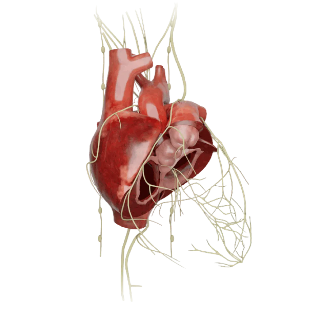


Test on the anatomy of the conducting system of heart
Evaluate the knowledge of the anatomy of the conducting system of heart. The test covers the topography of nodes, bundle of His, Purkinje fibers, their blood supply and innervation.
1/20
bold
text
1. Where is the topographic location of the sinuatrial node (nodus sinuatrialis)?
-
In the wall of the left atrium
The sinuatrial node (node of Keith and Flack) is located subepicardially in the wall of the right atrium between the opening of the superior vena cava and the right auricle.
-
In the interatrial septum above the oval fossa
The sinuatrial node (node of Keith and Flack) is located subepicardially in the wall of the right atrium between the opening of the superior vena cava and the right auricle.
-
In the area of the Koch's triangle
The sinuatrial node (node of Keith and Flack) is located subepicardially in the wall of the right atrium between the opening of the superior vena cava and the right auricle.
-
In the wall of the right atrium between the superior vena cava and the right atrial appendage
The sinuatrial node (node of Keith and Flack) is located subepicardially in the wall of the right atrium between the opening of the superior vena cava and the right auricle.
-
I find it difficult to answer
The sinuatrial node (node of Keith and Flack) is located subepicardially in the wall of the right atrium between the opening of the superior vena cava and the right auricle.
2. Which structure does NOT participate in forming the boundaries of the Koch's triangle where the atrioventricular node is located?
-
Tendon of Todaro
Boundaries of the Koch's triangle are formed by the tendon of Todaro, base of the septal leaflet of the tricuspid valve, and the coronary sinus opening. The Eustachian valve does not form its boundaries.
-
Base of the septal leaflet of the tricuspid valve
Boundaries of the Koch's triangle are formed by the tendon of Todaro, base of the septal leaflet of the tricuspid valve, and the coronary sinus opening. The Eustachian valve does not form its boundaries.
-
The Eustachian valve of the inferior vena cava (valvula venae cavae inferioris)
Boundaries of the Koch's triangle are formed by the tendon of Todaro, base of the septal leaflet of the tricuspid valve, and the coronary sinus opening. The Eustachian valve does not form its boundaries.
-
Opening of the coronary sinus
Boundaries of the Koch's triangle are formed by the tendon of Todaro, base of the septal leaflet of the tricuspid valve, and the coronary sinus opening. The Eustachian valve does not form its boundaries.
-
I find it difficult to answer
Boundaries of the Koch's triangle are formed by the tendon of Todaro, base of the septal leaflet of the tricuspid valve, and the coronary sinus opening. The Eustachian valve does not form its boundaries.
3. Which artery in most cases (approximately 60%) provides a branch to the sinoatrial node (r. nodi sinuatrialis)?
-
Right coronary artery (a. coronaria dextra)
In most individuals (around 60%), the branch to the sinoatrial node originates from the initial segment of the right coronary artery.
-
Circumflex branch (r. circumflexus)
In most individuals (around 60%), the branch to the sinoatrial node originates from the initial segment of the right coronary artery.
-
Anterior interventricular branch
In most individuals (around 60%), the branch to the sinoatrial node originates from the initial segment of the right coronary artery.
-
Posterior interventricular branch
In most individuals (around 60%), the branch to the sinoatrial node originates from the initial segment of the right coronary artery.
-
I find it difficult to answer
In most individuals (around 60%), the branch to the sinoatrial node originates from the initial segment of the right coronary artery.
4. From which blood vessel does the artery supplying the atrioventricular node (r. nodi atrioventricularis) predominantly originate?
-
From the anterior interventricular artery
The branch to the atrioventricular node originates from the right coronary artery in the region of the crux of the heart (crux cordis) in 80-90% of cases.
-
From the circumflex artery
The branch to the atrioventricular node originates from the right coronary artery in the region of the crux of the heart (crux cordis) in 80-90% of cases.
-
From the right coronary artery (in the region of the crux of the heart)
The branch to the atrioventricular node originates from the right coronary artery in the region of the crux of the heart (crux cordis) in 80-90% of cases.
-
From the left coronary artery (main stem)
The branch to the atrioventricular node originates from the right coronary artery in the region of the crux of the heart (crux cordis) in 80-90% of cases.
-
I find it difficult to answer
The branch to the atrioventricular node originates from the right coronary artery in the region of the crux of the heart (crux cordis) in 80-90% of cases.
5. Through which anatomical structure does the trunk of the bundle of His (fasciculus atrioventricularis) penetrate from the atria into the ventricles?
-
Through the left fibrous ring
The trunk of the atrioventricular bundle passes from the right atrium into the ventricles through an opening in the right fibrous triangle, part of the fibrous skeleton of the heart.
-
Through the muscular part of the interventricular septum
The trunk of the atrioventricular bundle passes from the right atrium into the ventricles through an opening in the right fibrous triangle, part of the fibrous skeleton of the heart.
-
Through the right fibrous body (trigonum fibrosum dextrum)
The trunk of the atrioventricular bundle passes from the right atrium into the ventricles through an opening in the right fibrous triangle, part of the fibrous skeleton of the heart.
-
Through the tendinous center of the diaphragm
The trunk of the atrioventricular bundle passes from the right atrium into the ventricles through an opening in the right fibrous triangle, part of the fibrous skeleton of the heart.
-
I find it difficult to answer
The trunk of the atrioventricular bundle passes from the right atrium into the ventricles through an opening in the right fibrous triangle, part of the fibrous skeleton of the heart.
6. Within which anatomical structure does the part of the right ventricular trunk (crus dextrum) pass before branching into Purkinje fibers?
-
In the septomarginal trabecula (trabecula septomarginalis)
The right bundle branch of the His bundle directs towards the base of the anterior papillary muscle of the right ventricle, passing through the thickness of the septomarginal trabecula.
-
In the anterior papillary muscle of the left ventricle
The right bundle branch of the His bundle directs towards the base of the anterior papillary muscle of the right ventricle, passing through the thickness of the septomarginal trabecula.
-
In the tendinous cords of the tricuspid valve
The right bundle branch of the His bundle directs towards the base of the anterior papillary muscle of the right ventricle, passing through the thickness of the septomarginal trabecula.
-
In the wall of the pulmonary trunk
The right bundle branch of the His bundle directs towards the base of the anterior papillary muscle of the right ventricle, passing through the thickness of the septomarginal trabecula.
-
I find it difficult to answer
The right bundle branch of the His bundle directs towards the base of the anterior papillary muscle of the right ventricle, passing through the thickness of the septomarginal trabecula.
7. Which interatrial conducting bundle transmits impulses from the sinoatrial node to the left atrium?
-
Wenckebach's bundle
The Bachmann's bundle (anterior internodal tract) provides a branch to the left atrium, ensuring its synchronous contraction with the right atrium.
-
Thorel's bundle
The Bachmann's bundle (anterior internodal tract) provides a branch to the left atrium, ensuring its synchronous contraction with the right atrium.
-
Bundle of His
The Bachmann's bundle (anterior internodal tract) provides a branch to the left atrium, ensuring its synchronous contraction with the right atrium.
-
Bachmann's interatrial bundle
The Bachmann's bundle (anterior internodal tract) provides a branch to the left atrium, ensuring its synchronous contraction with the right atrium.
-
I find it difficult to answer
The Bachmann's bundle (anterior internodal tract) provides a branch to the left atrium, ensuring its synchronous contraction with the right atrium.
8. In which layer of the heart wall are the subendocardial conducting branches (Purkinje fibers) mainly located?
-
In the subepicardial adipose tissue
Purkinje fibers (rami subendocardiales) form the terminal network of the conducting system and are located in the subendocardial layer of the ventricular myocardium.
-
Between the outer and middle layers of the myocardium
Purkinje fibers (rami subendocardiales) form the terminal network of the conducting system and are located in the subendocardial layer of the ventricular myocardium.
-
Directly beneath the endocardium of the ventricles
Purkinje fibers (rami subendocardiales) form the terminal network of the conducting system and are located in the subendocardial layer of the ventricular myocardium.
-
In the parietal layer of the pericardium
Purkinje fibers (rami subendocardiales) form the terminal network of the conducting system and are located in the subendocardial layer of the ventricular myocardium.
-
I find it difficult to answer
Purkinje fibers (rami subendocardiales) form the terminal network of the conducting system and are located in the subendocardial layer of the ventricular myocardium.
9. Into which main branches (bundles) is the left bundle branch of the His (crus sinistrum) traditionally divided within the myocardium of the left ventricle?
-
Medial and lateral
The left bundle branch of the bundle of His is traditionally divided into anterior (fasiculus anterior) and posterior (fasciculus posterior) branches; some authors also distinguish a median branch.
-
Superior and inferior
The left bundle branch of the bundle of His is traditionally divided into anterior (fasiculus anterior) and posterior (fasciculus posterior) branches; some authors also distinguish a median branch.
-
Longitudinal and transverse
The left bundle branch of the bundle of His is traditionally divided into anterior (fasiculus anterior) and posterior (fasciculus posterior) branches; some authors also distinguish a median branch.
-
Anterior and posterior (sometimes a septal branch is distinguished)
The left bundle branch of the bundle of His is traditionally divided into anterior (fasiculus anterior) and posterior (fasciculus posterior) branches; some authors also distinguish a median branch.
-
I find it difficult to answer
The left bundle branch of the bundle of His is traditionally divided into anterior (fasiculus anterior) and posterior (fasciculus posterior) branches; some authors also distinguish a median branch.
10. What do the internodal tracks of Bachmann, Wenckebach, and Thorel anatomically connect?
-
Right and left atria
Anterior (Bachmann), middle (Wenckebach), and posterior (Thorel) internodal tracts conduct impulses from the sinoatrial node to the atrioventricular node.
-
Sinoatrial and atrioventricular nodes
Anterior (Bachmann), middle (Wenckebach), and posterior (Thorel) internodal tracts conduct impulses from the sinoatrial node to the atrioventricular node.
-
Atria and ventricles
Anterior (Bachmann), middle (Wenckebach), and posterior (Thorel) internodal tracts conduct impulses from the sinoatrial node to the atrioventricular node.
-
Purkinje fibers of the left and right ventricles
Anterior (Bachmann), middle (Wenckebach), and posterior (Thorel) internodal tracts conduct impulses from the sinoatrial node to the atrioventricular node.
-
I find it difficult to answer
Anterior (Bachmann), middle (Wenckebach), and posterior (Thorel) internodal tracts conduct impulses from the sinoatrial node to the atrioventricular node.
11. At what level does the bifurcation of the trunk of the bundle of His into right and left branches occur?
-
At the border of the membranous and muscular parts of the interventricular septum
The trunk of the atrioventricular bundle divides into right and left branches (crura) at the level of the transition from the membranous part of the interventricular septum to the muscular part.
-
At the heart apex area
The trunk of the atrioventricular bundle divides into right and left branches (crura) at the level of the transition from the membranous part of the interventricular septum to the muscular part.
-
At the opening of the pulmonary trunk
The trunk of the atrioventricular bundle divides into right and left branches (crura) at the level of the transition from the membranous part of the interventricular septum to the muscular part.
-
At the base of the anterior papillary muscles
The trunk of the atrioventricular bundle divides into right and left branches (crura) at the level of the transition from the membranous part of the interventricular septum to the muscular part.
-
I find it difficult to answer
The trunk of the atrioventricular bundle divides into right and left branches (crura) at the level of the transition from the membranous part of the interventricular septum to the muscular part.
12. From which nerve do parasympathetic preganglionic fibers that reach the sinuatrial node come?
-
N. phrenicus
Parasympathetic innervation of the heart's conduction system, in particular the sinoatrial node, is provided by fibers of the vagus nerve (n. vagus), predominantly the right one.
-
Sympathetic trunk
Parasympathetic innervation of the heart's conduction system, in particular the sinoatrial node, is provided by fibers of the vagus nerve (n. vagus), predominantly the right one.
-
N. vagus
Parasympathetic innervation of the heart's conduction system, in particular the sinoatrial node, is provided by fibers of the vagus nerve (n. vagus), predominantly the right one.
-
N. laryngeus recurrens
Parasympathetic innervation of the heart's conduction system, in particular the sinoatrial node, is provided by fibers of the vagus nerve (n. vagus), predominantly the right one.
-
I find it difficult to answer
Parasympathetic innervation of the heart's conduction system, in particular the sinoatrial node, is provided by fibers of the vagus nerve (n. vagus), predominantly the right one.
13. Which morphological feature of the atrioventricular node accounts for the physiological delay in impulse conduction?
-
Presence of a thick myelin sheath
The atrioventricular delay is due to thinner fibers of the AV-node than those of the working myocardium, having fewer nexus and forming a complex network, which slows down impulse conduction.
-
Absence of blood capillaries in the node
The atrioventricular delay is due to thinner fibers of the AV-node than those of the working myocardium, having fewer nexus and forming a complex network, which slows down impulse conduction.
-
Location of the node within the fibrous ring
The atrioventricular delay is due to thinner fibers of the AV-node than those of the working myocardium, having fewer nexus and forming a complex network, which slows down impulse conduction.
-
Smaller diameter of the node's muscle fibers and their intricate interweaving (maze-like structure)
The atrioventricular delay is due to thinner fibers of the AV-node than those of the working myocardium, having fewer nexus and forming a complex network, which slows down impulse conduction.
-
I find it difficult to answer
The atrioventricular delay is due to thinner fibers of the AV-node than those of the working myocardium, having fewer nexus and forming a complex network, which slows down impulse conduction.
14. Which part of the nervous system anatomically predominates in the innervation of the ventricular part of the conducting system (Purkinje fibers)?
-
Parasympathetic (fibers of the vagus nerve)
While the atrial nodes (SA and AV) are richly innervated by parasympathetic and sympathetic fibers, the ventricular system is predominantly innervated by sympathetic nerves.
-
Sympathetic (fibers from cervical and thoracic ganglia)
While the atrial nodes (SA and AV) are richly innervated by parasympathetic and sympathetic fibers, the ventricular system is predominantly innervated by sympathetic nerves.
-
Somatic (branches of intercostal nerves)
While the atrial nodes (SA and AV) are richly innervated by parasympathetic and sympathetic fibers, the ventricular system is predominantly innervated by sympathetic nerves.
-
Metasympathetic (intramural atrial ganglia)
While the atrial nodes (SA and AV) are richly innervated by parasympathetic and sympathetic fibers, the ventricular system is predominantly innervated by sympathetic nerves.
-
I find it difficult to answer
While the atrial nodes (SA and AV) are richly innervated by parasympathetic and sympathetic fibers, the ventricular system is predominantly innervated by sympathetic nerves.
15. What closely adheres to the base of the atrioventricular node (nodus atrioventricularis) in the Koch's triangle?
-
To the membrane of the oval window
The AV-node is located subendocardially in the lower part of the interatrial septum, with its base closely adhering to the right fibrous body of the heart's fibrous skeleton.
-
To the opening of the superior vena cava
The AV-node is located subendocardially in the lower part of the interatrial septum, with its base closely adhering to the right fibrous body of the heart's fibrous skeleton.
-
To the right fibrous body (trigonum fibrosum dextrum)
The AV-node is located subendocardially in the lower part of the interatrial septum, with its base closely adhering to the right fibrous body of the heart's fibrous skeleton.
-
To the left auricle
The AV-node is located subendocardially in the lower part of the interatrial septum, with its base closely adhering to the right fibrous body of the heart's fibrous skeleton.
-
I find it difficult to answer
The AV-node is located subendocardially in the lower part of the interatrial septum, with its base closely adhering to the right fibrous body of the heart's fibrous skeleton.
16. Which arteries primarily supply blood to the right bundle branch and anterior branch of the left bundle branch of the His?
-
Circumflex branch of the left coronary artery
The right bundle branch and the anterior branch of the left bundle are primarily vascularized by the septal (interventricular) branches of the anterior interventricular branch of the left coronary artery.
-
Posterior interventricular branch of the right coronary artery
The right bundle branch and the anterior branch of the left bundle are primarily vascularized by the septal (interventricular) branches of the anterior interventricular branch of the left coronary artery.
-
Right marginal artery
The right bundle branch and the anterior branch of the left bundle are primarily vascularized by the septal (interventricular) branches of the anterior interventricular branch of the left coronary artery.
-
Septal branches of the anterior interventricular artery
The right bundle branch and the anterior branch of the left bundle are primarily vascularized by the septal (interventricular) branches of the anterior interventricular branch of the left coronary artery.
-
I find it difficult to answer
The right bundle branch and the anterior branch of the left bundle are primarily vascularized by the septal (interventricular) branches of the anterior interventricular branch of the left coronary artery.
17. Where does the left bundle branch (crus sinistrum) emerge beneath the endocardium of the left ventricle?
-
Near the ostium of the pulmonary veins
The left bundle branch emerges beneath the endocardium on the left side of the interventricular septum directly under the non-coronary and right coronary leaflets of the aortic valve.
-
At the apex of the left ventricle
The left bundle branch emerges beneath the endocardium on the left side of the interventricular septum directly under the non-coronary and right coronary leaflets of the aortic valve.
-
On the posterior wall of the left atrium
The left bundle branch emerges beneath the endocardium on the left side of the interventricular septum directly under the non-coronary and right coronary leaflets of the aortic valve.
-
Beneath the base of the non-coronary and the right coronary leaflets of the aortic valve
The left bundle branch emerges beneath the endocardium on the left side of the interventricular septum directly under the non-coronary and right coronary leaflets of the aortic valve.
-
I find it difficult to answer
The left bundle branch emerges beneath the endocardium on the left side of the interventricular septum directly under the non-coronary and right coronary leaflets of the aortic valve.
18. Which element of the conduction system is the primary pacemaker, generating electrical impulses with the highest frequency under normal conditions?
-
Sinuatrial node (Keith-Flack node)
The sinuatrial node has the highest automaticity (generating 60-80 impulses per minute) and is the principal center for the initiation of electrical impulses in the heart.
-
Atrioventricular node (Aschoff-Tawara node)
The sinuatrial node has the highest automaticity (generating 60-80 impulses per minute) and is the principal center for the initiation of electrical impulses in the heart.
-
Trunk of the Bundle of His
The sinuatrial node has the highest automaticity (generating 60-80 impulses per minute) and is the principal center for the initiation of electrical impulses in the heart.
-
Purkinje fibers
The sinuatrial node has the highest automaticity (generating 60-80 impulses per minute) and is the principal center for the initiation of electrical impulses in the heart.
-
I find it difficult to answer
The sinuatrial node has the highest automaticity (generating 60-80 impulses per minute) and is the principal center for the initiation of electrical impulses in the heart.
19. What macroscopic anatomical shape does the sinuatrial node have in an adult?
-
Spindle or crescent shape
Macroscopically, the sinuatrial node is a spindle-shaped or crescent cluster of specialized cardiomyocytes, measuring 10-20 mm in length.
-
Sphere shape
Macroscopically, the sinuatrial node is a spindle-shaped or crescent cluster of specialized cardiomyocytes, measuring 10-20 mm in length.
-
Ring shape
Macroscopically, the sinuatrial node is a spindle-shaped or crescent cluster of specialized cardiomyocytes, measuring 10-20 mm in length.
-
Pyramid shape
Macroscopically, the sinuatrial node is a spindle-shaped or crescent cluster of specialized cardiomyocytes, measuring 10-20 mm in length.
-
I find it difficult to answer
Macroscopically, the sinuatrial node is a spindle-shaped or crescent cluster of specialized cardiomyocytes, measuring 10-20 mm in length.
20. How do the specialized cardiomyocytes of the conduction system transmit impulses to the contractile (working) myocardium of the ventricles?
-
Through chemical synapses with the release of acetylcholine
The electrical impulse is transmitted directly from the subendocardial conduction fibers (Purkinje fibers) to the working cardiomyocytes via gap junctions (nexuses) in intercalated discs.
-
Through gap junctions (nexuses) in intercalated discs
The electrical impulse is transmitted directly from the subendocardial conduction fibers (Purkinje fibers) to the working cardiomyocytes via gap junctions (nexuses) in intercalated discs.
-
Via the nerve endings of the sympathetic trunk
The electrical impulse is transmitted directly from the subendocardial conduction fibers (Purkinje fibers) to the working cardiomyocytes via gap junctions (nexuses) in intercalated discs.
-
Through hormone release into the interstitial fluid
The electrical impulse is transmitted directly from the subendocardial conduction fibers (Purkinje fibers) to the working cardiomyocytes via gap junctions (nexuses) in intercalated discs.
-
I find it difficult to answer
The electrical impulse is transmitted directly from the subendocardial conduction fibers (Purkinje fibers) to the working cardiomyocytes via gap junctions (nexuses) in intercalated discs.
Retake this quiz?
Your current progress will be reset.