1/20
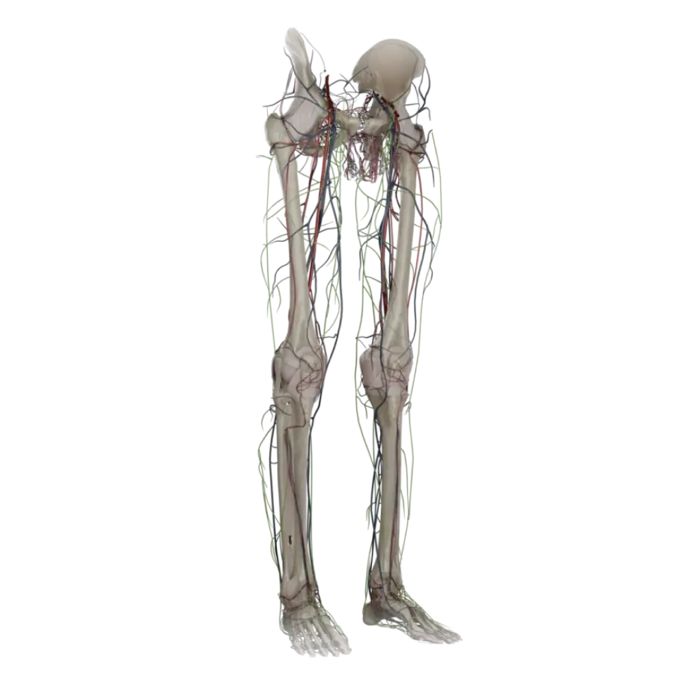


Anatomy test on the vessels of the lower limb
Evaluate the knowledge of lower limb vessel anatomy. The test strictly assesses the topography, branching, and supply areas of the arteries and veins of the legs.
1. The femoral artery (a. femoralis) is a continuation of:
-
External iliac artery
The femoral artery is the direct continuation of a. iliaca externa after it passes under the inguinal ligament through the vascular lacuna.
-
Internal iliac artery
The femoral artery is the direct continuation of a. iliaca externa after it passes under the inguinal ligament through the vascular lacuna.
-
Common iliac artery
The femoral artery is the direct continuation of a. iliaca externa after it passes under the inguinal ligament through the vascular lacuna.
-
The obturator artery
The femoral artery is the direct continuation of a. iliaca externa after it passes under the inguinal ligament through the vascular lacuna.
-
I find it difficult to answer
The femoral artery is the direct continuation of a. iliaca externa after it passes under the inguinal ligament through the vascular lacuna.
2. In the femoral triangle, the femoral artery is located:
-
Medially to the femoral vein
In the femoral triangle, the neurovascular bundle is positioned (medial to lateral): femoral vein, femoral artery, femoral nerve.
-
Posterior to the femoral vein
In the femoral triangle, the neurovascular bundle is positioned (medial to lateral): femoral vein, femoral artery, femoral nerve.
-
Laterally to the femoral vein
In the femoral triangle, the neurovascular bundle is positioned (medial to lateral): femoral vein, femoral artery, femoral nerve.
-
Anterior to the femoral vein
In the femoral triangle, the neurovascular bundle is positioned (medial to lateral): femoral vein, femoral artery, femoral nerve.
-
I find it difficult to answer
In the femoral triangle, the neurovascular bundle is positioned (medial to lateral): femoral vein, femoral artery, femoral nerve.
3. Which artery passes through the adductor canal (canalis adductorius)?
-
Deep femoral artery.
The femoral artery, together with the femoral vein and saphenous nerve, passes through the adductor canal (Hunter's canal) towards the posterior of the leg.
-
Obturator artery.
The femoral artery, together with the femoral vein and saphenous nerve, passes through the adductor canal (Hunter's canal) towards the posterior of the leg.
-
Anterior tibial artery.
The femoral artery, together with the femoral vein and saphenous nerve, passes through the adductor canal (Hunter's canal) towards the posterior of the leg.
-
Femoral artery.
The femoral artery, together with the femoral vein and saphenous nerve, passes through the adductor canal (Hunter's canal) towards the posterior of the leg.
-
I find it difficult to answer
The femoral artery, together with the femoral vein and saphenous nerve, passes through the adductor canal (Hunter's canal) towards the posterior of the leg.
4. The deep femoral artery (a. profunda femoris) typically arises from the femoral artery at a distance:
-
At the level of the inguinal ligament
A. profunda femoris branches off from the lateral circumference of the femoral artery in the femoral triangle, 3-4 cm distal to the inguinal ligament.
-
3-4 cm below the inguinal ligament
A. profunda femoris branches off from the lateral circumference of the femoral artery in the femoral triangle, 3-4 cm distal to the inguinal ligament.
-
In the adductor canal
A. profunda femoris branches off from the lateral circumference of the femoral artery in the femoral triangle, 3-4 cm distal to the inguinal ligament.
-
10 cm below the inguinal ligament
A. profunda femoris branches off from the lateral circumference of the femoral artery in the femoral triangle, 3-4 cm distal to the inguinal ligament.
-
I find it difficult to answer
A. profunda femoris branches off from the lateral circumference of the femoral artery in the femoral triangle, 3-4 cm distal to the inguinal ligament.
5. The medial femoral circumflex artery (a. circumflexa femoris medialis) predominantly supplies:
-
The anterior group of thigh muscles
The a. circumflexa femoris medialis moves medially, giving off branches to the hip joint and also to the muscles of the medial thigh group.
-
Lower leg muscles
The a. circumflexa femoris medialis moves medially, giving off branches to the hip joint and also to the muscles of the medial thigh group.
-
Knee joint.
The a. circumflexa femoris medialis moves medially, giving off branches to the hip joint and also to the muscles of the medial thigh group.
-
The hip joint and medial group of thigh muscles
The a. circumflexa femoris medialis moves medially, giving off branches to the hip joint and also to the muscles of the medial thigh group.
-
I find it difficult to answer
The a. circumflexa femoris medialis moves medially, giving off branches to the hip joint and also to the muscles of the medial thigh group.
6. The popliteal artery (a. poplitea) divides into its terminal branches (anterior and posterior tibial arteries) at the level:
-
At the upper edge of the popliteal muscle
The bifurcation of the popliteal artery occurs at a. tibialis anterior and posterior occurs at the lower edge of the popliteal muscle.
-
The knee joint gap
The bifurcation of the popliteal artery occurs at a. tibialis anterior and posterior occurs at the lower edge of the popliteal muscle.
-
At the lower edge of the popliteal muscle
The bifurcation of the popliteal artery occurs at a. tibialis anterior and posterior occurs at the lower edge of the popliteal muscle.
-
In the upper third of the lower leg
The bifurcation of the popliteal artery occurs at a. tibialis anterior and posterior occurs at the lower edge of the popliteal muscle.
-
I find it difficult to answer
The bifurcation of the popliteal artery occurs at a. tibialis anterior and posterior occurs at the lower edge of the popliteal muscle.
7. In the popliteal fossa, the topography of vessels and nerves from superficial to deep (posterior to anterior) is as follows:
-
Artery, vein, nerve
Syntopy of elements in the popliteal fossa (rule 'NeVA'): the most superficial is the tibial nerve, followed by the vein, and deepest is the artery.
-
Nerve, vein, artery
Syntopy of elements in the popliteal fossa (rule 'NeVA'): the most superficial is the tibial nerve, followed by the vein, and deepest is the artery.
-
Vein, artery, nerve
Syntopy of elements in the popliteal fossa (rule 'NeVA'): the most superficial is the tibial nerve, followed by the vein, and deepest is the artery.
-
Nerve, artery, vein
Syntopy of elements in the popliteal fossa (rule 'NeVA'): the most superficial is the tibial nerve, followed by the vein, and deepest is the artery.
-
I find it difficult to answer
Syntopy of elements in the popliteal fossa (rule 'NeVA'): the most superficial is the tibial nerve, followed by the vein, and deepest is the artery.
8. The anterior tibial artery (a. tibialis anterior) penetrates the anterior surface of the lower leg through:
-
The superior muscle-peroneal aperture
The a. tibialis anterior branches from the popliteal artery and emerges on the anterior surface of the lower leg through an opening in the upper part of the membrana interossea cruris.
-
Cruropopliteal canal
The a. tibialis anterior branches from the popliteal artery and emerges on the anterior surface of the lower leg through an opening in the upper part of the membrana interossea cruris.
-
The inferior muscle-peroneal aperture
The a. tibialis anterior branches from the popliteal artery and emerges on the anterior surface of the lower leg through an opening in the upper part of the membrana interossea cruris.
-
An opening in the upper part of the interosseous membrane
The a. tibialis anterior branches from the popliteal artery and emerges on the anterior surface of the lower leg through an opening in the upper part of the membrana interossea cruris.
-
I find it difficult to answer
The a. tibialis anterior branches from the popliteal artery and emerges on the anterior surface of the lower leg through an opening in the upper part of the membrana interossea cruris.
9. The dorsalis pedis artery (a. dorsalis pedis) is a continuation of:
-
The posterior tibial artery
The a. dorsalis pedis is a direct continuation of a. tibialis anterior after it passes under the retinacula of the extensor tendons.
-
The anterior tibial artery
The a. dorsalis pedis is a direct continuation of a. tibialis anterior after it passes under the retinacula of the extensor tendons.
-
The fibular artery
The a. dorsalis pedis is a direct continuation of a. tibialis anterior after it passes under the retinacula of the extensor tendons.
-
The medial plantar artery
The a. dorsalis pedis is a direct continuation of a. tibialis anterior after it passes under the retinacula of the extensor tendons.
-
I find it difficult to answer
The a. dorsalis pedis is a direct continuation of a. tibialis anterior after it passes under the retinacula of the extensor tendons.
10. In the crural-popliteal canal (canalis cruropopliteus) passes:
-
Anterior tibial artery.
Through the canalis cruropopliteus (Gruber's canal) pass the posterior tibial artery, accompanying veins, and the tibial nerve.
-
Popliteal artery.
Through the canalis cruropopliteus (Gruber's canal) pass the posterior tibial artery, accompanying veins, and the tibial nerve.
-
Posterior tibial artery.
Through the canalis cruropopliteus (Gruber's canal) pass the posterior tibial artery, accompanying veins, and the tibial nerve.
-
Fibular artery
Through the canalis cruropopliteus (Gruber's canal) pass the posterior tibial artery, accompanying veins, and the tibial nerve.
-
I find it difficult to answer
Through the canalis cruropopliteus (Gruber's canal) pass the posterior tibial artery, accompanying veins, and the tibial nerve.
11. The fibular artery (a. fibularis) branches from:
-
The posterior tibial artery
The a. fibularis is the largest branch of the posterior tibial artery, branching off from it in the upper third of the lower leg.
-
The anterior tibial artery
The a. fibularis is the largest branch of the posterior tibial artery, branching off from it in the upper third of the lower leg.
-
The popliteal artery
The a. fibularis is the largest branch of the posterior tibial artery, branching off from it in the upper third of the lower leg.
-
The femoral artery
The a. fibularis is the largest branch of the posterior tibial artery, branching off from it in the upper third of the lower leg.
-
I find it difficult to answer
The a. fibularis is the largest branch of the posterior tibial artery, branching off from it in the upper third of the lower leg.
12. The great saphenous vein (v. saphena magna) drains into:
-
The popliteal vein
The v. saphena magna passes through the saphenous hiatus in the femoral triangle and drains into the femoral vein.
-
The external iliac vein
The v. saphena magna passes through the saphenous hiatus in the femoral triangle and drains into the femoral vein.
-
The internal iliac vein
The v. saphena magna passes through the saphenous hiatus in the femoral triangle and drains into the femoral vein.
-
The femoral vein
The v. saphena magna passes through the saphenous hiatus in the femoral triangle and drains into the femoral vein.
-
I find it difficult to answer
The v. saphena magna passes through the saphenous hiatus in the femoral triangle and drains into the femoral vein.
13. The small saphenous vein (v. saphena parva) originates from:
-
The medial edge of the dorsal venous network of the foot
The v. saphena parva is formed from the lateral part of the rete venosum dorsale pedis and ascends behind the lateral malleolus.
-
The lateral edge of the dorsal venous network of the foot
The v. saphena parva is formed from the lateral part of the rete venosum dorsale pedis and ascends behind the lateral malleolus.
-
The plantar venous arch
The v. saphena parva is formed from the lateral part of the rete venosum dorsale pedis and ascends behind the lateral malleolus.
-
From the deep veins of the lower leg
The v. saphena parva is formed from the lateral part of the rete venosum dorsale pedis and ascends behind the lateral malleolus.
-
I find it difficult to answer
The v. saphena parva is formed from the lateral part of the rete venosum dorsale pedis and ascends behind the lateral malleolus.
14. The arterial network of the knee joint (rete articulare genus) does NOT receive branches directly from:
-
The popliteal artery
The external iliac artery does not participate in the formation of the knee joint's network; it involves branches of a. Femoralis, a. Poplitea, a. Tibialis anterior.
-
The femoral artery
The external iliac artery does not participate in the formation of the knee joint's network; it involves branches of a. Femoralis, a. Poplitea, a. Tibialis anterior.
-
The anterior tibial artery
The external iliac artery does not participate in the formation of the knee joint's network; it involves branches of a. Femoralis, a. Poplitea, a. Tibialis anterior.
-
External iliac artery
The external iliac artery does not participate in the formation of the knee joint's network; it involves branches of a. Femoralis, a. Poplitea, a. Tibialis anterior.
-
I find it difficult to answer
The external iliac artery does not participate in the formation of the knee joint's network; it involves branches of a. Femoralis, a. Poplitea, a. Tibialis anterior.
15. The medial plantar artery (a. plantaris medialis) passes on the foot in the corresponding groove along with:
-
The lateral plantar nerve.
The a. plantaris medialis topographically accompanies n. Plantaris medialis in the medial plantar groove of the foot.
-
The deep fibular nerve.
The a. plantaris medialis topographically accompanies n. Plantaris medialis in the medial plantar groove of the foot.
-
The medial plantar nerve.
The a. plantaris medialis topographically accompanies n. Plantaris medialis in the medial plantar groove of the foot.
-
The sural nerve.
The a. plantaris medialis topographically accompanies n. Plantaris medialis in the medial plantar groove of the foot.
-
I find it difficult to answer
The a. plantaris medialis topographically accompanies n. Plantaris medialis in the medial plantar groove of the foot.
16. The lateral plantar artery (a. plantaris lateralis) forms the plantar arch (arcus plantaris) by anastomosing with:
-
The deep plantar branch of the dorsalis pedis artery
The plantar arch results from the anastomosis of the terminal part of a. Plantaris lateralis with the ramus plantaris profundus of a. Dorsalis pedis.
-
The medial plantar artery
The plantar arch results from the anastomosis of the terminal part of a. Plantaris lateralis with the ramus plantaris profundus of a. Dorsalis pedis.
-
The perforating branch of the fibular artery
The plantar arch results from the anastomosis of the terminal part of a. Plantaris lateralis with the ramus plantaris profundus of a. Dorsalis pedis.
-
The posterior tibial artery
The plantar arch results from the anastomosis of the terminal part of a. Plantaris lateralis with the ramus plantaris profundus of a. Dorsalis pedis.
-
I find it difficult to answer
The plantar arch results from the anastomosis of the terminal part of a. Plantaris lateralis with the ramus plantaris profundus of a. Dorsalis pedis.
17. Perforating arteries (aa. perforantes) of the thigh originate from:
-
The femoral artery
Aa. Perforantes (usually three) originate from a. profunda femoris, penetrate the adductor muscles and supply the posterior group of the muscles of thigh.
-
The deep femoral artery
Aa. Perforantes (usually three) originate from a. profunda femoris, penetrate the adductor muscles and supply the posterior group of the muscles of thigh.
-
The obturator artery
Aa. Perforantes (usually three) originate from a. profunda femoris, penetrate the adductor muscles and supply the posterior group of the muscles of thigh.
-
The inferior gluteal artery
Aa. Perforantes (usually three) originate from a. profunda femoris, penetrate the adductor muscles and supply the posterior group of the muscles of thigh.
-
I find it difficult to answer
Aa. Perforantes (usually three) originate from a. profunda femoris, penetrate the adductor muscles and supply the posterior group of the muscles of thigh.
18. Which vein passes behind the lateral malleolus?
-
Great saphenous vein
The small saphenous vein (v. saphena parva) passes behind the lateral malleolus (malleolus lateralis), ascending along the posterior surface of the leg.
-
Anterior tibial vein
The small saphenous vein (v. saphena parva) passes behind the lateral malleolus (malleolus lateralis), ascending along the posterior surface of the leg.
-
Small saphenous vein
The small saphenous vein (v. saphena parva) passes behind the lateral malleolus (malleolus lateralis), ascending along the posterior surface of the leg.
-
Femoral vein
The small saphenous vein (v. saphena parva) passes behind the lateral malleolus (malleolus lateralis), ascending along the posterior surface of the leg.
-
I find it difficult to answer
The small saphenous vein (v. saphena parva) passes behind the lateral malleolus (malleolus lateralis), ascending along the posterior surface of the leg.
19. The deep vein of the thigh (v. profunda femoris) drains into the femoral vein, usually at the level of:
-
8-10 cm below the inguinal ligament
The deep vein of the thigh drains into v. femoralis distal to the origin of the deep femoral artery, approximately 8-10 cm below the inguinal ligament.
-
2-3 cm below the inguinal ligament
The deep vein of the thigh drains into v. femoralis distal to the origin of the deep femoral artery, approximately 8-10 cm below the inguinal ligament.
-
In the adductor canal
The deep vein of the thigh drains into v. femoralis distal to the origin of the deep femoral artery, approximately 8-10 cm below the inguinal ligament.
-
Directly under the inguinal ligament
The deep vein of the thigh drains into v. femoralis distal to the origin of the deep femoral artery, approximately 8-10 cm below the inguinal ligament.
-
I find it difficult to answer
The deep vein of the thigh drains into v. femoralis distal to the origin of the deep femoral artery, approximately 8-10 cm below the inguinal ligament.
20. The descending genicular artery (a. descendens genicularis) branches out from the femoral artery:
-
In the femoral triangle
The descending genicular artery (a. descendens genicularis) branches out from the femoral artery in the adductor canal and perforates its anterior wall (lamina vastoadductoria).
-
Under the inguinal ligament
The descending genicular artery (a. descendens genicularis) branches out from the femoral artery in the adductor canal and perforates its anterior wall (lamina vastoadductoria).
-
In the popliteal fossa
The descending genicular artery (a. descendens genicularis) branches out from the femoral artery in the adductor canal and perforates its anterior wall (lamina vastoadductoria).
-
In the adductor canal
The descending genicular artery (a. descendens genicularis) branches out from the femoral artery in the adductor canal and perforates its anterior wall (lamina vastoadductoria).
-
I find it difficult to answer
The descending genicular artery (a. descendens genicularis) branches out from the femoral artery in the adductor canal and perforates its anterior wall (lamina vastoadductoria).
Retake this quiz?
Your current progress will be reset.
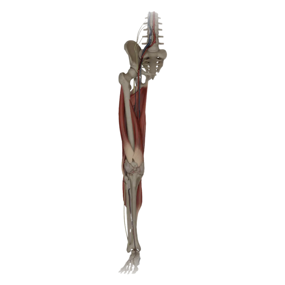
Vessels of the lower limb.
Popliteal artery.
0/20